

Three in-one fenestrated approaches of yolk-shell, silver-silica nanoparticles: A comparative study of antibacterial, antifungal and anti-cancerous applications
- PMID: 37576197
- PMCID: PMC10412894
- DOI: 10.1016/j.heliyon.2023.e18034
Three in-one fenestrated approaches of yolk-shell, silver-silica nanoparticles: A comparative study of antibacterial, antifungal and anti-cancerous applications
Abstract
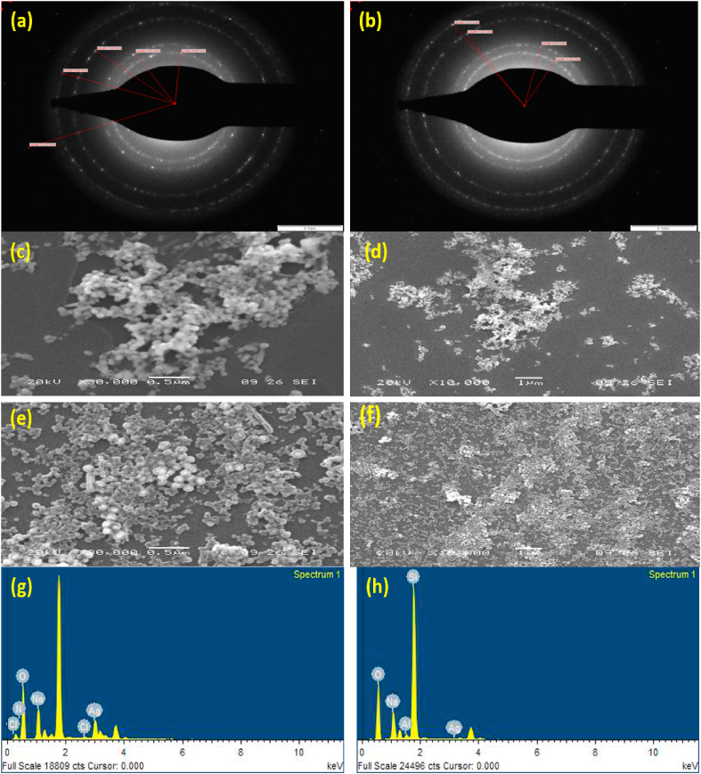
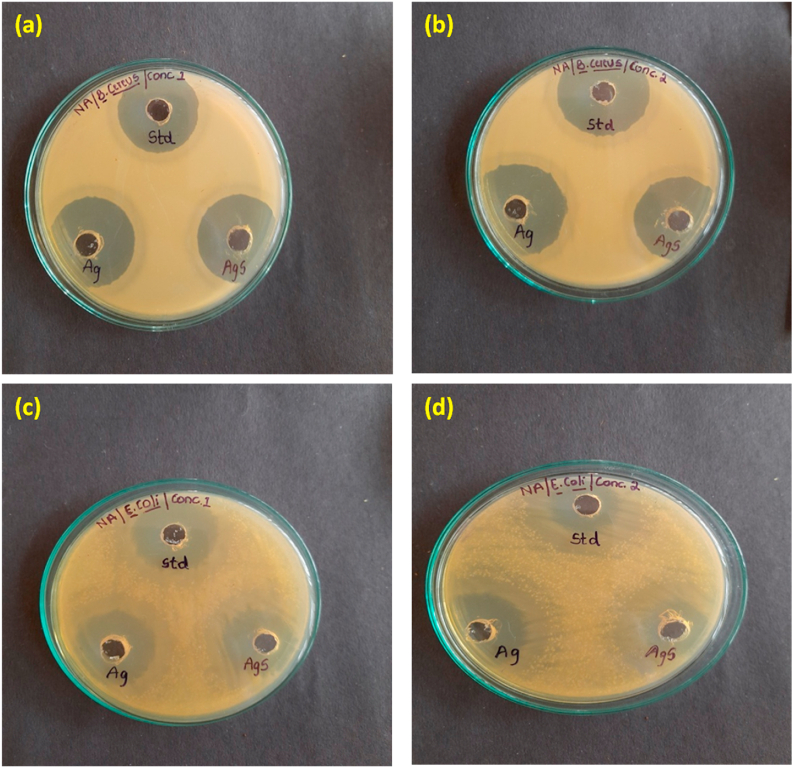
Yolk-shell-based silica-coated silver nanoparticles are prominently used in the biomedical field aas well as bare silver nanoparticles for various biological applications. The present work narrates the synthesis and silica coating of metallic silver nanoparticles and investigates their antibacterial, antifungal, and anticancerous activity. Both synthesized nanoparticles were characterized by TEM, and SEM-EDX. The average size of silver nanoparticles was 50 nm, while after coating with silica, the average size of silica-coated silver nanoparticles was 80 nm. The nanoparticles' antibacterial, antifungal, and anticancer properties were comparatively examined in vitro. Agar well diffusion method was employed to explore the antibacterial activity against gram-positive bacteria (Bacillus cereus) and gram-negative bacteria (Escherichia coli) at different concentrations and antifungal activity against Candida Albicans. To understand the minimum concentration of both nanoparticles, we employed the minimum inhibitory concentration (MIC) test, against bacterial and fungal strains, which was dose dependent. We learned that bare silver nanoparticles showed high antibacterial activity, whereas silica-coated silver nanoparticles surpassed their antifungal capability over bare silver nanoparticles against Candida albicans. The anticancer activity of the as-prepared nanoparticles was executed in opposition to the prostate cancer cell (PC-3) line by MTT assay, which showed meaningful activity. Following this, flow cytometry was also effectuated to learn about the number of apoptotic and necrotic cells. The results of this study demonstrate the dynamic anti-cancerous, antibacterial, and antifungal activities of bare silver nanoparticles and silica-coated silver nanoparticles for a long-lasting period.
Keywords: Energy-dispersive X-ray spectroscopy; MTT-assay; Minimum inhibitory concentration; Nanoparticles; Prostrate cancer.
© 2023 The Authors.
Conflict of interest statement
The authors declare that they have no known competing financial interests or personal relationships that could have appeared to influence the work reported in this paper.
Figures


Similar articles
-
Tailoring Mesoporous Silica-Coated Silver Nanoparticles and Polyurethane-Doped Films for Enhanced Antimicrobial Applications.Nanomaterials (Basel). 2024 Mar 2;14(5):462. doi: 10.3390/nano14050462. Nanomaterials (Basel). 2024. PMID: 38470791 Free PMC article.
-
Mycogenic Synthesis of Extracellular Zinc Oxide Nanoparticles from Xylaria acuta and Its Nanoantibiotic Potential.Int J Nanomedicine. 2020 Nov 2;15:8519-8536. doi: 10.2147/IJN.S271743. eCollection 2020. Int J Nanomedicine. 2020. PMID: 33173290 Free PMC article.
-
Synthesis of Functional Silver Nanoparticles and Microparticles with Modifiers and Evaluation of Their Antimicrobial, Anticancer, and Antioxidant Activity.J Funct Biomater. 2020 Oct 23;11(4):76. doi: 10.3390/jfb11040076. J Funct Biomater. 2020. PMID: 33113975 Free PMC article.
-
Green synthesis, characterization of silver nanoparticals for biomedical application and environmental remediation.J Microbiol Methods. 2022 Feb;193:106384. doi: 10.1016/j.mimet.2021.106384. Epub 2021 Nov 23. J Microbiol Methods. 2022. PMID: 34826520 Review.
-
Limitations of Recent Studies Dealing with the Antibacterial Properties of Silver Nanoparticles: Fact and Opinion.Nanomaterials (Basel). 2019 Dec 13;9(12):1775. doi: 10.3390/nano9121775. Nanomaterials (Basel). 2019. PMID: 31847133 Free PMC article. Review.
Cited by
-
Calcofluor White-Phosphatidylethanolamine Conjugate-Enhanced Ethosomal Delivery of Voriconazole for Targeting Candida albicans.Int J Nanomedicine. 2024 Dec 5;19:13047-13069. doi: 10.2147/IJN.S488456. eCollection 2024. Int J Nanomedicine. 2024. PMID: 39654804 Free PMC article.
-
Impacts of designed vanillic acid-polymer-magnetic iron oxide nanocomposite on breast cancer cells.Heliyon. 2024 Jun 12;10(12):e32863. doi: 10.1016/j.heliyon.2024.e32863. eCollection 2024 Jun 30. Heliyon. 2024. PMID: 38994094 Free PMC article.
References
-
- Tan W., Wang K., He X., Zhao X.J., Drake T., Wang L., et al. Bionanotechnology based on silica nanoparticles. Med. Res. Rev. 2004;24(5):621–638. - PubMed
-
- Dhanalekshmi K.I., Meena K.S. Comparison of antibacterial activities of Ag@TiO2 and Ag@SiO2 core-shell nanoparticles. Spectrochim. Acta Part A Mol Biomol Spectrosc. 2014;128:887–890. - PubMed
-
- Camporotondia D.E., Fogliaa M.L., Alvareza G.S., Meberta A.M., Diaza L.E., Coradinb T., et al. Antimicrobial properties of silica modified nanoparticles. Microb Pathog Strateg Combat them Sci Technol Educ. 2013;(January):283–290.
-
- Xiu Z., Zhang Q., Puppala H.L., Colvin V.L., Alvarez P.J.J. Negligible particle-specific antibacterial activity of silver nanoparticles. Nano Lett. 2012;12(8):4271–4275. - PubMed
LinkOut - more resources
Full Text Sources
