

Surface-Modified Biobased Polymeric Nanoparticles for Dual Delivery of Doxorubicin and Gefitinib in Glioma Cell Lines
- PMID: 37576633
- PMCID: PMC10413376
- DOI: 10.1021/acsomega.3c01375
Surface-Modified Biobased Polymeric Nanoparticles for Dual Delivery of Doxorubicin and Gefitinib in Glioma Cell Lines
Abstract
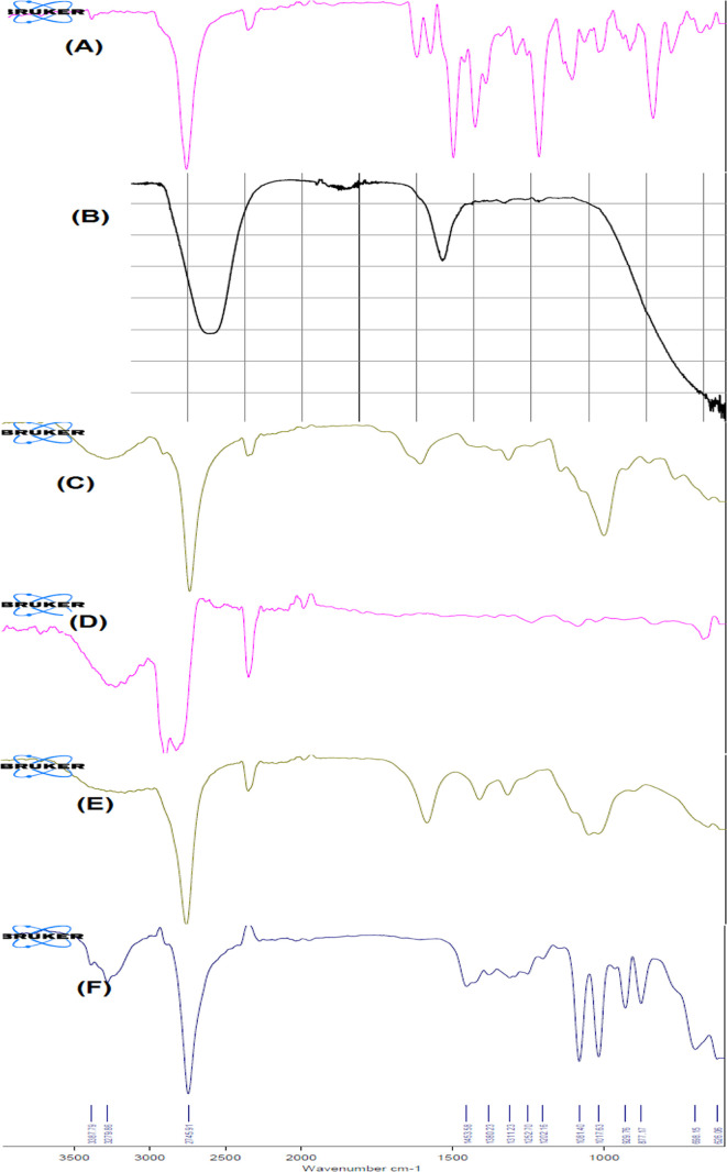
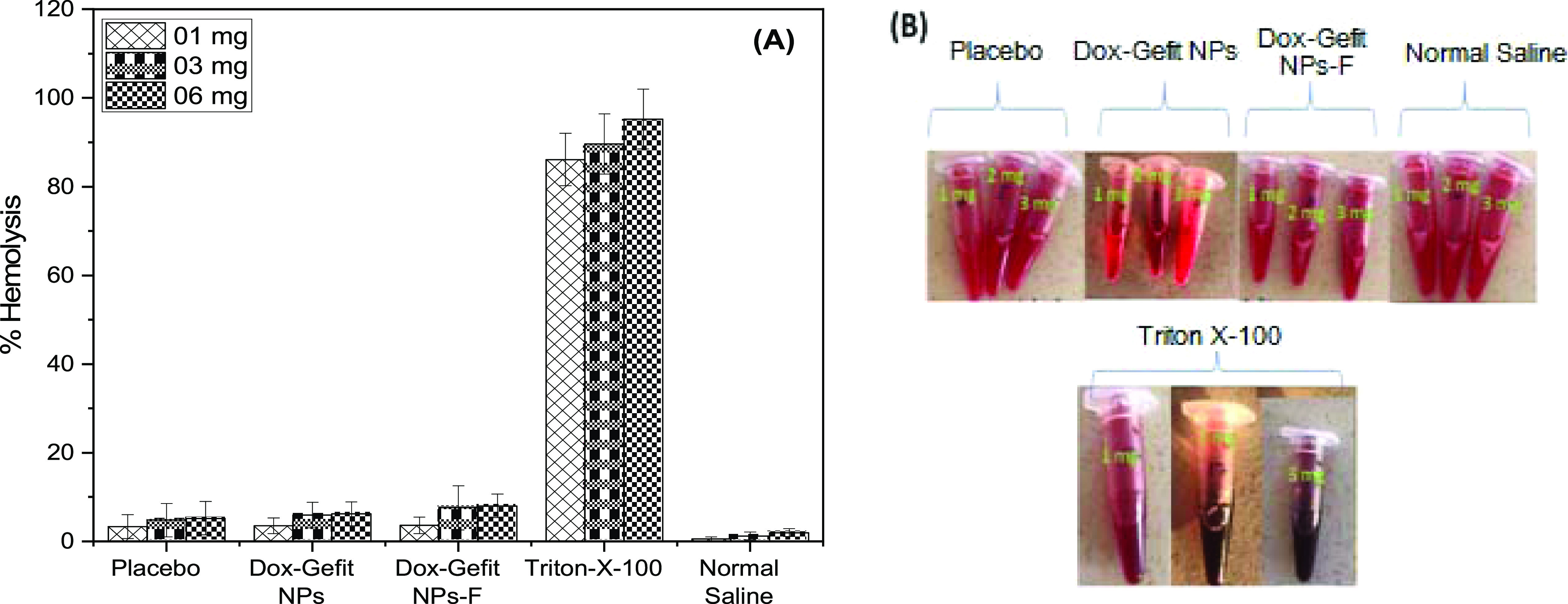
Glioma is a malignant form of brain cancer that is challenging to treat due to the progressive growth of glial cells. To target overexpressed folate receptors in glioma brain tumors, we designed and investigated doxorubicin-gefitinib nanoparticles (Dox-Gefit NPs) and folate conjugated Dox-Gefit NPs (Dox-Gefit NPs-F). Dox-Gefit NPs and Dox-Gefit NPs-F were characterized by multiple techniques including Fourier transform infrared spectroscopy (FT-IR), X-ray diffraction (XRD), differential scanning calorimetry (DSC), proton nuclear magnetic resonance (1H NMR), and transmission electron microscopy (TEM). In vitro release profiles were measured at both physiological and tumor endosomal pH. The cytotoxicity of the Dox-Gefit NP formulations was measured against C6 and U87 glioma cell lines. A hemolysis assay was performed to investigate biocompatibility of the formulations, and distribution of the drugs in different organs was also estimated. The Dox-Gefit NPs and Dox-Gefit NPs-F were 109.45 ± 7.26 and 120.35 ± 3.65 nm in size and had surface charges of -18.0 ± 3.27 and -20.0 ± 8.23 mV, respectively. Dox-Gefit NPs and Dox-Gefit NPs-F significantly reduced the growth of U87 cells, with IC50 values of 9.9 and 3.2 μM. Similarly, growth of the C6 cell line was significantly reduced, with IC50 values of 8.43 and 3.31 μM after a 24 h incubation, in Dox-Gefit NPs and Dox-Gefit NPs-F, respectively. The percentage drug releases of Dox and Gefit from Dox-Gefit NPs at pH 7.4 were 60.87 ± 0.59 and 68.23 ± 0.1%, respectively. Similarly, at pH 5.4, Dox and Gefit releases from NPs were 70.87 ± 0.28 and 69.24 ± 0.12%, respectively. Biodistribution analysis revealed that more Dox and Gefit were present in the brain than in the other organs. The functionalized NPs inhibited the growth of glioma cells due to high drug concentrations in the brain. Folate conjugated NPs of Dox-Gefit could be a treatment option in glioma therapy.
© 2023 The Authors. Published by American Chemical Society.
Conflict of interest statement
The authors declare no competing financial interest.
Figures


References
-
- Lin Y. K.; Wang S. W.; Lee R. S. Redox-responsive dasatinib-containing hyaluronic acid prodrug and co-delivery of doxorubicin for cancer therapy. Int. J. Polym. Mat. Poly. Biomat. 2021, 70, 1329–1343. 10.1080/00914037.2020.1798434. - DOI
-
- Henriksen O. M.; Del M. Á. M.; Figueiredo P.; Hangel G.; Keil V. C.; Nechifor R. E.; Riemer F.; Schmainda K. M.; Warnert E. A. H.; Wiegers E. C.; Booth T. C. High-Grade Glioma Treatment Response Monitoring Biomarkers: A Position Statement on the Evidence Supporting the Use of Advanced MRI Techniques in the Clinic, and the Latest Bench-to-Bedside Developments. Part 1: Perfusion and Diffusion Techniques. Front. Oncol. 2022, 12, 810263 10.3389/fonc.2022.810263. - DOI - PMC - PubMed
-
- Stephen J. B.; Shawn K.; Rifaquat R.; Eudocia Q.; Lee G. P.; Dunn E. G.; Susan M.; Chang L. B. N.; Manmeet S.; Ahluwalia R. S.; Minesh P.; Mehta D. A. Glioblastoma Clinical Trials: Current Landscape and Opportunities for Improvement. Clin. Cancer Res. 2022, 15, 594–602. 10.1158/1078-0432.CCR-21-2750. - DOI - PMC - PubMed
LinkOut - more resources
Full Text Sources
Miscellaneous
