Review
. 2023 Jul 21:22:100741.
doi: 10.1016/j.mtbio.2023.100741. eCollection 2023 Oct.

Microenvironment-targeted strategy steers advanced bone regeneration

Affiliations
Review

Microenvironment-targeted strategy steers advanced bone regeneration

Shuyue Hao et al. Mater Today Bio. .

Abstract

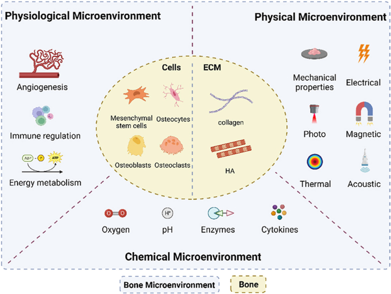
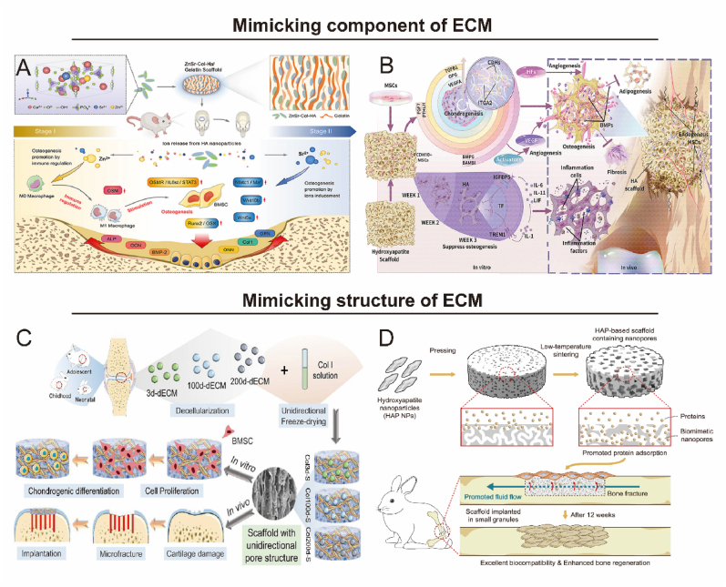
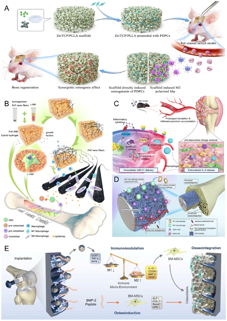
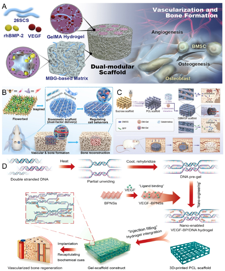
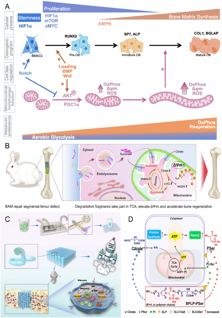
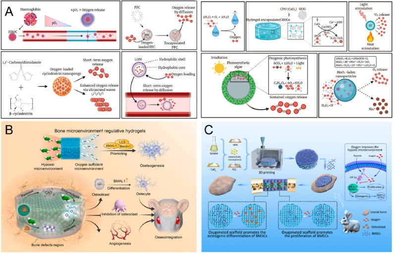
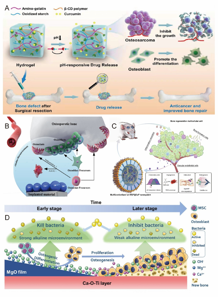
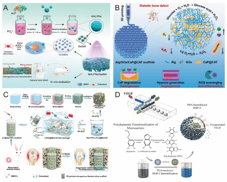
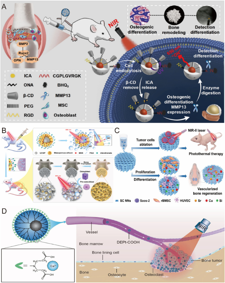
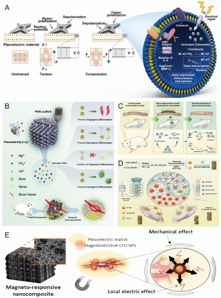
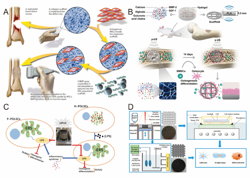
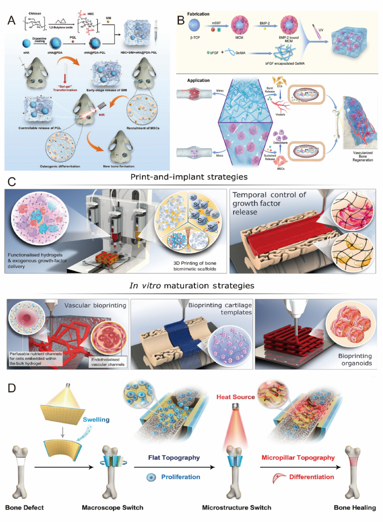
Treatment of large bone defects represents a great challenge in orthopedic and craniomaxillofacial surgery. Traditional strategies in bone tissue engineering have focused primarily on mimicking the extracellular matrix (ECM) of bone in terms of structure and composition. However, the synergistic effects of other cues from the microenvironment during bone regeneration are often neglected. The bone microenvironment is a sophisticated system that includes physiological (e.g., neighboring cells such as macrophages), chemical (e.g., oxygen, pH), and physical factors (e.g., mechanics, acoustics) that dynamically interact with each other. Microenvironment-targeted strategies are increasingly recognized as crucial for successful bone regeneration and offer promising solutions for advancing bone tissue engineering. This review provides a comprehensive overview of current microenvironment-targeted strategies and challenges for bone regeneration and further outlines prospective directions of the approaches in construction of bone organoids.

Keywords: Biomaterials; Bone regeneration; Chemical microenvironment; Physical microenvironment; Physiological microenvironment.


Conflict of interest statement

We declare that we have no financial and personal relationships with other people or organizations that can inappropriately influence our work, there is no professional or other personal interest of any nature or kind in any product, service, and/or company that could be construed as influencing the position presented in, or the review of, the manuscript entitled, “Microenvironment-Targeted Strategy Steers Advanced Bone Regeneration”.

Figures

Image 1
Graphical abstract
Fig. 1
Fig. 1
The diagram of bone tissue cell microenvironment. Bone microenvironment can be divided into three parts: physiological microenvironment, chemical microenvironment and physical microenvironment.
Fig. 2
Fig. 2
Biomaterials mimic the component and structure of ECM for bone repair. A) Collagen-HA co-assembled scaffolds loaded with immune ions are programmed to promote osteogenic gene expression in BMSCs. Reproduced and adapted with permission [25]. Copyright 2022, Elsevier. B) Bionic HA scaffold regulates the cellular activity of MSCs and enhances the ossification process to promote bone repair. Reproduced and adapted with permission [37]. Copyright 2021, Elsevier. C) Age-specific dECM provides different local environments for BMSCs and promotes microfracture repair in vivo. Reproduced and adapted with permission [40]. Copyright 2021, American Chemical Society. D) HA scaffold with natural bone-mimicking nanopores to improve bone regeneration efficiency. Reproduced and adapted with permission [41]. Copyright 2022, Springer Nature.
Fig. 3
Fig. 3
Biomaterials mimic the immune environment to promote bone repair. A) Sustained release of Zn2+ from bioactive ceramics induces M2 macrophages, further promoting periosteal-derived progenitor cells-mediated bone regeneration. Reproduced and adapted with permission [126]. Copyright 2021, Springer Nature. B) Blood-derived hybrid hydrogels promote bone healing by reprogramming the immune environment. Reproduced and adapted with permission [127]. Copyright 2022, American Chemical Society. C) Nanocomplexes deliver miR-21 and IL-4 in layers to promote macrophage polarization to the M2 phenotype. Reproduced and adapted with permission [128]. Copyright 2021, Wiley-VCH GmbH. D) Mg–Al laminated double hydroxide coating induces macrophage polarization to an M2 anti-inflammatory phenotype that exhibits good osteogenic and angiogenic potential. Reproduced and adapted with permission [129]. Copyright 2021, Elsevier. E) Dual-effect coated Ti screws modified with Zn2+ and BMP-2 co-regulate the bone immune microenvironment at the bone-implant interface. Reproduced and adapted with permission [130]. Copyright 2022, Springer Nature.
Fig. 4
Fig. 4
Biomaterials enhance angiogenesis to promote bone repair. A) 26SCS-functionalized bimodular scaffolds deliver BMP-2 and VEGF for synergistic osteogenesis and angiogenesis. Reproduced and adapted with permission [133]. Copyright 2020, Elsevier. B) 3D-printed two-factor delivery scaffolds sequentially release DMOG and Sr ions that match the angiogenic and osteogenic processes. Reproduced and adapted with permission [135]. Copyright 2023, American Chemical Society. C) Nanofibrous gelatin-silica hybrid scaffold with the spatiotemporal release of DMOG and bone-forming peptide to enhance angiogenesis and improve bone regeneration. Reproduced and adapted with permission [86]. Copyright 2022, Wiley-VCH GmbH. D) Dynamic DNA hydrogels loaded with black phosphorus nanosheets continuously release VEGF and promote mature vessel growth to induce osteogenesis. Reproduced and adapted with permission [136]. Copyright 2022, Elsevier.
Fig. 5
Fig. 5
Biomaterials improve energy metabolism for bone repair. A) Bioenergetic regulation and signaling during osteoblast differentiation. Reproduced and adapted with permission [139]. Copyright 2022, Elsevier. B) Bioenergetically active material scaffolds that accelerate bone formation by degradation-mediated increases in mitochondrial membrane potential. Reproduced and adapted with permission [87]. Copyright 2020, American Association for the Advancement of Science. C) Mg2+ energy drive improved low-dose BMP-2-induced bone regeneration. Reproduced and adapted with permission [88]. Copyright 2022, Elsevier. D) Citrate-based biomaterials support osteogenic differentiation by providing degradation products during degradation and regulating the metabolic pathway of energy production. Reproduced and adapted with permission [89]. Copyright 2018, National Academy of Sciences.
Fig. 6
Fig. 6
Biomaterials release oxygen to promote bone repair. A) Oxygen release mechanisms of various oxygen-producing biomaterials. Reproduced and adapted with permission [147]. Copyright 2021, Elsevier. B) A novel composite hydrogel scavenges ROS and prolongs oxygen production to reverse the hypoxic microenvironment in areas of bone defects. Reproduced and adapted with permission [90]. Copyright 2022, Elsevier. C) 3D-printed bionic oxygen-containing scaffolds enhance the expression of osteogenic regulatory transcription factors and accelerate osteogenesis. Reproduced and adapted with permission [91]. Copyright 2022, American Chemical Society.
Fig. 7
Fig. 7
pH-responsive biomaterials promote bone repair. A) pH-responsive hydrogel targets selective osteosarcoma cells to release pro-bone repair drugs. Reproduced and adapted with permission [150]. Copyright 2022, American Chemical Society. B) Schematic diagram of the microenvironment of nearby cells affected by the implanted biomaterial. Reproduced and adapted with permission [151]. Copyright 2019, American Chemical Society. C) A nanoscale drug delivery system that reduces pH and can actively build a bone regenerative repair microenvironment. Reproduced and adapted with permission [93]. Copyright 2020, Elsevier. D) Customizable alkaline surface to enhance the osteogenic properties of Ti implants. Reproduced and adapted with permission [94]. Copyright 2021, Elsevier.
Fig. 8
Fig. 8
Biomaterials release enzymes and cytokines to promote bone repair. A) Mineralase-based hydrogels promote BMSC osteogenic differentiation to induce in situ mineralization. Reproduced and adapted with permission [96]. Copyright 2022, American Chemical Society. B) Enzymatic multifunctional scaffold loaded with GOx and catalase alleviates hyperglycemic environment and promotes bone regeneration. Reproduced and adapted with permission [158]. Copyright 2021, Wiley-VCH GmbH. C) Nanoenzyme-enhanced hydrogel improves the hypoxic environment for osseointegration at the RA prosthesis interface. Reproduced and adapted with permission [97]. Copyright 2022, Springer Nature Limited. D) Multifunctional microcarriers with sequential delivery of BMP-2 and VEGF. Reproduced and adapted with permission [168]. Copyright 2020, Springer Nature Limited.
Fig. 9
Fig. 9
Mechanical properties of biomaterials promote bone regeneration. A) Effect of different arrangements of collagen fibers on bone stiffness. Reproduced and adapted with permission [173]. Copyright 2021, American Chemical Society. B) Comparison of bone formation under different shear stresses. Reproduced and adapted with permission [174]. Copyright 2019, Biomedical Engineering Society. C) Microfluidic chip generates constant shear stress to verify the effect of shear force on osteogenic differentiation and shear stress distribution schematic. Reproduced and adapted with permission [178]. Copyright 2014, plos.org. D) Pressure clouds and flow distribution of spiral structure brackets with the different modulus of elasticity. Reproduced and adapted with permission [183]. Copyright 2019, Elsevier.
Fig. 10
Fig. 10
Biomaterials respond to photo and thermal stimulation to promote bone repair. A) Upconversion nanoparticle loaded with epimedium promotes MSC osteogenic differentiation. Reproduced and adapted with permission [207]. Copyright 2022, American Chemical Society. B) Targeted treatment of osteoporosis by upconversion nanoparticles releasing NO. Reproduced and adapted with permission [208]. Copyright 2021, American Chemical Society. C) Wesselsite nanosheet functionalized scaffold repairs tumor-induced bone defects. Reproduced and adapted with permission [209]. Copyright 2021, Wiley-VCH GmbH. D) Carboxy-capped dendrimer-mediated photothermal response inhibits bone tumor growth and tumor-associated osteolysis. Reproduced and adapted with permission [210]. Copyright 2018, American Chemical Society.
Fig. 11
Fig. 11
Biomaterials respond to electrical and magnetic stimulation to promote bone repair. A) Schematic diagram of piezoelectric material surface mechanical strain induced charge generation triggering cell signaling pathway. Reproduced and adapted with permission [218]. Copyright 2020, WILEY-VCH Verlag GmbH. B) Composite scaffold with piezoelectric properties provides an endogenous electric field to promote bone regeneration in bone defects. Reproduced and adapted with permission [104]. Copyright 2022, Elsevier. C) Bionic piezoelectric bone membranes doped with polydopamine-modified hydroxyapatite (PHA) and barium titanate (PBT) synergistically promote osteogenesis and immunity. Reproduced and adapted with permission [220]. Copyright 2023, American Chemical Society. D) Schematic diagram of magnetron degradation in polymer implants. Reproduced and adapted with permission [106]. Copyright 2021, Wiley-VCH GmbH. E) Electromagnetic co-stimulation of bionic 3D scaffolds synergistically promotes bone repair. Reproduced and adapted with permission [226]. Copyright 2019, American Chemical Society.
Fig. 12
Fig. 12
Biomaterials respond to ultrasound stimulation to promote bone repair. A) Schematic diagram of ultrasound-mediated targeted gene delivery to the fracture site. Reproduced and adapted with permission [108]. Copyright 2017, American Association for the Advancement of Science. B) Schematic diagram of endogenous cell recruitment by pulsed ultrasound remote stimulation. Reproduced and adapted with permission [109]. Copyright 2022, Elsevier. C) Schematic diagram of LIPUS stimulation of periodontal stem cells to promote osteogenic differentiation. Reproduced and adapted with permission [238]. Copyright 2020, Springer Nature. D) Schematic diagram of LIPUS stimulated micro-arc oxidized Ti implants for bone repair. Reproduced and adapted with permission [239]. Copyright 2019, American Chemical Society.
Fig. 13
Fig. 13
Programmable design of biomaterials for bone repair. A) Thermally responsive hydrogels are combined with light-responsive nanoparticles to construct programmed delivery systems for on-demand growth factor release. Reproduced and adapted with permission [240]. Copyright 2022, Elsevier. B) Composite hydrogel that mimics the natural cascade reaction of bone healing for spatiotemporally regulated release of cytokines. Reproduced and adapted with permission [111]. Copyright 2021, Springer Nature. C) Multiple bioprinting technologies for bone repair. Reproduced and adapted with permission [244]. Copyright 2021, Elsevier. D) Constructing multi-responsive bilayers using 4D printing technology to precisely regulate stem cell fate and optimize bone repair. Reproduced and adapted with permission [258]. Copyright 2021, Wiley-VCH GmbH.
Fig. 14
Fig. 14
Engineered bone microenvironments assist in building bone organoids. A) Schematic diagram of the process of building bone organoids. Reproduced and adapted with permission [268]. Copyright 2022, Elsevier. B) The hydrogel-based construction of bone organoids bionically mimics the physiological microenvironment and promotes cell adhesion, proliferation and differentiation. Reproduced and adapted with permission [270]. Copyright 2022, Elsevier. C) BMSC and EC cells can form a pseudo-vascularized network within the mesenchymal compartment to generate bone marrow-like organs with in situ functional characteristics. Reproduced and adapted with permission [272]. Copyright 2022, American Institute of Physics. D) Composite hydrogel sensory electrical stimulation reconstructs the bone's physical microenvironment to promote bone regeneration. Reproduced and adapted with permission [274]. Copyright 2022, Elsevier.

References

    1. Campana V., Milano G., Pagano E., Barba M., Cicione C., Salonna G., Lattanzi W., Logroscino G. Bone substitutes in orthopaedic surgery: from basic science to clinical practice. J. Mater. Sci. Mater. Med. 2014;25(10):2445–2461. - PMC - PubMed
    1. Chen X.N., Fan H.Y., Deng X.W., Wu L.N., Yi T., Gu L.X., Zhou C.C., Fan Y.J., Zhang X.D. Scaffold structural microenvironmental cues to guide tissue regeneration in bone tissue applications. Nanomaterials. 2018;8(11):960. - PMC - PubMed
    1. Alonzo M., Primo F.A., Kumar S.A., Mudloff J.A., Dominguez E., Fregoso G., Ortiz N., Weiss W.M., Joddar B. Bone tissue engineering techniques, advances, and scaffolds for treatment of bone defects. Curr. Opinion in Biomed. Eng. 2021;17 - PMC - PubMed
    1. Bian Y., Hu T., Lv Z., Xu Y., Wang Y., Wang H., Zhu W., Feng B., Liang R., Tan C., Weng X. Bone tissue engineering for treating osteonecrosis of the femoral head. Exploration. 2023;3(2) - PMC - PubMed
    1. Mansour A., Mezour M.A., Badran Z., Tamimi F. Extracellular matrices for bone regeneration: a literature review. Tissue. Eng. Pt. A. 2017;23(23–24):1436–1451. - PubMed

LinkOut - more resources