

Preoperative MRI accuracy after neoadjuvant chemoradiation for locally advanced rectal cancer
- PMID: 37577010
- PMCID: PMC10419690
- DOI: 10.15386/mpr-2542
Preoperative MRI accuracy after neoadjuvant chemoradiation for locally advanced rectal cancer
Abstract
Background and aims: To evaluate the performance of magnetic resonance imaging (MRI) in restaging locally advanced rectal cancers (LARC) after neoadjuvant chemoradiotherapy (nCRT), with pathologic correlation.
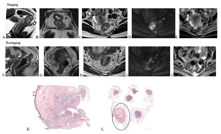
Methods: 80 patients with LARC treated with neoadjuvant therapy, with restaging MRI and surgery, were enrolled and prospectively reviewed. The diagnostic accuracy of the restaging MRI was assessed for tumor (ymrT), nodal status (ymrN), circumferential resection margin (ymrCRM), extramural vascular invasion (ymrEMVI) and tumoral deposits (ymrN1c) by calculating the sensitivity (Se), specificity (Sp), negative predictive values (NPV) and positive predictive values (PPV). Response to treatment was classified as good response (complete/near complete) vs. poor response (poor/partial response). The agreement between the tumor regression grade at MRI (mrTRG) and pathology (pTRG) was reported, as well the performance of mrTRG to identify good responders. The correlation between restaging MRI and histopathology was assessed by Spearman correlation coefficient.
Results: The MRI accuracy ranged between 63.8% and 92.5% for T stage and was 81.3% for N stage. All MRI parameters evaluated at restaging were statistically significant correlated with histopathology evaluation, but EMVI. There was moderate correlation for N and N1c and a positive strong correlation for T, CRM and TRG (Spearman correlation coefficient of 0.390 for mrN1c-pN1c, 0.428 for mrN-pN, 0.522 for mrCRM-pCRM, 0.550 for mrT-pT and 0.731 for mrTRG-pTRG). Diagnostic accuracy of anal sphincter invasion was 91.3%, with a negative predictive value (NPV) of 100%. Accuracy rate varied between 70% for partial response to 93.75% for complete response after nCRT.
Conclusions: MR imaging had good accuracy in restaging LARCs after nCRT. Our results showed high MRI accuracy in detecting anal sphincter involvement for low rectal tumors, with high NPV to exclude tumoral invasion. Restaging MRI predicted well the tumor regression grade, with good diagnostic performance in differentiating good responders from poor/partial responders. The accuracy was high for detecting complete response.
Keywords: magnetic resonance imaging; neoadjuvant chemoradiotherapy; rectal neoplasms; tumor response.
Figures

References
-
- Benson AB, Venook AP, Al-Hawary MM, Arain MA, Chen YJ, Ciombor KK, et al. NCCN Guidelines Insights: Rectal Cancer, Version 6.2020. J Natl Compr Canc Netw. 2020;18:806–815. - PubMed
-
- van de Velde CJ, Boelens PG, Borras JM, Coebergh JW, Cervantes A, Blomqvist L, et al. EURECCA colorectal: multidisciplinary management: European consensus conference colon & rectum. Eur J Cancer. 2014;50:1e1–1.e34. - PubMed
-
- Maas M, Beets-Tan RG, Lambregts DM, Lammering G, Nelemans PJ, Engelen SM, et al. Wait-and-see policy for clinical complete responders after chemoradiation for rectal cancer. J Clin Oncol. 2011;29:4633–4640. - PubMed
-
- Beets-Tan RGH, Lambregts DMJ, Maas M, Bipat S, Barbaro B, Curvo-Semedo L, et al. Magnetic resonance imaging for clinical management of rectal cancer: Updated recommendations from the 2016 European Society of Gastrointestinal and Abdominal Radiology (ESGAR) consensus meeting. Eur Radiol. 2018;28:1465–1475. - PMC - PubMed
LinkOut - more resources
Full Text Sources