. 2023 Dec;28(12):121206.
doi: 10.1117/1.JBO.28.12.121206. Epub 2023 Aug 12.

Iterative prototyping based on lessons learned from the falloposcope in vivo pilot study experience

Affiliations

Iterative prototyping based on lessons learned from the falloposcope in vivo pilot study experience

Andrew D Rocha et al. J Biomed Opt. 2023 Dec.

Abstract

Significance: High grade serous ovarian cancer is the most deadly gynecological cancer, and it is now believed that most cases originate in the fallopian tubes (FTs). Early detection of ovarian cancer could double the 5-year survival rate compared with late-stage diagnosis. Autofluorescence imaging can detect serous-origin precancerous and cancerous lesions in ex vivo FT and ovaries with good sensitivity and specificity. Multispectral fluorescence imaging (MFI) can differentiate healthy, benign, and malignant ovarian and FT tissues. Optical coherence tomography (OCT) reveals subsurface microstructural information and can distinguish normal and cancerous structure in ovaries and FTs.

Aim: We developed an FT endoscope, the falloposcope, as a method for detecting ovarian cancer with MFI and OCT. The falloposcope clinical prototype was tested in a pilot study with 12 volunteers to date to evaluate the safety and feasibility of FT imaging prior to standard of care salpingectomy in normal-risk volunteers. In this manuscript, we describe the multiple modifications made to the falloposcope to enhance robustness, usability, and image quality based on lessons learned in the clinical setting.

Approach: The 0.8 mm diameter falloposcope was introduced via a minimally invasive approach through a commercially available hysteroscope and introducing a catheter. A navigation video, MFI, and OCT of human FTs were obtained. Feedback from stakeholders on image quality and procedural difficulty was obtained.

Results: The falloposcope successfully obtained images in vivo. Considerable feedback was obtained, motivating iterative improvements, including accommodating the operating room environment, modifying the hysteroscope accessories, decreasing endoscope fragility and fiber breaks, optimizing software, improving fiber bundle images, decreasing gradient-index lens stray light, optimizing the proximal imaging system, and improving the illumination.

Conclusions: The initial clinical prototype falloposcope was able to image the FTs, and iterative prototyping has increased its robustness, functionality, and ease of use for future trials.

Keywords: endoscopic optical coherence tomography; fallopian tube; in vivo imaging; microendoscope iterative design and prototyping; multispectral fluorescence imaging; ovarian cancer.


Figures

Fig. 1
Fig. 1
Clinical prototype falloposcope. (a) Before enveloping the working length of the falloposcope in heat shrink, the distal tip ferrule holds the fiber bundle, OCT fiber probe, illumination fiber, and pull wires that are threaded through the MLE. (b) The falloposcope is designed to be small enough to fit through a 5.5 Fr introducing catheter.
Fig. 2
Fig. 2
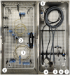
Left: sterilization case with disposable instrumentation, including the (1) falloposcope, (2) introducing catheter, and (3) introducing catheter components. Right: sterilization case with reusable instrumentation, including (4) hysteroscope camera, (5) light guide cable, (6) hysteroscope sheath, (7) hysteroscope telescope, and (8) additional necessary components.
Fig. 3
Fig. 3
Attending physician performing a hysterectomy; the research team observes after our first successful 15-min pilot study procedure was completed. Sources and detectors are located in the black medical rack.
Fig. 4
Fig. 4
Introducing catheter components. Top: original configuration of a Tuohy Borst (Qosina 80402); Male Luer Lock Tubing Connector (Qosina 71637); and hemostasis valve y-connector (Qosina 80325), which connects to the 5.5 fr SSG catheter (Cook J-SSG-554086). Bottom: current configuration of a Tuohy Borst Adapter from the modified Novy cornual cannulation set, a female Luer lock to male Luer lock connector (Qosina 17656), two female Luer locks to male Luer lock connectors (Qosina 80379) connect to the 5.5 fr modified Novy cornual cannulation catheter (Cook J-NCS-504070) with included y-connector, inner catheter, and 0.35” guide wire.
Fig. 5
Fig. 5
Close up of the protected proximal fibers bridging the medical rack and the endoscope’s vinyl tubing.
Fig. 6
Fig. 6
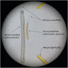
Standard splice protector has a long rigid rod creating a rigid to flexible transition that may become a breaking point at the splice junction. The custom splice protector is semi-rigid and shorter and has eliminated breakage at the splice junction.
Fig. 7
Fig. 7
Top: the falloposcope’s proximal working length was initially designed with a SS hypotube to provide a surface for catheter components to slide onto. This created a rigid to flexible transition that was a kink point. Bottom: the new version has a two-layered approach to go from a more rigid braided PEBA elastomer to a semi-rigid braided polyimide section serving the same function while also acting as a crush-resistant strain relief for the fiber.
Fig. 8
Fig. 8
(a) The raw image is of an opal diffusing glass backed Ronchi ruling. (b) The flat field image of the fiber bundle shows that the relative illumination from core to core is not uniform. Using a flat field corrected test image (c), panels (a)–(c) are normalized for easier viewing.
Fig. 9
Fig. 9
(a) Light leaks out of the thin cladding bare fiber and into fluorescent PEEK MLE. (b) Our solution was to replace the clear heat shrink covering the OD of the GRIN lens with black heat shrink.
Fig. 10
Fig. 10
Simplified representation of the proximal imaging system in the medical cart to show how light from the imaging fiber bundle is collimated, filtered, and then focused onto an image sensor.
Fig. 11
Fig. 11
(a) Example reflectance image obtained with 488 nm laser illumination. The lumen (arrow) as well as a protrusion in the wall (arrowhead) are shown. The high reflectance streak in the center is specular reflectance. Gaussian blur is applied to reduce the honeycomb artifact. (b) Example OCT image, with overlapping FT plicae (arrows show examples). Histogram adjusted to increase contrast.

References

    1. American Cancer Society, “Survival rates for ovarian cancer,” 2022, https://www.cancer.org/cancer/ovarian-cancer/detection-diagnosis-staging....
    1. Siegel R. L., et al. , “Cancer statistics, 2022,” CA Cancer J. Clin. 72, 7–33 (2022). 10.3322/caac.21708 - DOI - PubMed
    1. Vang R., Shih I., Kurman R. J., “Fallopian tube precursors of ovarian low- and high-grade serous neoplasms,” Histopathology 62(1), 44–58 (2013). 10.1111/his.12046 - DOI - PubMed
    1. Shih I., Wang Y., Wang T. L., “The origin of ovarian cancer species and precancerous landscape,” Am. J. Pathol. 191(1), 26–39 (2021). 10.1016/j.ajpath.2020.09.006 - DOI - PMC - PubMed
    1. Chan A., et al. , “New insights into the pathogenesis of ovarian carcinoma: time to rethink ovarian cancer screening,” Obstet. Gynecol. 120(4), 935–940 (2012). 10.1097/AOG.0b013e318269b8b1 - DOI - PubMed

Publication types