. 2023 Aug 10;10(8):ofad369.
doi: 10.1093/ofid/ofad369. eCollection 2023 Aug.

Delayed and Attenuated Antibody Responses to Coronavirus Disease 2019 Vaccination With Poor Cross-Variant Neutralization in Solid-Organ Transplant Recipients-A Prospective Longitudinal Study

Affiliations

Delayed and Attenuated Antibody Responses to Coronavirus Disease 2019 Vaccination With Poor Cross-Variant Neutralization in Solid-Organ Transplant Recipients-A Prospective Longitudinal Study

May Y Liew et al. Open Forum Infect Dis. .

Abstract

Background: Therapeutically immunosuppressed transplant recipients exhibit attenuated responses to severe acute respiratory syndrome coronavirus 2 (SARS-CoV-2) vaccines. To elucidate the kinetics and variant cross-protection of vaccine-induced antibodies in this population, we conducted a prospective longitudinal study in heart and lung transplant recipients receiving the SARS-CoV-2 messenger RNA (mRNA) 3-dose vaccination series.

Methods: We measured longitudinal serum antibody and neutralization responses against the ancestral and major variants of SARS-CoV-2 in SARS-CoV-2-uninfected lung (n = 18) and heart (n = 17) transplant recipients, non-lung-transplanted patients with cystic fibrosis (n = 7), and healthy controls (n = 12) before, during, and after the primary mRNA vaccination series.

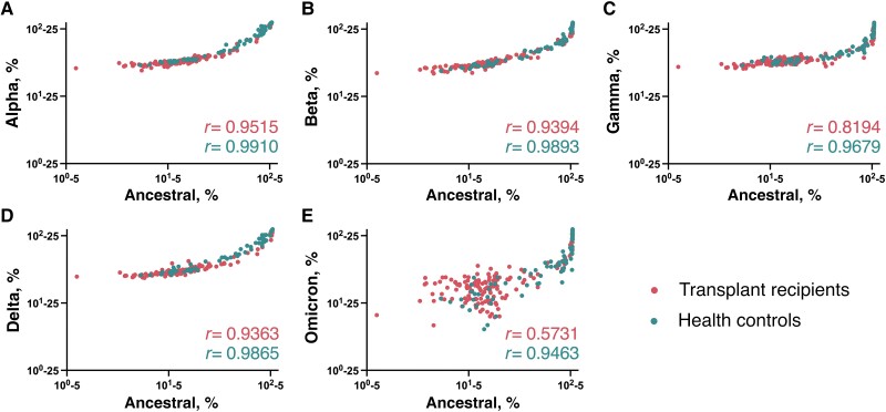
Results: Among healthy controls, strong anti-spike responses arose immediately following vaccination and displayed cross-neutralization against all variants. In contrast, among transplant recipients, after the first 2 vaccine doses, increases in antibody concentrations occurred gradually, and cross-neutralization was completely absent against the Omicron B.1.1.529 variant. However, most (73%) of the transplant recipients had a significant response to the third vaccine dose, reaching levels comparable to those of healthy controls, with improved but attenuated neutralization of immune evasive variants, particularly Beta, Gamma, and Omicron. Responses in non-lung-transplanted patients with cystic fibrosis paralleled those in healthy controls.

Conclusions: In this prospective, longitudinal analysis of variant-specific antibody responses, lung and heart transplant recipients display delayed and defective responses to the first 2 SARS-CoV-2 vaccine doses but significantly augmented responses to a third dose. Gaps in antibody-mediated immunity among transplant recipients are compounded by decreased neutralization against Omicron variants, leaving many patients with substantially weakened immunity against currently circulating variants.

Keywords: COVID-19 vaccination; cross-variant neutralization; cystic fibrosis; longitudinal antibody responses; solid-organ transplant recipients.


Conflict of interest statement

Potential conflicts of interest. All authors: No reported conflicts.

Figures

Figure 1.
Figure 1.
Anti–severe acute respiratory syndrome coronavirus 2 (SARS-CoV-2) spike receptor-binding domain serum antibodies in lung and heart transplant recipients through to after the third vaccine dose. Serum antibody concentrations in lung (n = 16) and heart (n = 15) transplant recipients are compared with those in healthy controls (n = 12). A, Immunoglobulin levels in transplant recipients and healthy controls at each time point. Pairwise comparisons were performed using Mann-Whitney tests. B, C, Immunoglobulin levels in transplant recipients (B) or healthy controls (C) across time points. Box plots show 25th, 50th, and 75th percentiles; whiskers, maximum and minimum. Multiple-group comparisons were performed using Kruskal-Wallis tests. *P < .05; **P < .01; ***P < .001; NS, not significant. D, E, Longitudinal progression of antibody concentrations, displayed as line plots for each transplant recipient and healthy control as a function of days since receipt of first vaccine dose (D) or relative to each vaccine dose received (E). All data points are shown; y-axes are in logarithmic scale.
Figure 2.
Figure 2.
Anti–severe acute respiratory syndrome coronavirus 2 (SARS-CoV-2) spike receptor-binding domain serum antibody responses and neutralizing antibody levels among participants with cystic fibrosis without lung transplantation (n = 5), lung transplant recipients (n = 16), and healthy controls (n = 9). A, Immunoglobulin levels in patients with cystic fibrosis, lung transplant, and healthy controls at each time point. B, Neutralization activity by pseudovirus neutralization assay of serum at 24 weeks after primary 2-dose vaccination series. Abbreviation: ID50, median infective dose. C–H, Neutralization activity in patients with cystic fibrosis, lung transplant recipients, and healthy controls at each time point against the ancestral (D614) SARS-CoV-2 strain (C) and the variants Alpha (B.1.1.7) (D), Beta (B.1.351) (E), Gamma (P1) (F), Delta (B.1.617.2) (G), and Omicron (B.1.1.529) (H). Pairwise comparisons were performed using Mann-Whitney tests. Box plots show 25th, 50th, and 75th percentiles; whiskers, maximum and minimum. *P < .05; **P < .01; ***P < .001; NS, not significant. All data points are shown; y-axes are in logarithmic scale.
Figure 3.
Figure 3.
Neutralization activity of serum in lung and heart transplant recipients through to after the third vaccine dose. Pseudovirus neutralization assay was used to measure neutralization activity in serum samples from lung (n = 16) and heart (n = 15) transplant recipients compared with healthy controls (n = 12). A, Neutralization activity in transplant recipients and healthy controls at each time point. Pairwise comparisons were performed using Mann-Whitney tests. B, C, Neutralization activity in transplant recipients (B) and healthy controls (C) across time points. Box plots show 25th, 50th, and 75th percentiles; whiskers, maximum and minimum. Multiple-group comparisons were performed using Kruskal-Wallis tests. *P < .05; **P < .01; ***P < .001; NS, not significant. D, E, Longitudinal progression of neutralizing antibody titers as a function of days since receipt of first vaccine dose (D) or relative to each vaccine dose (E). All data points are shown; y-axes are in logarithmic scale. Abbreviation: ID50, median infective dose.
Figure 4.
Figure 4.
Neutralizing activity of antibodies against severe acute respiratory syndrome coronavirus 2 (SARS-CoV-2) spike S1 protein in serum samples from lung and heart transplant recipients through to after the third vaccine dose. Neutralizing activity against the ancestral SARS-CoV-2 strain and 5 variants was measured in serum samples from lung (n = 16) and heart (n = 15) transplant recipients compared with healthy controls (n = 12). A–F, Neutralization activity in transplant recipients and healthy controls at each time point against the ancestral strain (A) and the variants Alpha (B.1.1.7) (B), Beta (B.1.351) (C), Gamma (P1) (D), Delta (B.1.617.2) (E), and Omicron (B.1.1.529) (F). Pairwise comparisons were performed using Mann-Whitney tests. Box plots show 25th, 50th, and 75th percentiles; whiskers, maximum and minimum. *P < .05; **P < .01; ***P < .001; NS, not significant. All data points are shown; y-axes are in logarithmic scale.
Figure 5.
Figure 5.
Neutralizing activity of antibodies against severe acute respiratory syndrome coronavirus 2 (SARS-CoV-2) spike S1 protein in serum samples from lung and heart transplant recipients, for SARS-CoV-2 variants compared with the ancestral strain. Correlation plots compare neutralizing activity against the ancestral SARS-CoV-2 spike S1 protein (x-axes) with that against the variants Alpha (B.1.1.7) (A), Beta (B.1.351) (B), Gamma (P1) (C), Delta (B.1.617.2) (D), and Omicron (B.1.1.529) (E) (y-axes). Antibodies were measured in serum samples from lung (n = 16) and heart (n = 15) transplant recipients and healthy controls (n = 12). Spearman rank correlation coefficients are provided. All data points are shown; both axes are in logarithmic scale. As noted on the x-axis and y-axis scale bars, a standardized offset of each data point of 5% (x-axis) and 25% (y-axis) for clarity in plotting.

References

    1. Zhou P, Yang XL, Wang XG, et al. . A pneumonia outbreak associated with a new coronavirus of probable bat origin. Nature 2020; 579:270–3. - PMC - PubMed
    1. Shostak Y, Shafran N, Heching M, et al. . Early humoral response among lung transplant recipients vaccinated with BNT162b2 vaccine. Lancet Respir Med 2021; 9:e52–3. - PMC - PubMed
    1. Marinaki S, Adamopoulos S, Degiannis D, et al. . Immunogenicity of SARS-CoV-2 BNT162b2 vaccine in solid organ transplant recipients. Am J Transplant 2021; 21:2913–5. - PMC - PubMed
    1. Boyarsky BJ, Werbel WA, Avery RK, et al. . Antibody response to 2-dose SARS-CoV-2 mRNA vaccine series in solid organ transplant recipients. JAMA 2021; 325:2204–6. - PMC - PubMed
    1. Benotmane I, Gautier-Vargas G, Cognard N, et al. . Weak anti-SARS-CoV-2 antibody response after the first injection of an mRNA COVID-19 vaccine in kidney transplant recipients. Kidney Int 2021; 99:1487–9. - PMC - PubMed