

Multiple bronchiolar adenomas/ciliated muconodular papillary tumors of the bilateral lung with tumor budding and potential malignant transformation into squamous cell carcinoma: a case report and literature review
- PMID: 37577314
- PMCID: PMC10413017
- DOI: 10.21037/tlcr-23-374
Multiple bronchiolar adenomas/ciliated muconodular papillary tumors of the bilateral lung with tumor budding and potential malignant transformation into squamous cell carcinoma: a case report and literature review
Abstract
Background: Bronchiolar adenoma (BA)/ciliated muconodular papillary tumor (CMPT) is a rare lung tumor characterized by ciliated, mucous and basal cells. Recently, some cases of driver mutations or malignant transformations have been reported. However, the nature of BA/CMPT remains controversial. Here, we report a case of bilateral pulmonary multiple BAs with tumor budding and squamous metaplasia.
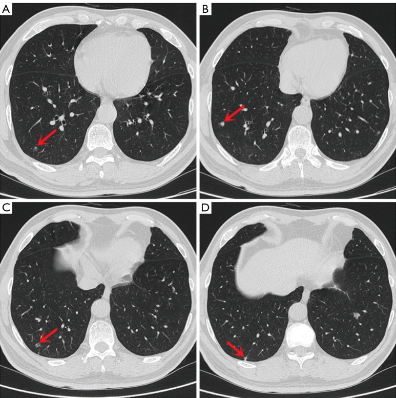
Case description: A 55-year-old man presented with multiple small nodules in the lower lobes of the bilateral lungs on physical examination 7 years prior. During the past 3 years of regular follow-up, some nodules had slightly enlarged. Because the nodules were mostly solid, the patient underwent video-assisted thoracoscopic segmentectomy of the left lower lung. A postoperative pathological diagnosis of BA was made. In all lesions, the fusion and mutation of major driver genes were not detected by next-generation sequencing (NGS). No recurrence or metastasis was observed after 37 months of follow-up. Notably, all five resected lesions were BA/CMPT, and one lesion was accompanied by squamous metaplasia and tumor budding.
Conclusions: Our report found that BA/CMPT with squamous metaplasia and tumor budding has the potential to transform into lung squamous cell carcinoma, expanding its connection with malignant transformation. Smoking may be one of the risk factors. We also found that BA/CMPT can be multiple lesions rather than a solitary lesion.
Keywords: Bronchiolar adenoma (BA); case report; ciliated muconodular papillary tumor (CMPT); malignant transformation; tumor budding.
2023 Translational Lung Cancer Research. All rights reserved.
Conflict of interest statement
Conflicts of Interest: All authors have completed the ICMJE uniform disclosure form (available at https://tlcr.amegroups.com/article/view/10.21037/tlcr-23-374/coif). The authors have no conflicts of interest to declare.
Figures


Similar articles
-
Case Report: Two cases of bronchiolar adenoma/ciliated muconodular papillary tumor characterized by significant basal cell hyperplasia and squamous metaplasia.Front Oncol. 2025 Jul 25;15:1617720. doi: 10.3389/fonc.2025.1617720. eCollection 2025. Front Oncol. 2025. PMID: 40786515 Free PMC article.
-
Bronchiolar adenoma/ciliated muconodular papillary tumor mixed with adenocarcinoma in situ in the same tumor.Thorac Cancer. 2023 Feb;14(4):427-431. doi: 10.1111/1759-7714.14784. Epub 2022 Dec 28. Thorac Cancer. 2023. PMID: 36578104 Free PMC article.
-
Clinicopathological characteristics and molecular analysis of lung cancer associated with ciliated muconodular papillary tumor/bronchiolar adenoma.Pathol Int. 2023 May;73(5):188-197. doi: 10.1111/pin.13316. Epub 2023 Mar 9. Pathol Int. 2023. PMID: 36896472
-
Bronchiole adenoma/pulmonary ciliated mucinous nodular papillary tumor: Case series and literature review.Medicine (Baltimore). 2023 Dec 15;102(50):e36559. doi: 10.1097/MD.0000000000036559. Medicine (Baltimore). 2023. PMID: 38115282 Free PMC article. Review.
-
Multiple bronchiolar adenomas with malignant transformation and CCNE1 mutation: a case report and literature review.J Cardiothorac Surg. 2021 Oct 18;16(1):307. doi: 10.1186/s13019-021-01687-5. J Cardiothorac Surg. 2021. PMID: 34663408 Free PMC article. Review.
Cited by
-
Bronchiolar adenoma/ciliated muconodular papillary tumor complicated by lymphoid interstitial pneumonia in a patient with Sjögren's disease: A case report and systematic review.Thorac Cancer. 2024 Oct;15(28):1975-1988. doi: 10.1111/1759-7714.15420. Epub 2024 Aug 18. Thorac Cancer. 2024. PMID: 39155148 Free PMC article.
-
Discriminating bronchiolar adenoma from peripheral lung cancer by thin-section computed tomography (CT): a 2-center study.Quant Imaging Med Surg. 2024 Oct 1;14(10):7086-7097. doi: 10.21037/qims-24-687. Epub 2024 Aug 19. Quant Imaging Med Surg. 2024. PMID: 39429574 Free PMC article.
-
Case Report: Two cases of bronchiolar adenoma/ciliated muconodular papillary tumor characterized by significant basal cell hyperplasia and squamous metaplasia.Front Oncol. 2025 Jul 25;15:1617720. doi: 10.3389/fonc.2025.1617720. eCollection 2025. Front Oncol. 2025. PMID: 40786515 Free PMC article.
References
-
- Ishikawa Y. Ciliated muconodular papillary tumor of the peripheral lung: benign or malignant. Pathol Clin Med 2002;20:964-5.
-
- Chang JC, Montecalvo J, Borsu L, et al. Bronchiolar Adenoma: Expansion of the Concept of Ciliated Muconodular Papillary Tumors With Proposal for Revised Terminology Based on Morphologic, Immunophenotypic, and Genomic Analysis of 25 Cases. Am J Surg Pathol 2018;42:1010-26. 10.1097/PAS.0000000000001086 - DOI - PMC - PubMed
-
- WHO Classification of Tumours Editorial Board. WHO classification of tumours Thoracic Tumours 5th ed Lyon: IARC Press; 2021.
-
- Harada T, Akiyama Y, Ogasawara H, et al. Ciliated muconodular papillary tumor of the peripheral lung: A newly defined rare tumor. Respiratory Medicine CME 2008;1:176-8. 10.1016/j.rmedc.2008.04.005 - DOI
Publication types
LinkOut - more resources
Full Text Sources
Miscellaneous