[Preprint]. 2023 Aug 6:2023.08.04.552048.
doi: 10.1101/2023.08.04.552048.

A molecular atlas of adult C. elegans motor neurons reveals ancient diversity delineated by conserved transcription factor codes

Affiliations

A molecular atlas of adult C. elegans motor neurons reveals ancient diversity delineated by conserved transcription factor codes

Jayson J Smith et al. bioRxiv. .

Update in

Abstract

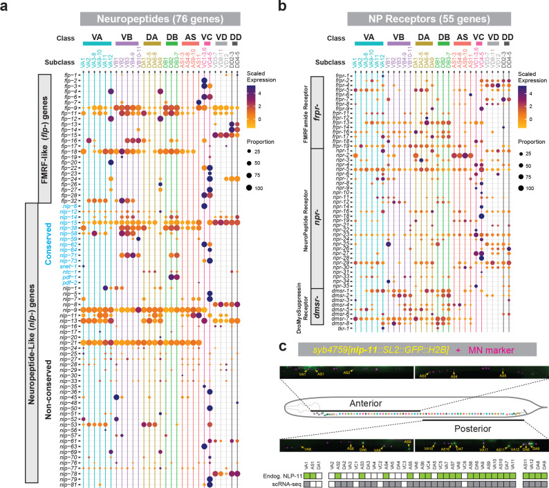
Motor neurons (MNs) constitute an ancient cell type targeted by multiple adult-onset diseases. It is therefore important to define the molecular makeup of adult MNs in animal models and extract organizing principles. Here, we generated a comprehensive molecular atlas of adult Caenorhabditis elegans MNs and a searchable database (http://celegans.spinalcordatlas.org). Single-cell RNA-sequencing of 13,200 cells revealed that ventral nerve cord MNs cluster into 29 molecularly distinct subclasses. All subclasses are delineated by unique expression codes of either neuropeptide or transcription factor gene families. Strikingly, we found that combinatorial codes of homeodomain transcription factor genes define adult MN diversity both in C. elegans and mice. Further, molecularly defined MN subclasses in C. elegans display distinct patterns of connectivity. Hence, our study couples the connectivity map of the C. elegans motor circuit with a molecular atlas of its constituent MNs, and uncovers organizing principles and conserved molecular codes of adult MN diversity.

Keywords: C. elegans; Hox genes; adult motor neurons; neuropeptides; scRNA-seq; transcription factors.


Conflict of interest statement

ETHICS DECLARATIONS The authors declare no competing interests.

Figures

Figure 1:
Figure 1:. Strategy for single-cell RNA-seq of adult C. elegans motor neurons.
(a) Schematic of cardinal motor neuron (MN) classes in the adult C. elegans retrovesicular ganglion (RVG), ventral nerve cord (VNC), and preanal ganglion (PAG) arranged anatomically from anterior to posterior. (b) Fluorescence micrographs depicting expression of acr-2p::GFP and lin-39p::RFP transgenes used for scRNA-seq. (c) Single neuron reporter data of each transgenic strain depicted in b. (d) Overview of the workflow for scRNA-seq. (e) UMAP plot showing molecular separation of all eight cardinal MN classes. (f) Dot plot showing log expression and percent of cells expressing known MN class-specific genes. (g) UMAP plot in e, but colors depict strain of origin. (h) Dot plot showing log expression and percent of cells expressing neurotransmitter identity genes.
Figure 2:
Figure 2:. scRNA-seq identifies striking MN diversity and a subclass-specific TF code.
(a) UMAP and table showing 24 cholinergic motor neuron (MN) subclasses (left) and 5 GABAergic MN subclasses (right) in adult C. elegans. (b) Plot depicting gene ontology (GO) categories (wormcat.com) that are over-represented (Fisher’s exact test, p-value < 0.01) in adult MN subclasses. Genes involved in transcription/gene regulation are in green. (c) Chart depicting the number of transcription factors (TF) detected in adult MNs that belong to each TF family. (d) Dot plots showing scaled expression and proportion of cells expressing MN subclass-specific transcription factors. TFs families are indicated to the left of genes.
Figure 3:
Figure 3:. Hox gene expression delineates adult motor neuron subclasses.
(a) The C. elegans Hox cluster. (b) Expression matrix of endogenous fluorescent reporters for all 6 Hox genes (rows) in every MN (columns) in the adult RVG, VNC, and PAG. MN-specific reporters used for unambiguous identification of each cell (see Methods). (c) Tables showing strong concordance for (rows) Hox gene expression between (columns) scRNA-seq analysis and endogenous reporters in cholinergic and GABAergic MN subclasses. Colored boxes indicate only scRNA-seq expression pattern. ‘ScRNA-seq + Reporter’ below tables indicates where expression of all Hox genes in the MN subclass are in complete agreement between the two methods (+) or where is there is disagreement (−). Asterisks (*) indicate anatomical MN classes for which all subclasses can be defined using Hox expression codes. (d) Feature plots showing log expression of Hox genes in VA MNs. (e) UMAP showing all VA MNs with color-coded subclasses and subclass-specific Hox expression (left) and schematic of Hox expression in VA MNs in anatomical context (right). (f) UMAP of elt-1/GATA1–3 expression in VA MNs. (g) Fluorescence micrographs of endogenous elt-1::mNG expression and NeuroPAL transgene (middle) in RVG and anterior VNC MNs, accompanied by elt-1/GATA1–3 single-cell expression data (bottom).
Figure 4:
Figure 4:. Combinatorial expression of extra synaptic signaling genes delineates adult motor neuron subclasses.
(a, b) Dot plots showing scaled expression and proportion of cells expressing genes encoding a neuropeptides or b neuropeptide receptors in cholinergic and GABAergic MN subclasses. Individual genes are represented as rows and MN subclasses are represented as columns. (c) Fluorescence micrographs, and single-cell fluorescent reporter expression data for nlp-11(syb4759).
Figure 5:
Figure 5:. DA and DB MN subclasses display unique connectivity patterns.
(a, c, e) Schematics depicting soma locations for DA (a), DB (c), and VC (e) MNs in the RVG, VNC, and PAG and their respective morphologies (bottom). Morphologies are derived from electron microscopy (EM) reconstructions summarized at wormatlas.org and wormwiring.org. vBWMs = ventral body wall muscle; dBWMs = dorsal body wall muscles. (b, d, f) Neural network diagrams of DA (b), DB (d), and VC (f) MNs. Network diagrams were generated using the Nematode Neural Network Viewer (nemanode.org) using the complete adult dataset derived from previous studies ,. A hierarchical network layout depicts all connections with at least one chemical synapse or gap junction. Colors indicate neurotransmitter identity: red = cholinergic; Blue = GABAergic; Yellow = glutamatergic; Gray = unknown.
Figure 6:
Figure 6:. Homeodomain (HD) transcription factor genes delineate adult mouse motor neuron diversity.
(a) Schematic depicting cholinergic MN populations captured in single-nucleus RNA-seq of the adult mouse spinal cord, including visceral (dark blue) and skeletal (green) MNs . (b) Pie chart depicting the number of homeodomain (HD) transcription factor genes detected in adult mouse MNs that belong to each HD TF family based on mammalian HD TF classifications . HD TF families with only one gene detected or HD TF genes that were unassigned to any family are combined into one slice, and collectively account for 13.2% (10/76). (c) Dot plot showing scaled expression and proportion of cells expressing the 76 HD TF genes detected in either visceral or skeletal MNs. HD TF families are color-coded and indicated to the left of the genes. In situ hybridization data derived from Allen Brain Atlas for each HD TF gene assessed by expression in the ventral surface of cross sections of the spinal cord. YES (black), clear expression of indicated gene; NA (gray), no data available at Allen Brain Atlas for this gene; NO (red), no obvious expression in ventral surface of mouse spinal cord. (d) Clustering dendrogram based on averaged expression of each HD TF gene across all visceral and skeletal MN subclasses.

References

    1. Dalla Torre di Sanguinetto S. A., Dasen J. S. & Arber S. Transcriptional mechanisms controlling motor neuron diversity and connectivity. Curr Opin Neurobiol 18, 36–43, doi:S0959-4388(08)00026-3 [pii] 10.1016/j.conb.2008.04.002 (2008). - DOI - PubMed
    1. Dasen J. S. Transcriptional networks in the early development of sensory-motor circuits. Curr Top Dev Biol 87, 119–148, doi: 10.1016/S0070-2153(09)01204-6 (2009). - DOI - PubMed
    1. Lee S. K. & Pfaff S. L. Transcriptional networks regulating neuronal identity in the developing spinal cord. Nat Neurosci 4 Suppl, 1183–1191, doi: 10.1038/nn750nn750 nn750 [pii] (2001). - DOI - PubMed
    1. Catela C. & Kratsios P. Transcriptional mechanisms of motor neuron development in vertebrates and invertebrates. Dev Biol, doi: 10.1016/j.ydbio.2019.08.022 (2019). - DOI - PubMed
    1. Hobert O., Glenwinkel L. & White J. Revisiting Neuronal Cell Type Classification in Caenorhabditis elegans. Curr Biol 26, R1197–R1203, doi: 10.1016/j.cub.2016.10.027 (2016). - DOI - PubMed

Publication types