[Preprint]. 2023 Aug 2:2023.08.02.551666.
doi: 10.1101/2023.08.02.551666.

Molecular profiling of sponge deflation reveals an ancient relaxant-inflammatory response

Affiliations

Molecular profiling of sponge deflation reveals an ancient relaxant-inflammatory response

Fabian Ruperti et al. bioRxiv. .

Update in

Abstract

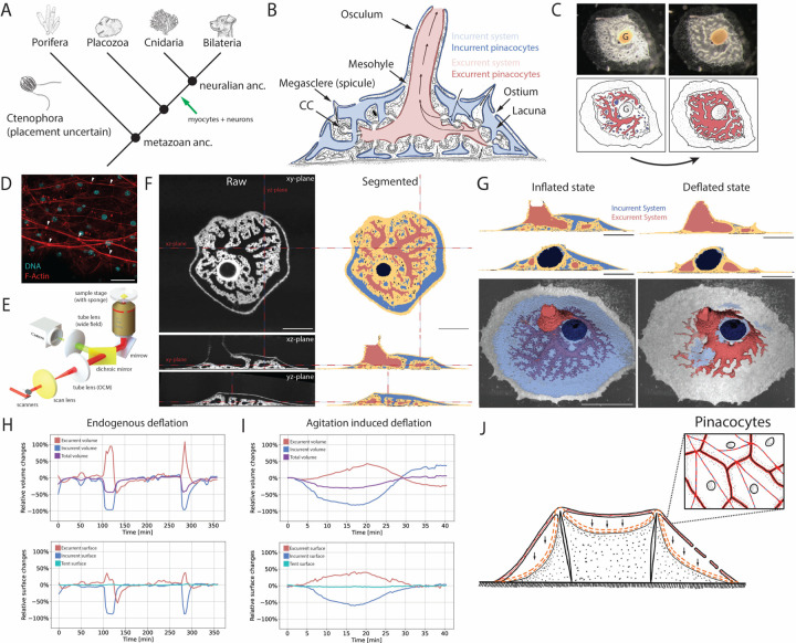
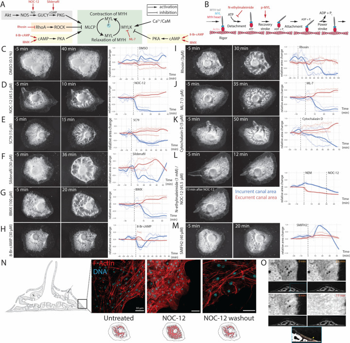
A hallmark of animals is the coordination of whole-body movement. Neurons and muscles are central to this, yet coordinated movements also exist in sponges that lack these cell types. Sponges are sessile animals with a complex canal system for filter-feeding. They undergo whole-body movements resembling "contractions" that lead to canal closure and water expulsion. Here, we combine 3D optical coherence microscopy, pharmacology, and functional proteomics to elucidate anatomy, molecular physiology, and control of these movements. We find them driven by the relaxation of actomyosin stress fibers in epithelial canal cells, which leads to whole-body deflation via collapse of the incurrent and expansion of the excurrent system, controlled by an Akt/NO/PKG/A pathway. A concomitant increase in reactive oxygen species and secretion of proteinases and cytokines indicate an inflammation-like state reminiscent of vascular endothelial cells experiencing oscillatory shear stress. This suggests an ancient relaxant-inflammatory response of perturbed fluid-carrying systems in animals.

Keywords: OCM; Spongilla; cell type evolution; functional proteomics; inflammation; sponge movment; vascular system.


Figures

Figure 1.
Figure 1.. 3D in vivo optical coherence microscopy (OCM) of juvenile Spongilla.
(A) Animal phylogeny marking proposed origin of neurons and myocytes. (B) Illustration of juvenile Spongilla lacustris specimen showing incurrent (blue) and excurrent (red) canal systems. Epithelial pinacocytes are highlighted by darker color tones. CC: Choanocyte chamber. (C) Photograph and illustration of endogenous deflation of S. lacustris highlighting incurrent (blue) and excurrent (red) systems. G: Gemmule. (D) Confocal max intensity projection of actomyosin stress fibers of the tent pinacocytes stained for F-actin (phalloidin, red) and DNA (DAPI, cyan). White arrowheads depict the cell-cell junctions connecting neighboring stress fibers. Scale bar, 30 um. (E) Schematic of the microscope body part of the optical coherence microscope (OCM). (F) OCM volumes showing orthogonal views of S. lacustris in inflated state. Segmented incurrent (blue), excurrent (red) systems, mesohyl/tent (yellow) and gemmule (dark blue). Scale bar, 500 µm. See also Table S1. (G) 3D reconstruction of inflated and deflated states. 2D Scale bar, 500 µm. 3D Scale bar, 1000 µm. See also Figure S1. (H) Volume and surface area changes measured from segmented OCM volumes during two cycles of endogenous sponge whole-body deflations over 6 hours (f = 1/3 min-1). Excurrent system (red), incurrent system (blue), total sponge (purple), tent (turquoise). (I) Relative volume and surface area changes measured from segmented OCM volumes during one cycle of whole-body deflation induced by mechanical agitation (f = 1/30 s-1). (J) Illustration of epithelial tent collapse and tent pinacocytes highlighting cell traversing actomyosin stress fibers.
Figure 2.
Figure 2.. Pharmacological perturbation of actomyosin activity.
(A) Smooth/non-muscle actomyosin regulatory pathways with pharmacological compounds highlighted in red. MYL: myosin light chain, MYH: myosin heavy chain, MYLK: myosin light chain kinase, MYLP: myosin light chain phosphatase, NOS: nitric oxide synthase, NO: nitric oxide, GUCY: guanylate cyclase, PKG: protein kinase G, ROCK: RhoA/Rho-associated protein kinase, PKA: protein kinase A, CaM: calmodulin. Background colors relate to Figure S2A. See also Figure S2B. (B) Actomyosin cross-bridge cycle showing states of myosin conformation and actin binding. N-ethylmaleimide (NEM) treatment blocks ATP binding and myosin release, stabilizing a rigor state. (C-M) Time-lapse imaging (f = 1/30 s-1) of S. lacustris during the treatment with pharmacological compounds. Overhead images show sponges 5 min pre-treatment (left) and during respective behavior (right). Plots show average change in relative areas across replicates of segmented incurrent (dark blue) and excurrent (dark red) canal areas. See also Figure S2C-N. Dashed line marks application of compound. Individual replicates shown in light tones. (N) Confocal max intensity projections of actomyosin stress fibers of the tent pinacocytes stained for F-actin (red, phalloidin) and DNA (cyan, DAPI) in an untreated sponge, NOC-12 treated sponge (deflated) and sponge after NOC-12 washout and return to inflated state. Scale bars, 30 um. See also Figure S2O. (O) Ostia closure during agitation induced deflation. Simultaneous tent collapse and ostia closure is visible in the overlay section (blue square). Annotated tent colors correspond to time points in upper panels. Ostium: yellow arrow head. See also Figure S2.
Figure 3.
Figure 3.. Phosphoproteomics and thermal proteome profiling of sponge deflation.
(A) Experimental setup of sponge functional proteomics. S. lacustris deflation was induced by circular agitation or NO treatment. (B) Volcano plots of TPP and quantitative phosphoproteomic results of NO treated and agitated sponges. TPP results show individual protein changes in stability and abundance. Phosphorylation differences are quantified by unique phosphopeptides. Proteins related to stress fibers with significant changes are individually colored: actomyosin contractility (red), actin (rose), actin bundling (green), actin dynamics (blue), membrane attachment or cell junction (yellow). FLNA/B/C: filamin. GSN: gelsolin. ITGA/B: integrin alpha/beta. LIMA1: LIM-domain and actin-binding protein 1. PDEs: phosphodiesterases. PLS: plastin. PCDH15: protocadherin 15. SVIL: supervillin. TLN: talin. (C) Illustration of actomyosin proteins significantly changing stability, abundance or phosphorylation state. Colors relate to (B). Proteins with asterisk relate to Figure S4A. (D) Quantification of S. lacustris Akt kinase 1/2/3 phosphopeptide corresponding to H. sapiens Akt1 T443. Quantification of S. lacustris FOXO 1/2/3 phosphopeptide with phosphorylated serine corresponding to H. sapiens FOXO3 S253. The barplot shows normalized single-cell RNAseq expression of Spongilla Foxo1/2/3. Incurrent pinacocytes 1 (incPin1) and 2 (incPin2) are highlighted in blue. Excurrent/Apendopinacocytes 1 (apnPin1) and 2 (apnPin2) are highlighted in red. Lph: Lophocytes, basPin: Basopinacocytes, Scp: Sclerophorocytes, Met1: Metabolocytes 1, Met2: Metabolocytes 2, Chb1: Choanoblasts 1, Chb2: Choanoblasts 2, Cho: Choanocytes, Apo: Apopylar cells, Myp1: Myopeptidocytes 1, Myp2: Myopeptidocytes 2, Amb: Amoebocytes, Grl: Granulocytes, Nrd: Neuroid cells, Mes1: Mesocytes 1, Mes2: Mesocytes 2, Mes3: Mesocytes 3, Arc: Archaeocytes, Scl: Sclerocytes. (E) Correlation of phosphopeptide fold changes of NO treated and agitated sponges. Peptides of proteins highlighted in (B) and (C) are highlighted in the same colors. (F) Enrichment of kinase family activity after NO treatment and agitation (prediction by GPS 5.0 (63)). (G) Correlation of kinase activity enrichment between agitated and NO-treated sponges.
Figure 4.
Figure 4.. Secretomic and metabolomic profiling of agitation-induced deflated sponges.
(A) GO term enrichment of significant protein stability changes (relates to Figure 3A). Green dot highlights GO term for proteins in volcano plot (relates to Figure 3B). SCAMP2/3/4/5: secretory carrier-associated membrane protein 2/3/4/5, VAMP4: vesicle associated membrane protein 4, DNAJC5/DNAJC5B: DnaJ homolog subfamily C member 5/B (B) Secretomics experimental setup. (C) Secreted protein fold changes, highlighting proteinases (pink) and ROS catabolizing enzymes (yellow). See also Figure S5A, C. (D) Quantification of secreted proteinases (pink) and ROS catabolizing enzymes (yellow). P-values were calculated using the Wilcox rank sum test. Colors relate to (C). (E) Relative protein abundance of PI16, Granulin, and MIF in sponge medium after agitation-induced deflation. See also Figure S5B. (F) PI16 normalized expression from S. lacustris single-cell RNAseq atlas. (G) ColabFold structural prediction (79, 80) of Spongilla granulin domain (beige) aligned with its best morpholog (Danio rerio granulin domain, UniprotID: Q7T3M4, aa 95 – 147). The peptide detected in the secretomics experiment highlighted in red. Conserved disulfide bonds are shown in yellow. (H) Quantification of pyridoxamine (power = 0.99, precision = 15.5%) and hypotaurine (power = 0.87, precision = 9.2%) in the sponge body after agitation-induced deflation by manual integration of TIC normalized areas. (I) Schematic of proposed relaxant-inflammatory reaction of Spongilla in response to (oscillatory) shear stress.
Figure 5.
Figure 5.. Comparison of oscillatory shear stress reactions in vertebrate vasculature and S. lacustris pinacoderm.
Cellular components of the vertebrate vascular wall include vascular endothelial cells with actomyosin stress fibers (actin: red, myosin: light blue) oriented parallel to blood flow and connected by adherens junctions (green) (109, 110), smooth myocytes, and fibroblasts. Endothelial cells are connected to the ECM via focal adhesions (111) In the vascular system, endothelial cells adopt a sensory and signaling role whereas the smooth muscle cells are primarily responsible for the maintenance and regulation of vascular tone. Spongilla pinacoderm apparently assume both roles. Pinacocytes have actomyosin stress fibers connected by cell-cell junctions composed of focal adhesion and adherens junction proteins (19) Alteration of flow by oscillatory shear stress leads to Akt kinase activation and NO production, resulting in tension release or relaxation of actin stress fibers and smooth myocytes. In both systems, pro-inflammatory reactions are mediated by ROS increase and secretion of granulin, proteinases, and MIF.

References

    1. Brunet Thibaut and King Nicole. The origin of animal multicellularity and cell differentiation. Dev. Cell, 43(2):124–140, October 2017. - PMC - PubMed
    1. Simpson T L. The Cell Biology of Sponges. Springer Science & Business Media, December 2012.
    1. Asadzadeh Seyed Saeed, Larsen Poul S, Riisgård Hans Ulrik, and Walther Jens H. Hydrodynamics of the leucon sponge pump. J. R. Soc. Interface, 16(150):20180630, January 2019. - PMC - PubMed
    1. Goldstein Josephine, Bisbo Nicklas, Funch Peter, and Riisgård Hans Ulrik. Contraction-Expansion and the effects on the aquiferous system in the demosponge halichondria panicea. Front. Mar. Sci., 7, February 2020.
    1. Reiswig H M. In situ pumping activities of tropical demospongiae. Mar. Biol., 9(1):38–50, April 1971.

Publication types