[Preprint]. 2023 Aug 2:2023.08.02.551707.
doi: 10.1101/2023.08.02.551707.

Requirement of GrgA for Chlamydia infectious progeny production, optimal growth, and efficient plasmid maintenance

Affiliations

Requirement of GrgA for Chlamydia infectious progeny production, optimal growth, and efficient plasmid maintenance

Bin Lu et al. bioRxiv. .

Update in

Abstract

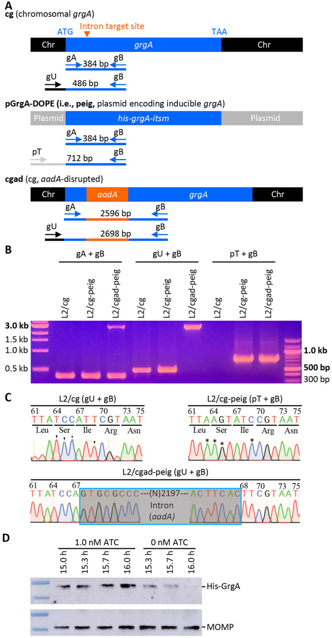
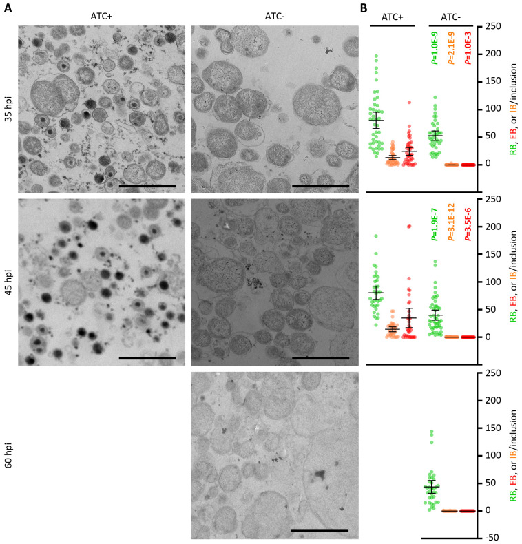
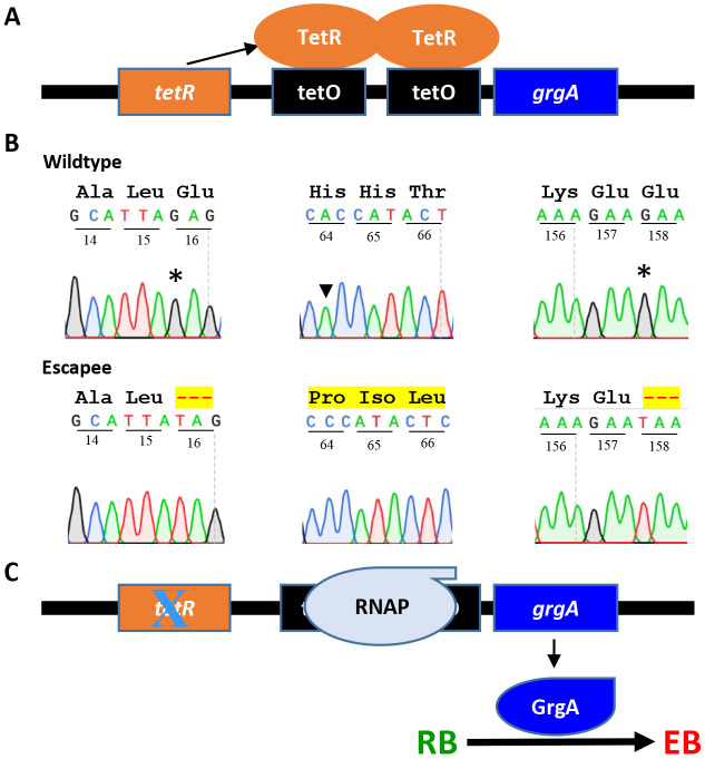
Chlamydia, an obligate intracellular bacterial pathogen, has a unique developmental cycle involving the differentiation of invading elementary bodies (EBs) to noninfectious reticulate bodies (RBs), replication of RBs, and redifferentiation of RBs into progeny EBs. Progression of this cycle is regulated by three sigma factors, which direct the RNA polymerase to their respective target gene promoters. We hypothesized that the Chlamydia-specific transcriptional regulator GrgA, previously shown to activate σ66 and σ28, plays an essential role in chlamydial development and growth. To test this hypothesis, we applied a novel genetic tool known as dependence on plasmid-mediated expression (DOPE) to create Chlamydia trachomatis with conditional GrgA-deficiency. We show that GrgA-deficient C. trachomatis RBs have a growth rate that is approximately half of the normal rate and fail to transition into progeny EBs. In addition, GrgA-deficient C. trachomatis fail to maintain its virulence plasmid. Results of RNA-seq analysis indicate that GrgA promotes RB growth by optimizing tRNA synthesis and expression of nutrient-acquisition genes, while it enables RB-to-EB conversion by facilitating the expression of a histone and outer membrane proteins required for EB morphogenesis. GrgA also regulates numerous other late genes required for host cell exit and subsequent EB invasion into host cells. Importantly, GrgA stimulates the expression of σ54, the third and last sigma factor, and its activator AtoC, and thereby indirectly upregulating the expression of σ54-dependent genes. In conclusion, our work demonstrates that GrgA is a master transcriptional regulator in Chlamydia and plays multiple essential roles in chlamydial pathogenicity.


Conflict of interest statement

COMPETING INTERESTS STATEMENT The authors declare no conflict of interest.

Figures

Figure 1.
Figure 1.. Confirmation of the disruption of the chromosome-encoded grgA by group II-intron and re-expression of GrgA from a transformed plasmid in the DOPE system.
(A) Schematic drawings of grgA alleles, locations of intron-target site, diagnostic primers, and sizes of PCR products obtained with different sets of primers. Abbreviations: itsm, intron target site mutated; Chr, chromosome. (B) Gel image of PCR products amplified with DNA of wildtype C. trachomatis L2 with intact chromosomal grgA (L2/cg), L2/cg transformed with the his-grgA-itsm expression plasmid pGrgA-DOPE (L2/cg-peig), and L2 with aadA-disrupted chromosomal grgA complemented with pGrgA-DOPE (L2/cgad-peig) using primer sets shown in (A). (C) Sanger sequencing tracings of PCR products showing the intron-target site in L2/cg, mutations surrounding this site conferring resistance to intron targeting in peig, and grgA-intron joint regions in the chromosome of L2/cg-peig. Wildtye bases and corresponding mutated bases are shown with arrowheads and asterisks, respectively. (D) Western blotting showing time-dependent loss of His-GrgA in L2/cgad-peig upon ATC withdrawal. The membrane was first probed with an anti-major outer membrane protein antibody L2-5, striped, and then reprobed with an anti-GrgA antibody.
Figure 2.
Figure 2.. GrgA deficiency slows RB growth and disables the formation of infectious progeny.
L2/cg-peig-infected HeLa cells were cultured in the presence or absence of 1 nM ATC. At indicated hpi, cultures were terminated for immunofluorescence assay (A), genome copy quantification (B), or quantification of inclusion-forming unt (C). (B, D) Data represent averages ± standard deviations of triplicate cultures.
Figure 3.
Figure 3.. Lack of EB formation in GrgA-deficent cultures.
(A) Representative electron microscopic (EM) images of L2/cgad-peig cultured in ATC-containing and ATC-free media at indicated hpi. 60 h was not processed for EM because most inclusion already burst by that point. Note that EBs are ~400 nm diameter cellular forms with high electron density found in ATC-containing cultures. Small irregularly-shaped electron-dense particles in both ATC-containing and ATC-free cultures are glycogen particles. Cell types are color-encoded as shown in the Y-axis labels. Size bar equals 2 μm. (B) Scattergraph of RBs, EBs, and intermediate bodies (IBs) counted from multiple inclusions.
Figure 4.
Figure 4.. Failed grgA repression in pGrgA-DOPE is responsible for EB escape in ATC-free cultures of L2/cgad-peig.
(A) Schematic shows mechanism for TetR-mediated grgA transcription repression in L2/cgad-peig cultured in the absence of ATC. (B) Mutations identified in tetR in pGrgA-DOPE recovered from L2/cg-peig EBs formed in the absence of ATC lead to premature translation termination or frameshift. Codon positions in tetR are numbered. Wild-type amino acid sequences are shown in black. Highlighted hyphens and amino acids indicate protein sequence truncation and alteration, respectively. (C) Mutations detected in (B) result in a loss in TetR-mediated grgA repression, leading to GrgA expression in the absence of ATC and consequent EB formation.
Figure 5.
Figure 5.. GrgA deficiency causes plasmid loss.
(A) Representiative images of live cultures of L2/cgad-peig labeled with C6-NBD-ceramide (a green fluorescence lipid) in ampicillin-free media with or without 1 nM ATC. Note that the red fluorescence protein mKate2 is expressed by pGrgA-DOPE, which also encode a β-lactamase rendering ampicillin resistance. (B) Scattergraph of percentages of inclusions with broad color shift after merging green and red chanels from multiple images from cultures described in A. (C) Kinetics of plasmids per chromosome in ATC-containing and ATC-free cultures of L2/cgad-peig. Quantifiication of the pGrgA-DOPE and chromosome was carried out with qPCR as descrited in Materials and Methods. Data represent averages ± standard deviation from biological triplicates. (D) Representiative images of live cultures of L2/cgad-peig labeled with C6-NBD-ceramide in ampicillin-containing media with or without 1 nM ATC. (E) Scattergraph of percentages of inclusions with broad color shift after merging green and red chanels from multiple images from cultures described in A. (F) Kinetics of plasmids per chromosome in ATC-containing and ATC-free cultures of L2/cgad-peig. Data represent averages ± standard deviation from biological triplicates.
Figure 6.
Figure 6.. GrgA deficiency disrupts late gene activation.
(A) Timing of RNA extraction for RNA-Seq analysis of ATC-containing and ATC-free cultures of L2/cgad-peig and equivalent chrosmosome copies in two types cultures at the defined midcycle points as well as the early late cycle points. (B) High intragroup consistency of RNA-Seq data revealed by pincipal component analysis. (C) Most of late genes with ≥ 5-fold increases in ATC-containing cultures had lower degree of increases in ATC-free cultures in RNA-Seq analysis. (D) Confirmation of insufficient activation of four late genes in ATC-free cultures by qRT-PCR analysis. Data were averages ± standard deviations of biological triplicates.
Figure 7.
Figure 7.. Confirmation of downregulated expression of rpoN and its regulators atoC and ato.
Presented are qRT-PCR data (averages ± standard deviations) obtained from biological triplicates.
Figure 8.
Figure 8.. Proposed mechanisms for regulation of σ66, σ28, and σ54 target genes by GrgA.
Distinct regions of GrgA interact with σ66 and σ28 to directly regulate the transcription from their target gene promoters by the RNA polymerase core enzyme (comprised of α, β, β’, and ω subunits). Among σ66-dependent genes are rpoN (σ54) and atoC, which are upregulated by GrgA. Accordingly, σ54 target genes are indirectly upregulated by GrgA. Figure was generated using paid subscription to Biorender.

Similar articles

References

    1. Abdelrahman YM, Belland RJ. 2005. The chlamydial developmental cycle. FEMS Microbiology Reviews 29:949–59. - PubMed
    1. Hackstadt T, Baehr W, Ying Y. 1991. Chlamydia trachomatis developmentally regulated protein is homologous to eukaryotic histone H1. PNAS 88:3937–3941. - PMC - PubMed
    1. Lambden PR, Everson JS, Ward ME, Clarke IN. 1990. Sulfur-rich proteins of Chlamydia trachomatis: developmentally regulated transcription of polycistronic mRNA from tandem promoters. Gene 87:105–12. - PubMed
    1. Lee JK, Enciso GA, Boassa D, Chander CN, Lou TH, Pairawan SS, Guo MC, Wan FYM, Ellisman MH, Sutterlin C, Tan M. 2018. Replication-dependent size reduction precedes differentiation in Chlamydia trachomatis. Nat Commun 9:45. - PMC - PubMed
    1. Belland RJ, Zhong G, Crane DD, Hogan D, Sturdevant D, Sharma J, Beatty WL, Caldwell HD. 2003. Genomic transcriptional profiling of the developmental cycle of Chlamydia trachomatis. Proc Natl Acad Sci USA 100:8478–83. - PMC - PubMed

Publication types