[Preprint]. 2023 Aug 1:2023.08.01.551478.
doi: 10.1101/2023.08.01.551478.

A feedback control mechanism governs the synthesis of lipid-linked precursors of the bacterial cell wall

Affiliations

A feedback control mechanism governs the synthesis of lipid-linked precursors of the bacterial cell wall

Lindsey S Marmont et al. bioRxiv. .

Update in

Abstract

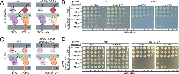
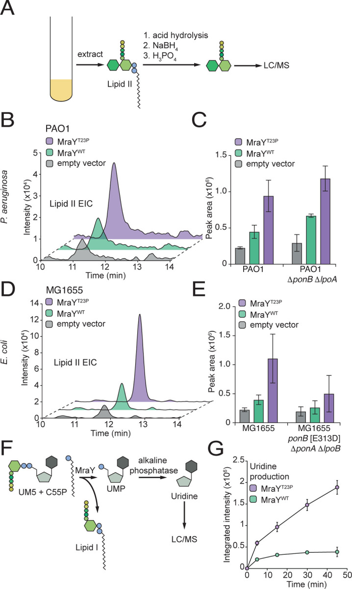
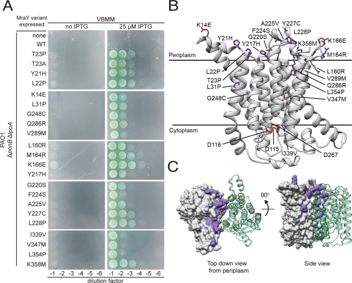
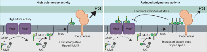
Many bacterial surface glycans such as the peptidoglycan (PG) cell wall, O-antigens, and capsules are built from monomeric units linked to a polyprenyl lipid carrier. How this limiting lipid carrier is effectively distributed among competing pathways has remained unclear for some time. Here, we describe the isolation and characterization of hyperactive variants of Pseudomonas aeruginosa MraY, the essential and conserved enzyme catalyzing the formation of the first lipid-linked PG precursor called lipid I. These variants result in the elevated production of the final PG precursor lipid II in cells and are hyperactive in a purified system. Amino acid substitutions within the activated MraY variants unexpectedly map to a cavity on the extracellular side of the dimer interface, far from the active site. Our structural evidence and molecular dynamics simulations suggest that the cavity is a binding site for lipid II molecules that have been transported to the outer leaflet of the membrane. Overall, our results support a model in which excess externalized lipid II allosterically inhibits MraY, providing a feedback mechanism to prevent the sequestration of lipid carrier in the PG biogenesis pathway. MraY belongs to the broadly distributed polyprenyl-phosphate N-acetylhexosamine 1-phosphate transferase (PNPT) superfamily of enzymes. We therefore propose that similar feedback mechanisms may be widely employed to coordinate precursor supply with demand by polymerases, thereby optimizing the partitioning of lipid carriers between competing glycan biogenesis pathways.

Keywords: Major: Biological Sciences; Minor: Microbiology; bacteriology; biochemistry; peptidoglycan; transferase.


Figures

Figure 1.
Figure 1.. MraY(T23P) restores growth to strains defective in PG biosynthesis.
Schematic representation of the aPBPs and their outer membrane lipoprotein activators in Pseudomonas aeruginosa (A) and Escherichia coli (C). Ten-fold serial dilutions of cells of the indicated P. aeruginosa (B) or E. coli (D) strains harboring expression plasmids producing the indicated MraY variant. Dilutions were plated on the indicated medium with or without IPTG to induce the production of MraY variants as indicated. Abbreviations: OM, outer membrane; PG, peptidoglycan; IM, inner membrane; GT, glycosyltransferase; TP, transpeptidase; LB, lysogeny broth; LBNS, LB with no added NaCl; VBMM, Vogel-Bonner minimal medium; IPTG, isopropyl-B-D-1-thiogalactopyranoside.
Figure 2.
Figure 2.. Cells expressing MraY(T23P) accumulate lipid II.
(A) Schematic representation of the method used to isolate and analyze lipid II from bacterial cells. (B-D) Representative extracted ion chromatograms of lipid II (EIC) and quantification of EICs for P. aeruginosa (B,C) or E. coli (D,E) strains expressing the indicated MraY variant. Three independent replicates of the extractions were performed and lipid II levels quantified using the area of the peak from the extracted ion chromatogram using the Agilent software. Error bars represent the standard deviation. For MraY(T23P) vs MraY(WT) in PAO1 P<0.05 PAO1 ΔponB ΔlpoA P<0.01, in PAO1, MG1655 P<0.05, MG1655 ΔponA ΔlpoB ponB[E313D], not significant. (F). Schematic representation of the MraY enzyme assay. (G) Representative time course showing the production of uridine in assays containing purified MraY or MraY(T23P) as indicated. The assay was repeated at least twice with two independent preparations of protein. Error bars represent standard deviation of reactions performed in duplicate. Abbreviations: NaBH4, sodium borohydride; H3PO4, phosphoric acid; LC/MS, liquid chromatography mass spectrometry; UM5, UDP-MurNAc-pentapeptide; C55P, undecaprenylphosphate.
Figure 3.
Figure 3.. Amino acid substitutions in hyperactive MraY variants localize to the extracytoplasmic surface of the dimer interface.
(A) Ten-fold serial dilutions of P. aeruginosa ΔponB ΔlpoA cells harboring expression plasmids producing the indicated MraY variant were plated on VBMM with or without IPTG to induce the MraY variants as indicated. (B) Structural model of P. aeruginosa MraY created using AlphaFold in cartoon viewed from the plane of the membrane. Residues altered in hyperactive variants tested in (A) are shown in stick representation (purple), while those residues previously implicated in catalysis are shown in red. (C) Structural model of the MraY dimer created using AlphaFold. Surface representation of one protomer is shown in grey with the residues altered in hyperactive variants colored in purple. The other protomer is shown in cartoon representation (green) for simplicity. Left, the periplasmic view of the dimer; Right, view from the plane of the membrane.
Figure 4.
Figure 4.. Identification of a potential lipid II binding site in MraY.
(A) Periplasmic view of the cryo-EM structure of the EcMraY(T23P) dimer within the YES complex shown in surface representation, with the unmodeled electron density shown (green). (B) As in A, membrane view of the electron density within the dimer interface of the EcMraY dimer with the foreground MraY removed. (C) Top view of the MraY dimer in a mixed CG membrane. Two lipid II molecules (highlighted as green, gold and pink spheres) freely enter the MraY cavity during unbiased MD simulations. In 8/9 repeats, 2 or 3 lipid I or II molecules bind the cavity. In the last repeat, one lipid II and one C55P molecule bind. (D) Lipid II contacts with MraY residues that interact with lipid II for over 60% of atomistic MD simulations. Error bars represent standard error from 5 repeats. Darker green bars represent residues altered in hyperactive variants. (E) Lipid II contacts with MraY residues by part of lipid II that is interacting (tail & phosphate, MurNAc, GlcNAc or pentapeptide). Residues shown are same as those in Fig 4D. Darker bars represent residues altered in hyperactive variants. (F) Average density of lipid II molecules (green) from atomistic MD simulations of MraY (grey) bound to lipid II. Shown as inside view of dimer interface, where only one monomer of MraY is shown and residues altered in hyperactive variants are colored in purple.
Figure 5.
Figure 5.. Model for feedback regulation of MraY by flipped lipid II.
Shown are schematics summarizing the model for MraY regulation. Left: When PG polymerase activity is high, flipped lipid II is consumed at a rate proportional to its production such that steady-state levels of the precursor remains low and MraY activity is unimpeded. Right: When PG polymerase activity is reduced due to changes in growth conditions or other perturbations, lipid II will be produced faster than it is consumed, resulting in the accumulation of elevated levels of flipped lipid II. Higher levels of the precursor promote its binding to MraY dimers, reducing their activity in order to bring lipid II supply back in balance with demand by the polymerases. See text for details. Abbreviations: C55P, undecaprenylphosphate; UM5, UDP-MurNAc-pentapeptide; UG, UDP-GlcNAc; PG, peptidoglycan.

References

    1. May K. L. & Silhavy T. J. Making a membrane on the other side of the wall. Biochimica Et Biophysica Acta Bba - Mol Cell Biology Lipids 1862, 1386–1393 (2017). - PMC - PubMed
    1. Silhavy T. J., Kahne D. & Walker S. The Bacterial Cell Envelope. Csh Perspect Biol 2, a000414 (2010). - PMC - PubMed
    1. Huszczynski S. M., Lam J. S. & Khursigara C. M. The Role of Pseudomonas aeruginosa Lipopolysaccharide in Bacterial Pathogenesis and Physiology. Pathogens 9, 6 (2019). - PMC - PubMed
    1. Rohs P. D. A. & Bernhardt T. G. Growth and Division of the Peptidoglycan Matrix. Annu. Rev. Microbiol. 75, 1–22 (2021). - PubMed
    1. Piepenbreier H., Diehl A. & Fritz G. Minimal exposure of lipid II cycle intermediates triggers cell wall antibiotic resistance. Nat Commun 10, 2733 (2019). - PMC - PubMed

Publication types