[Preprint]. 2023 Aug 6:2023.08.04.552072.
doi: 10.1101/2023.08.04.552072.

The cell adhesion molecule Echinoid promotes tissue survival and separately restricts tissue overgrowth in Drosophila imaginal discs

Affiliations

The cell adhesion molecule Echinoid promotes tissue survival and separately restricts tissue overgrowth in Drosophila imaginal discs

Danielle C Spitzer et al. bioRxiv. .

Update in

Abstract

The interactions that cells in Drosophila imaginal discs have with their neighbors are known to regulate their ability to survive. In a screen of genes encoding cell surface proteins for gene knockdowns that affect the size or shape of mutant clones, we found that clones of cells with reduced levels of echinoid (ed) are fewer, smaller, and can be eliminated during development. In contrast, discs composed mostly of ed mutant tissue are overgrown. We find that ed mutant tissue has lower levels of the anti-apoptotic protein Diap1 and has increased levels of apoptosis which is consistent with the observed underrepresentation of ed mutant clones and the slow growth of ed mutant tissue. The eventual overgrowth of ed mutant tissue results not from accelerated growth, but from prolonged growth resulting from a failure to arrest growth at the appropriate final size. Ed has previously been shown to physically interact with multiple Hippo-pathway components and it has been proposed to promote Hippo pathway signaling, to exclude Yorkie (Yki) from the nucleus, and restrain the expression of Yki-target genes. We did not observe changes in Yki localization in ed mutant tissue and found decreased levels of expression of several Yorkie-target genes, findings inconsistent with the proposed effect of Ed on Yki. We did, however, observe increased expression of several Yki-target genes in wild-type cells neighboring ed mutant cells, which may contribute to elimination of ed mutant clones. Thus, ed has two distinct functions: an anti-apoptotic function by maintaining Diap1 levels, and a function to arrest growth at the appropriate final size. Both of these are unlikely to be explained by a simple effect on the Hippo pathway.


Figures
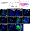
Figure 1:
Figure 1:. Clonal phenotypes observed in RNAi screen
(A) Summary of the screen where genes encoding cell-surface proteins were individually knocked down using RNAi transgenes (B-L) Phenotypes of imaginal discs containing clones generated with a FLP-out Gal4 and UAS-RNAi transgenes, Clones are marked by the inclusion of a UAS-GFP or UAS-RFP transgene as indicated. A cyst-like clone in (K) is shown at higher magnification in (L) and an orthogonal view in (L’). (B-K) are shown at the same magnification; the scale bar, 50 μm is shown in panel (B). (L, L’) are shown at a higher magnification: scale bar is 25 μm.
Figure 2:
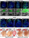
Figure 2:. Clones of echinoid mutant cells are eliminated when surrounded by wild-type cells.
(A-D) Imaginal discs containing GFP-marked clones of cells that express an RNAi directed against ed. Four different UAS-RNAi transgenes were used. Panels (A’-D’) and (A”-D”) show GFP-marked clones at higher magnification that are stained with an anti-Ed antibody. The magnified regions are indicated with dashed lines in (A-D). (E-H) GFP-marked clones of cells that are homozygous for the chromosome arm bearing FRT40A alone (E) or FRT40A and an allele of ed (F-H). Note the near absence of clones in the epithelium with the two null alleles of ed (F, G) and the presence of myoblast clones underlying the notum (indicated by an arrowheads). An orthogonal view is shown in (F’) with the disc epithelium outlined; the GFP-marked myoblasts are located basal to the epithelium. Both epithelial and myoblast clones are observed with the hypomorphic allele (H). (I-K) Clones of cells homozygous for either a wild-type chromosome arm distal to FTR40A (I) or edIF20, FRT40A (J) marked white, generated using eyFLP. The tester chromosome carries a recessive cell lethal resulting in the absence of wild-type twin spots. An overlay of the overgrown eye containing ed clones and the normally sized eye containing wild-type clones is shown in (K). (L, M) Clones generated using eyFLP using a wild-type tester chromosome that does not carry a recessive cell lethal mutation. FRT40A clones (L) or ed FRT40A clones (M) are white while the wild-type twin spots appear red in both panels. Note the almost complete absence of homozygous ed/ed tissue in eyes that contain wild-type twin spots.
Figure 3:
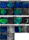
Figure 3:. echinoid clones are generated and then die, especially when more wild-type tissue is present.
(A-F) Wing discs containing clones expressing either UAS-w-RNAi (A, C, E) or UAS-ed-RNAi (B, D, F). Clones also express GFP. The clone density was varied by increasing the duration of the heat shock at 37° C during which hs-FLP is expressed. Thus, heat shocks of 12 min (A, B), 15 min (C, D) and 30 min (E, F) generate clones at progressively higher density. (G-K) An experiment to examine ed/ed clones 24 h and 72 h after generation by mitotic recombination. The design of the experiment is shown in (G). Discs were dissected 120 h AED and clones were induced either 24 h (H, I) or 72 h (J, K) prior to dissection. Clones generated using the FRT40A chromosome (H, J) are compared to clones homozygous for edIF20 (I, K).
Figure 4:
Figure 4:. echinoid mutant tissue has lower Diap1 levels and has higher levels of apoptosis
(A-D) Wing discs containing GFP-marked clones of cells that express either UAS-w-RNAi (A, B) or UAS-ed-RNAi (C, D). Clones that also express UAS-p35 are shown in (B, D). (E, F) Imaginal discs containing GFP-marked clones expressing UAS-w-RNAi (E) or UAS-ed-RNAi (F). Anti Dcp-1 antibody staining of the same discs is shown in (E’, F’). Images were taken at a basal Z-plane where Dcp-1 staining was most prominent. (G, H) Effect of reducing ed function in the entire wing pouch on levels of apoptosis. nub-Gal4 drives expression of UAS-GFP in (G, G’) or UAS-ed-RNAi in (H, H’). The pouch is visualized by GFP expression in (G) and with anti-Nubbin in (H). Apoptotic cells are visualized with anti-Dcp1. Note increased levels of apoptosis at the periphery of the pouch where nb-Gal4 expression is higher. (I, J) Discs expressing ed-RNAi in clones (I) or in the entire posterior compartment (J) stained with an anti-Diap1. The insets in (I, I’) show a clone at higher magnification. (K) Discs with GFP-marked clones that express both UAS-ed-RNAi and UAS-diap1 which results in an increase in both the number and size of clones. Staining with anti-Diap1 is shown in (K’). Note that the “rescued” clones still have smooth borders consistent with the propensity of cells with decreased ed to sort away from wild-type cells.
Figure 5:
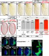
Figure 5:. Characteristics of overgrowth exhibited by echinoid mutant tissue
(A-I) Effect of reducing ed function on size of adult wings. (A-F) show adult wings of the indicated genotypes. An overlay of a wild-type wing and an overgrown wing of a heteroallelic combination of ed mutations is shown in (G). An overlay of nub-Gal4, UAS-w-RNAi and nub-Gal4, UAS-ed-RNAi is shown in (H). Quantification of wing areas is shown in (I). n=10 wings (+/+ [Oregon-R]; edsIH8/+; edIF20/+; edsIH8/edIF20; nub-Gal4, >GFP, >w-RNAi), 9 wings (nub-Gal4, >GFP, >ed-RNAi). “ns” indicates p> 0.05, ** indicates p< 0.01, calculated using ANOVA with post-hoc Tukey’s HSD test. Error bars indicate standard deviation. (J-P) A time course of growth of imaginal discs expressing either w-RNAi (J, K) or ed-RNAi (L-P) in the posterior compartment. All larvae expressing w-RNAi have pupariated soon after 120 h. However, much older larvae are observed in the population expressing ed-RNAi. Examples of discs from larvae that have delayed their pupariation are shown. All images are shown at the same scale (scale bar in panel J).
Figure 6:
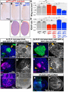
Figure 6:. Characterizations of mechanisms that function downstream of echinoid
(A-E) Effect of expressing full length ed (edFull) and a version where the cytoplasmic domain has been replaced by GFP (edΔC-GFP) on wing size and wing shape. (A) Wings of a heteroallelic combination of ed alleles that generate viable adults together with nub-Gal4. Inclusion of UAS-edFull (B) and UAS-edΔC-GFP (C) reduces wing size and brings the aspect ratio closer to wild-type. (B’) and (C’) show overlays of (B) and (C) over (A) respectively. For (D), n=10 wings (edsIH8/+; edIF20/+; edsIH8/edIF20; same as shown in Fig 5I), 4 wings (edsIH8/edIF20, nub-Gal4; edsIH8/edIF20, nub-Gal4, UAS-edFull), 6 wings (edsIH8/edIF20, nub-Gal4, UAS-edΔC-GFP). For (E), n=10 wings (edsIH8/+; edIF20/+), 9 wings (edsIH8/edIF20, edsIH8/edIF20, nub-Gal4, UAS-edΔC-GFP), 4 wings (edsIH8/edIF20, nub-Gal4; edsIH8/edIF20, nub-Gal4, UAS-edFull). The same wings were used in (D) and (E); number of wings differ if damage or mounting artifacts prevented measurement of both wing area and aspect ratio. “ns” indicates p> 0.05, ** indicates p< 0.01, calculated using ANOVA with post-hoc Tukey’s HSD test. Error bars indicate standard deviation. (F, F’) GFP-marked clones expressing ed-RNAi stained with anti-Diap1 (F’). Yellow arrowhead indicates wild type cells immediately adjacent to the clone. (G, G’) RFP marked clone expressing ed-RNAi shows decreased expression of a diap1 transcriptional reporter generated using 8 copies of the Hippo-response element (HRE) from the diap1 promoter (G’). (H, H’) RFP-labeled clones expressing ed-RNAi show no obvious alteration in the localization of a GFP-tagged Yki. The region of the disc enclosed by the dashed line is shown at higher magnification in the insets. (I, I’) GFP-labeled clone expressing ed-RNAi shows decreased expression of a ban-lacZ reporter (I’). The yellow arrowhead indicates wild-type cells adjacent to the clone that show increased expression of ban-lacZ. (J, J’) RFP-labeled clone expressing ed-RNAi shows decreased expression of a fj-lacZ reporter (J’). The yellow arrowhead indicates wild-type cells adjacent to the clone that show increased expression of ban-lacZ. (K, K’) Expression of ed-RNAi in the posterior compartment of the wing disc results in increased ex-lacZ expression (K’). The posterior compartment is identifiable because it does not express Ci (K).
Figure 7:
Figure 7:. echinoid mutant cells could be eliminated by a mechanism similar to cell competition at clonal boundaries,
(A, A’) GFP-marked clones expressing ed-RNAi (A) stained with an anti-Fat antibody. The inset shows a close-up of the boundary of the clone. (B) A model figure showing the effect of ed loss in ed cells and wild-type neighbors. Within ed clones, Fat levels are increased and Diap1 levels are decreased; the opposite is true in the wild-type neighbors. These differences may confer a competitive advantage to the wild-type neighbors, which could facilitate the elimination of ed clones from mosaic tissue.

References

    1. Adachi-Yamada T., and O’Connor M. B., 2002. Morphogenetic Apoptosis: A Mechanism for Correcting Discontinuities in Morphogen Gradients. Dev. Biol. 251: 74–90. 10.1006/dbio.2002.0821 - DOI - PubMed
    1. Adachi-Yamada T., Harumoto T., Sakurai K., Ueda R., Saigo K., et al. , 2005. Wing-to-Leg Homeosis by Spineless Causes Apoptosis Regulated by Fish-lips, a Novel Leucine-Rich Repeat Transmembrane Protein. Mol. Cell. Biol. 25: 3140–3150. 10.1128/MCB.25.8.3140-3150.2005 - DOI - PMC - PubMed
    1. Ahmed A., Chandra S., Magarinos M., and Vaessin H., 2003. Echinoid mutants exhibit neurogenic phenotypes and show synergistic interactions with the Notch signaling pathway. Development 130: 6295–6304. 10.1242/dev.00796 - DOI - PubMed
    1. Alpar L., Bergantiños C., and Johnston L. A., 2018. Spatially Restricted Regulation of Spätzle/Toll Signaling during Cell Competition. Dev. Cell 46: 706–719.e5. 10.1016/j.devcel.2018.08.001 - DOI - PMC - PubMed
    1. Amoyel M., and Bach E. A., 2014. Cell competition: how to eliminate your neighbours. Development 141: 988–1000. 10.1242/dev.079129 - DOI - PMC - PubMed

Publication types