. 2023 Aug 31;97(8):e0024623.
doi: 10.1128/jvi.00246-23. Epub 2023 Aug 14.

Discontinuous L-binding motifs in the transactivation domain of the vesicular stomatitis virus P protein are required for terminal de novo transcription initiation by the L protein

Affiliations

Discontinuous L-binding motifs in the transactivation domain of the vesicular stomatitis virus P protein are required for terminal de novo transcription initiation by the L protein

Nirmala Gupta et al. J Virol. .

Abstract

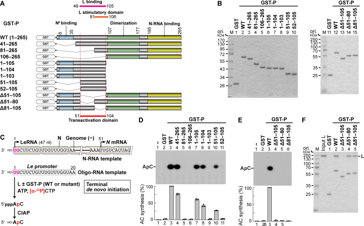
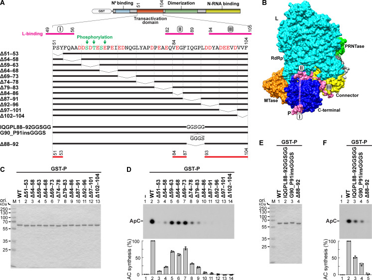
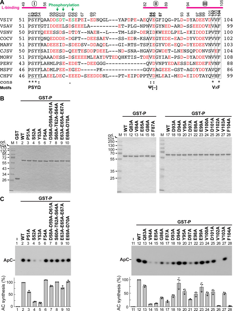
The phospho- (P) protein, the co-factor of the RNA polymerase large (L) protein, of vesicular stomatitis virus (VSV, a prototype of nonsegmented negative-strand RNA viruses) plays pivotal roles in transcription and replication. However, the precise mechanism underlying the transcriptional transactivation by the P protein has remained elusive. Here, using an in vitro transcription system and a series of deletion mutants of the P protein, we mapped a region encompassing residues 51-104 as a transactivation domain (TAD) that is critical for terminal de novo initiation, the initial step of synthesis of the leader RNA and anti-genome/genome, with the L protein. Site-directed mutagenesis revealed that conserved amino acid residues in three discontinuous L-binding sites within the TAD are essential for the transactivation activity of the P protein or important for maintaining its full activity. Importantly, relative inhibitory effects of TAD point mutations on synthesis of the full-length leader RNA and mRNAs from the 3'-terminal leader region and internal genes, respectively, of the genome were similar to those on terminal de novo initiation. Furthermore, any of the examined TAD mutations did not alter the gradient pattern of mRNAs synthesized from internal genes, nor did they induce the production of readthrough transcripts. These results suggest that these TAD mutations impact mainly terminal de novo initiation but rarely other steps (e.g., elongation, termination, internal initiation) of single-entry stop-start transcription. Consistently, the mutations of the essential or important amino acid residues within the P TAD were lethal or deleterious to VSV replication in host cells. IMPORTANCE RNA-dependent RNA polymerase L proteins of nonsegmented negative-strand RNA viruses belonging to the Mononegavirales order require their cognate co-factor P proteins or their counterparts for genome transcription and replication. However, exact roles of these co-factor proteins in modulating functions of L proteins during transcription and replication remain unknown. In this study, we revealed that three discrete L-binding motifs within a transactivation domain of the P protein of vesicular stomatitis virus, a prototypic nonsegmented negative-strand RNA virus, are required for terminal de novo initiation mediated by the L protein, which is the first step of synthesis of the leader RNA as well as genome/anti-genome.

Keywords: L protein; Mononegavirales; P protein; RNA-dependent RNA polymerase; replication; rhabdovirus; transcription; transcription factor; transcription initiation; vesicular stomatitis virus.


Conflict of interest statement

The authors declare no conflict of interest.

Figures

Fig 1
Fig 1
Mapping of a transcriptional transactivation domain in the VSV P protein. (A) The wild-type VSV P protein (265 amino acids) and its deletion mutants were expressed as glutathione S-transferase (GST) fusion proteins. The positions of the known functional domains and transactivation domain (mapped in this study) in the P protein are indicated on the top and bottom, respectively. (B) The indicated proteins (1 µg) were analyzed by 10% SDS-PAGE followed by staining with Coomassie Brilliant Blue or One-Step Blue. M lanes show marker proteins with the indicated molecular masses. (C) The VSV genome is wrapped with the N proteins to form a nucleocapsid (called N-RNA template). The 3′-terminal and internal initiation sites for synthesis of LeRNA/anti-genome and N mRNA, respectively, in the genome are indicated by bent arrows (upper). In vitro AC dinucleotide synthesis was performed with a naked 20-nt oligo-RNA template with the 3′-terminal genome sequence (Le promoter, middle), ATP, [α-32P]CTP, and the L protein in the presence or absence of GST-P (WT or mutant). The substrates and product (5′-pppApC) were digested with calf intestine alkaline phosphatase (CIAP). (D and E) After AC synthesis with the indicated samples, CIAP-resistant products (5′-HO-ApC) were analyzed by 20% urea-PAGE followed by autoradiography. The graphs show relative AC synthesis activities for respective reactions, in which radioactivities of respective products without (lane 1) and with (lane 3) of GST-P WT were set to 0% and 100%, respectively. The dot plots, columns, and error bars represent the individual normalized values, means, and standard deviations, respectively, from three independent experiments (n = 3). (F) A GST pull-down assay was carried out to examine the interaction between the L and P proteins. The L protein was incubated with GST or GST-P proteins (WT or deletion mutant) pre-bound to GSH beads. Bound proteins were analyzed along with input L (lane 1) by 10% SDS-PAGE followed by staining with One-Step Blue.
Fig 2
Fig 2
Three discontinuous regions in the P TAD are required for efficient transcription initiation with the L protein. (A) The amino acid sequence of the P TAD (residues 51–104) is shown below the schematic diagram of GST-P. Acidic and phosphoacceptor residues are highlighted in red and green, respectively. The locations of L-binding sites I–III are indicated above the TAD sequence by magenta lines. The indicated deletion, substitution, and insertion mutations were introduced into the TAD of GST-P. The positions of three discontinuous regions required for the transactivation activity of the P protein are indicated by red lines at the bottom. (B) A surface view of the 3D structure of the VSV L protein complexed with a P fragment (PDB id: 6U1X) is shown. L-binding sites I–III connected by unstructured linker regions (dashed lines) in the P fragment are colored in magenta. The RNA-dependent RNA polymerase, GDP polyribonucleotidyltransferase, connector, methyltransferase, and C-terminal domains of the L protein are colored in cyan, green, yellow, orange, and blue, respectively. The image was generated with the UCSF Chimera program (45). (CF) The indicated proteins were subjected to SDS-PAGE (C and E) and in vitro AC synthesis (D and F) as in Fig. 1.
Fig 3
Fig 3
Conserved amino acid residues in the P TAD are required for terminal de novo initiation. (A) The amino acid sequence of the P TAD (residues 51–104) of VSV (VSIV, vesicular stomatitis Indiana virus, GenBank (https://www.ncbi.nlm.nih.gov/genbank/) accession no.: FJ478454) is aligned with those of other vesiculoviral P proteins using the PSI-Coffee program (46). Virus names (GenBank accession nos. in brackets) are as follows: VSAV, vesicular stomatitis Alagoas virus (EU373658); VSNV, vesicular stomatitis New Jersey virus (M29788); COCV, vesicular stomatitis Cocal virus (EU373657); MARAV, Maraba virus (HQ660076); CJSV, Carajas virus (FW339542); MORV, Morreton virus (KM205007); PERV, Perinet virus (HM566195); MSPV, Malpais Spring virus (KC412247); CHPV, Chandipura virus (AJ810083). The numbers on the left and right of the sequences indicate the amino acid positions in respective P proteins. Identical (*), conserved (:), and semi-conserved (.) amino acid residues are indicated. Conserved motifs are shown at the bottom (Ω, Ψ, [–], and x indicate aromatic, aliphatic, negatively charged, and any amino acids, respectively). (B and C) Alanine-scanning mutagenesis was performed on the indicated amino acid residues in the TAD of GST-P. The names of the point mutants include the original amino acid (one-letter code) at the indicated position followed by the replacement amino acid. The indicated proteins were subjected to SDS-PAGE (B) and in vitro AC synthesis (C) as in Fig. 1.
Fig 4
Fig 4
The effects of substitution of closely related amino acids for the critical amino acid residues in the P TAD on the transactivation activity. The important or essential amino acid residues within the P TAD were replaced with their closely related amino acids. The indicated proteins were subjected to SDS-PAGE (A) and in vitro AC synthesis (B) as in Fig. 1.
Fig 5
Fig 5
Conserved amino acid residues in the P TAD are required for synthesis of LeRNA and mRNAs. (A) Nontagged P proteins (WT and mutants) were prepared by proteolytic removal of the N-terminal GST portions from the GST-P fusion proteins and analyzed by SDS-PAGE. (B) In vitro transcription was carried out with the L protein and N-RNA template in the presence or absence of the P protein (WT or mutant). The L-P RdRp complex initiates stop-start transcription at the 3′-end of the genome, and sequentially transcribes the leader region (Le) and internal genes (N, P, M, G, and L) into the leader RNA (LeRNA) and mRNAs with a 5′-cap structure and 3′-poly(A) tail, respectively. (C) 32P-labeled LeRNA and mRNAs (deadenylated) were analyzed by 20% (upper) and 5% (middle) urea-PAGE, respectively, followed by autoradiography. The graph shows relative LeRNA and mRNA synthesis activities for respective reactions, in which radioactivities of respective products without (lane 1) and with (lane 2) of the P protein WT were set to 0% and 100%, respectively. The dot plots, columns, and error bars represent the individual normalized values, means, and standard deviations, respectively, from three independent experiments (n = 3).
Fig 6
Fig 6
Conserved amino acid residues in the P TAD are required for efficient VSV replication in infected cells. (A and B) rVSVs were generated from a cDNA encoding the VSV anti-genome (WT) or those with the indicated mutations in the P gene using the reverse genetics system and amplified once. The indicated volumes (10−7–10−1 mL) of the culture supernatants were subjected to a plaque assay. (C) At least three virus clones generated from the respective cDNAs with the indicated mutations were plaque isolated and amplified. In case of V102A, viruses were amplified from three large (see ix) and eight pinpoint plaques (see Fig. 7). Representative images of plaques formed by one of the virus clones were shown. The P genes of all the clones were sequenced. cDNA sequences of respective mutation sites in the P genes of representative clones are shown above electropherograms. There were no other nucleotide changes in the P genes.
Fig 7
Fig 7
Emergence of revertant viruses from the V102A mutant. rVSVs were generated from the cDNA encoding the VSV anti-genome with the V102A mutant P gene as described in Fig. 6 and amplified from eight pinpoint plaques for the indicated incubation time. The indicated volumes (10−7–10−1 mL) of samples #1–5 were subjected to the plaque assay. Similar to sample #5, samples #6–8 did not form detectable plaques (not shown). cDNA sequences of the mutation site are shown above electropherograms.
Fig 8
Fig 8
Roles of the P protein in terminal de novo initiation with the L protein. The L protein possesses the N-terminal superdomain [composed of the RNA-dependent RNA polymerase (RdRp, cyan) and GDP polyribonucleotidyltransferase (PRNTase, green) domains] and C-terminal appendages [connector (yellow), methyltransferase (MTase, orange), and C-terminal (blue) domains]. The P protein has the N-terminal N0-binding, transactivation (this study), dimerization, and C-terminal N-RNA-binding domains. In the absence of the P protein (L alone), the C-terminal domains of the L protein are mobile. Binding of three L-binding sites (I–III) within the transactivation domain of the P protein to the C-terminal, RdRp, and connector domains, respectively, of the L protein induces conformational compaction of the L protein by tethering the C-terminal domains to the RdRp domain (21), resulting in the formation of the L-P complex that is fully competent for terminal de novo initiation (this study). The L-P complex interacts with the N-RNA template via the C-terminal N-RNA-binding domain of the P protein and forms a transcription initiation complex on the 3′-terminal of the genome with the initiator (ATP) and incoming (CTP) nucleotides. The conserved tryptophan residue (W) on the priming-capping loop extended from the PRNTase domain into the RdRp active site stabilizes the terminal initiation complex to conduct the first phosphodiester bond formation (AC dinucleotide synthesis) (18).
Fig 9
Fig 9
Models of WT and single-mutant P proteins in complex with L. In each panel, the L (cyan) and P (yellow) proteins are shown in cartoon representation. The P amino acid residue of interest is shown in stick representation, with the residue number noted in the top left corner of each panel. Panel (i) represents wild-type sequences [(A) S52; (B) Y53; (C) V102; (D) F104] and structures, while (ii) and (iii) show the selected mutations. L protein residues within 5Å of any atom in the noted P residue are also shown in sticks. Individual residues are labeled. Dashed lines represent hydrogen bonds. Orange residues in panel D note residues that form hydrogen bonds between L and P. Movement of these residues allows the bonds to be altered or new bonds to be formed.

Similar articles

Cited by

References

    1. Ogino T, Green TJ. 2019. RNA synthesis and capping by non-segmented negative strand RNA viral polymerases: lessons from a prototypic virus. Front Microbiol 10:1490. doi:10.3389/fmicb.2019.01490 - DOI - PMC - PubMed
    1. Green TJ, Zhang X, Wertz GW, Luo M. 2006. Structure of the vesicular stomatitis virus nucleoprotein-RNA complex. Science 313:357–360. doi:10.1126/science.1126953 - DOI - PubMed
    1. Ge P, Tsao J, Schein S, Green TJ, Luo M, Zhou ZH. 2010. Cryo-EM model of the bullet-shaped vesicular stomatitis virus. Science 327:689–693. doi:10.1126/science.1181766 - DOI - PMC - PubMed
    1. Emerson SU, Wagner RR. 1972. Dissociation and reconstitution of the transcriptase and template activities of vesicular stomatitis B and T virions. J Virol 10:297–309. doi:10.1128/JVI.10.2.297-309.1972 - DOI - PMC - PubMed
    1. Emerson SU, Yu Y. 1975. Both NS and L proteins are required for in vitro RNA synthesis by vesicular stomatitis virus. J Virol 15:1348–1356. doi:10.1128/JVI.15.6.1348-1356.1975 - DOI - PMC - PubMed

Publication types

MeSH terms

LinkOut - more resources