. 2023 Aug 31;97(8):e0050923.
doi: 10.1128/jvi.00509-23. Epub 2023 Aug 14.

Reverse genetics of parrot bornavirus 4 reveals a unique splicing of the glycoprotein gene that affects viral propagation

Affiliations

Reverse genetics of parrot bornavirus 4 reveals a unique splicing of the glycoprotein gene that affects viral propagation

Ryo Komorizono et al. J Virol. .

Abstract

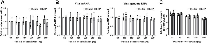
Viruses can utilize host splicing machinery to enable the expression of multiple genes from a limited-sized genome. Orthobornaviruses use alternative splicing to regulate the expression level of viral proteins and achieve efficient viral replication in the nucleus. Although more than 20 orthobornaviruses have been identified belonging to eight different viral species, virus-specific splicing has not been demonstrated. Here, we demonstrate that the glycoprotein (G) transcript of parrot bornavirus 4 (PaBV-4; species Orthobornavirus alphapsittaciforme), a highly virulent virus in psittacines, undergoes mRNA splicing and expresses a soluble isoform termed sGP. Interestingly, the splicing donor for sGP is not conserved in other orthobornaviruses, including those belonging to the same orthobornavirus species, suggesting that this splicing has evolved as a PaBV-4-specific event. We have also shown that exogenous expression of sGP does not affect PaBV-4 replication or de novo virion infectivity. In this study, to investigate the role of sGP in viral replication, we established a reverse genetics system for PaBV-4 by using avian cell lines and generated a recombinant virus lacking the spliced mRNA for sGP. Using the recombinant viruses, we show that the replication of the sGP-deficient virus is significantly slower than that of the wild-type virus and that the exogenous expression of sGP cannot restore its propagation efficiency. These results suggest that autologous or controlled expression of sGP by splicing may be important for PaBV-4 propagation. The reverse genetics system for avian bornaviruses developed here will be a powerful tool for understanding the replication strategies and pathogenesis of avian orthobornaviruses. IMPORTANCE Parrot bornavirus 4 (PaBV-4) is the dominant cause of proventricular dilatation disease, a severe gastrointestinal and central nervous system disease among avian bornaviruses. In this study, we discovered that PaBV-4 expresses a soluble isoform of glycoprotein (G), called sGP, through alternative splicing of the G mRNA, which is unique to this virus. To understand the role of sGP in viral replication, we generated recombinant PaBV-4 lacking the newly identified splicing donor site for sGP using a reverse genetics system and found that its propagation was significantly slower than that of the wild-type virus, suggesting that sGP plays an essential role in PaBV-4 infection. Our results provide important insights not only into the replication strategy but also into the pathogenesis of PaBV-4, which is the most prevalent bornavirus in captive psittacines worldwide.

Keywords: RNA splicing; avian virus; bornavirus; proventricular dilatation disease; reverse genetic analysis.


Conflict of interest statement

The authors declare no conflict of interest.

Figures

Fig 1
Fig 1
Identification of an alternative splicing event in the PaBV-4 G gene. (A) Read coverage of viral RNA in brain tissue of three PaBV-4-infected macaws. Viral reads acquired by RNA-seq were mapped to the PaBV-4 reference genome (accession number JX065209). (B) Viral transcripts and splicing junction in the G gene of PaBV-4. Splicing junctions were identified by STAR and visualized by Integrative Genomics Viewer (IGV). A schematic diagram of PaBV-4 G protein is indicated. SP, the signal peptide; GP-N, ectodomain subunit region (gp27); CS, the cleavage site by furin; GP-C, the transmembrane subunit region (gp29); TM, the transmembrane region; CT, the cytoplasmic tail; N-SD, newly identified splicing donor site; SA2, splicing acceptor 2. (C) Detection of novel G splicing in PaBV-4-infected cells by RT-PCR using different primer pairs. Agarose gel images of RT-PCR are shown. Two different primer pairs (primer pair 1 and 2; see Materials and Methods) were used. The directions and positions of each primer are shown in Fig. 1B. The bands marked with a triangle indicate amplicons of the identified G splice. (D and E) Read coverage of newly identified splicing junction in the G gene in PaBV-4 strain AR18A-infected QT6 cells. The splicing donor (G) and acceptor (T) sites were defined as position 0, and the relative sequence positions are shown.
Fig 2
Fig 2
The newly identified splicing is found only in PaBV-4. (A) Phylogenetic analysis of orthobornavirus G genes using the neighbor-joining method. NCBI accession numbers are shown next to each taxon. The surrounding nucleotide sequences of the splicing donor and acceptor sites are shown for each isolate. (B–F) Read coverage of the relative splicing donor site achieved from cell cultures persistently infected with different orthobornaviruses: (B) PaBV-4 strain 7I6, (C) PaBV-2 strain KOKO, (D) PaBV-7 strain KU2020, (E) MuBV-1, and (F) BoDV-1 strain He/80.
Fig 3
Fig 3
The newly identified splicing event produces an extracellular isoform of G. (A) Schematic representation of the full-length PaBV-4 G gene and the spliced isoform of G, sGP. While the full-length gene encodes for the signal peptide sequence (SP), the GP-N ectodomain subunit region (gp27), the cleavage site by furin (CS), the GP-C transmembrane subunit region (gp29), the transmembrane region (TM), and the cytoplasmic tail (CT), the spliced product encodes only for SP, GP-N, and the N-terminal part of CT. (B) Detection of recombinant G isoforms: Western blot analysis was performed on HEK293T cells transfected with full-length G or sGP expression plasmids using anti-BoDV-1 G or anti-FLAG antibodies. (C) The mCherry-fused signal peptide (SP-mCherry), full-length G (G-mCherry), and sGP (sGP-mCherry) were observed in transfected QT6-T cells. (D) Subcellular localization of FLAG-fused full-length G and sGP in transfected QT6-T cells was analyzed by IFA using anti-BoDV-1 G or anti-FLAG antibodies. Cell nuclei were counterstained with 4′,6-diamidino-2-phenylindole (DAPI). Intracellular (E) and extracellular (F) expression of HiBit-labeled sGP. HiBit expression was detected by chemiluminescence assay from cell lysate (E, intracellular) or cell culture supernatant (F, extracellular) at 48 h after the transfection of each construct into uninfected (white column) or PaBV-4-infected (gray column) QT6-T cells. Individual dots represent three independent experiments. Bars indicate the arithmetic mean ± standard error (SE).
Fig 4
Fig 4
The overexpression of sGP does not affect viral replication. (A) Effect of sGP expression on viral polymerase activity. PaBV-4 minigenome assays were performed in sGP-overexpressing DF-1T cells. The empty plasmid as a control (white column) or sGP expression plasmid (gray column) was transfected with minigenome and helper plasmids. Luciferase activity was measured 72 h after transfection. The amount of transfected plasmid is indicated. Results are shown as relative polymerase activity normalized to the sample with the lowest concentration of the empty vector (Control, 50 ng). (B–D) Effects of sGP expression on viral transcription, replication, and particle production. (B) PaBV-4-infected QT6 cells were transfected with empty plasmid as a control (white column) or sGP expression plasmid (gray column), and qRT-PCR was performed to quantify viral mRNA or genomic RNA at 48 h post transfection. (C) The supernatant of sGP-transfected PaBV-4-infected QT6 cells was collected at 48 h post transfection and dilution series of the supernatant were inoculated into uninfected QT6 cells to determine viral titers by IFA using an anti-PaBV-4 P antibody 3 days after inoculation. Dots indicate three independent experiments. Bars represent arithmetic mean ± standard error (SE).
Fig 5
Fig 5
Development of a PaBV-4 reverse genetics system and rescue of sGP-deficient PaBV-4. Schematic representation of the genome structure of rPaBV-4 p/m GFP (A) and the PaBV-4 reverse genetics procedure (B). (C and D) QT6 or DF-1 cells were transfected with expression plasmids encoding for different variants of rPaBV-4 p/m GFP together with helper plasmids. (−)AACA and (+)AACA indicate the modification of the terminal sequence of the PaBV-4 cDNA plasmid. At 96 h after transfection, the transfected cells were cocultured with fresh blasticidin-resistant QT6 cells and blasticidin treatment was started to remove the transfected cells. The rescue efficiency was assessed by determining the number of GFP-positive cells at 6 days (C) or 30 days (D) after transfection. Individual dots represent six independent experiments. Bars indicate the arithmetic mean ± standard error (SE). Statistical analysis was performed by Tukey’s multiple comparison test. **, P < 0.01. (E) Detection of PaBV-4 P and GFPs in QT6 cells infected with the indicated viruses by Western blot analysis. Antibodies used in this analysis are indicated on the left side of the panels. (F) Representative bright field image (top) and GFP fluorescence (bottom) from rPaBV-4 p/m GFP persistently infected cells 60 days after transfection. Scale bar: 100 µm.
Fig 6
Fig 6
rPaBV-4 without the G splicing donor reduces viral propagation. (A) Schematic diagram of the PaBV-4 G and the sequence design of the G splicing donor mutant for MutΔsGP. The nucleotide sequences of the splicing donor and acceptor sites in the G gene are shown in red letters. SP, signal peptide; GP-N, ectodomain subunit region (gp27); CS, the cleavage site by furin; GP-C, transmembrane subunit region (gp29); TM, transmembrane region; CT, cytoplasmic region. (B) Read coverage of RNA-seq data around the G splicing donor site. The splicing donor (G) was defined as 0, and the relative sequence positions are shown with the read coverage in WT (left) and MutΔsGP-infected QT6 cells (right). (C) Localization of N and P proteins in MutΔsGP-infected QT6 cells. IFA was performed using anti-BoDV-1 N and anti-BoDV-1 P antibodies. Nuclei were counterstained with DAPI. Scale bars: 10 µm. (D) Growth kinetics of rPaBV-4 MutΔsGP. QT6 cells were inoculated with WT and MutΔsGP viruses at a multiplicity of infection (MOI) of 0.05, and the percentage of GFP-positive cells was measured every 3 days. **, P < 0.01. (E) Competition assay between WT and MutΔsGP viruses. rPaBV-4 WT- and MutΔsGP-infected QT6 cells were cocultured with uninfected QT6 cells at a ratio of 1:1:8, and the population percentages of WT and MutΔsGP viruses in the culture were determined by sequencing of 25 cloned RT-PCR products every 3 days. D, days after coculture. Three independent experiments were performed and the respective mean values are shown. (F) The propagation of MutΔsGP virus in sGP-expressing QT6 cells. Parental (empty) and sGP-expressing (sGP) QT6 cells were inoculated with rPaBV-4 WT or MutΔsGP at an MOI of 0.05. Levels of viral genomic RNA (left) and P mRNA (right) were measured by qRT-PCR on the indicated days. dpi, day after inoculation. Means ± standard error (SE) of relative viral RNA levels normalized to GAPDH of three independent experiments are shown. The amount of viral RNA at 3 dpi was defined as 1.0, and the values for each time point (dpi) are shown as relative ratios. Significance was analyzed by one-way analysis of variance (ANOVA) followed by Dunnett’s multiple comparison test. Different letters (a, b, c) indicate statistically significant differences at P < 0.05.

Similar articles

References

    1. Kistler AL, Gancz A, Clubb S, Skewes-Cox P, Fischer K, Sorber K, Chiu CY, Lublin A, Mechani S, Farnoushi Y, Greninger A, Wen CC, Karlene SB, Ganem D, DeRisi JL. 2008. Recovery of divergent avian bornaviruses from cases of proventricular dilatation disease: identification of a candidate etiologic agent. Virol J 5:88. doi:10.1186/1743-422X-5-88 - DOI - PMC - PubMed
    1. Honkavuori KS, Shivaprasad HL, Williams BL, Quan PL, Hornig M, Street C, Palacios G, Hutchison SK, Franca M, Egholm M, Briese T, Lipkin WI. 2008. Novel borna virus in psittacine birds with proventricular dilatation disease. Emerg Infect Dis 14:1883–1886. doi:10.3201/eid1412.080984 - DOI - PMC - PubMed
    1. Sassa Y, Horie M, Fujino K, Nishiura N, Okazaki S, Furuya T, Nagai M, Omatsu T, Kojima A, Mizugami M, Ueda K, Iki H, Ebisawa K, Tomonaga K, Mizutani T. 2013. Molecular epidemiology of avian bornavirus from pet birds in Japan. Virus Genes 47:173–177. doi:10.1007/s11262-013-0913-3 - DOI - PubMed
    1. Heffels-Redmann U, Enderlein D, Herzog S, Herden C, Piepenbring A, Neumann D, Müller H, Capelli S, Müller H, Oberhäuser K, Gerlach H, Kaleta EF, Lierz M. 2011. Occurrence of avian bornavirus infection in captive psittacines in various European countries and its association with proventricular dilatation disease. Avian Pathol 40:419–426. doi:10.1080/03079457.2011.589825 - DOI - PubMed
    1. Weissenböck H, Bakonyi T, Sekulin K, Ehrensperger F, Doneley RJT, Dürrwald R, Hoop R, Erdélyi K, Gál J, Kolodziejek J, Nowotny N. 2009. Avian bornaviruses in psittacine birds from Europe and Australia with proventricular dilatation disease. Emerg Infect Dis 15:1453–1459. doi:10.3201/eid1509.090353 - DOI - PMC - PubMed

Publication types

LinkOut - more resources