. 2023 Aug 31;97(8):e0081523.
doi: 10.1128/jvi.00815-23. Epub 2023 Aug 14.

Nuclear proinflammatory cytokine S100A9 enhances expression of human papillomavirus oncogenes via transcription factor TEAD1

Affiliations

Nuclear proinflammatory cytokine S100A9 enhances expression of human papillomavirus oncogenes via transcription factor TEAD1

Seiichiro Mori et al. J Virol. .

Erratum in

Abstract

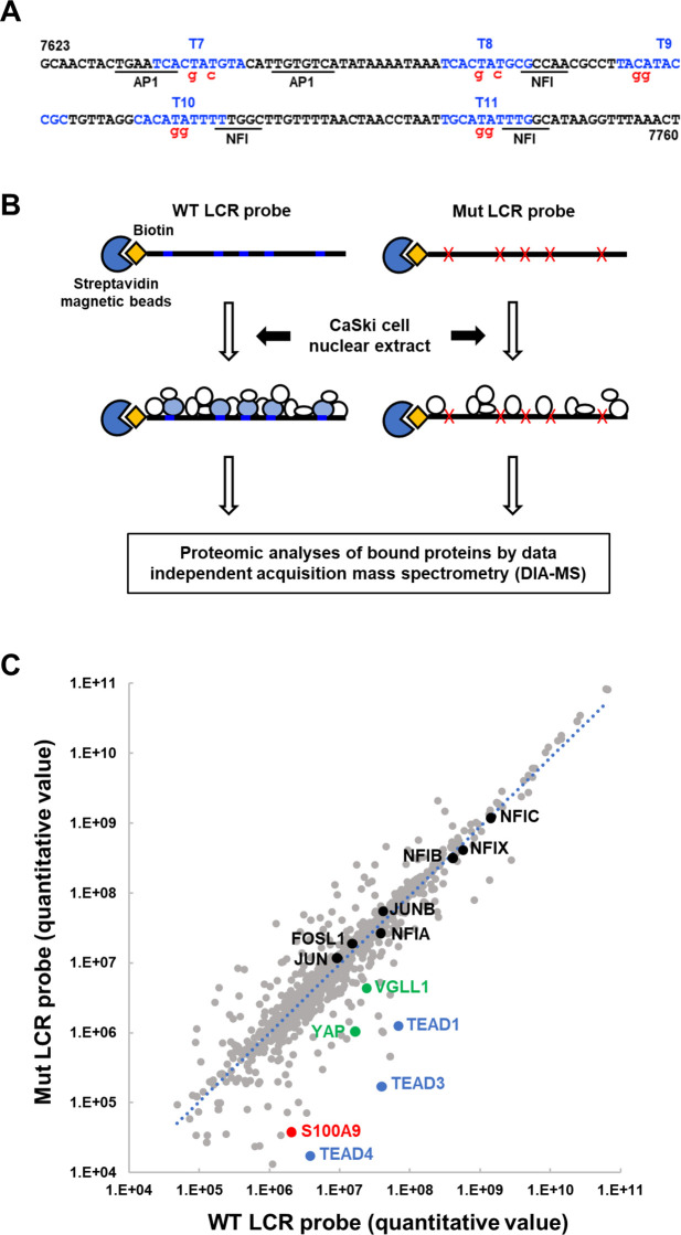
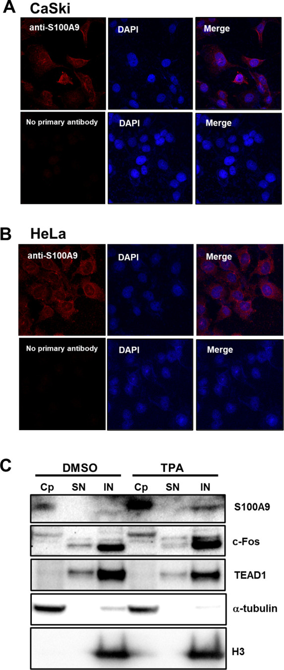
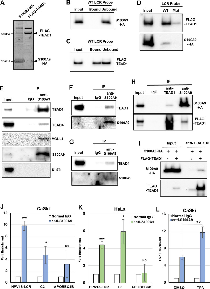
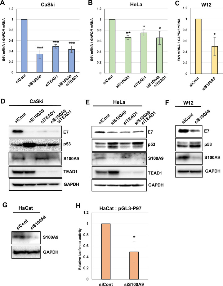
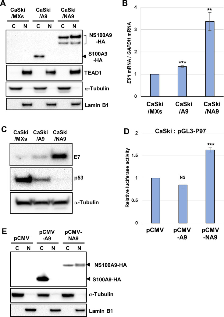
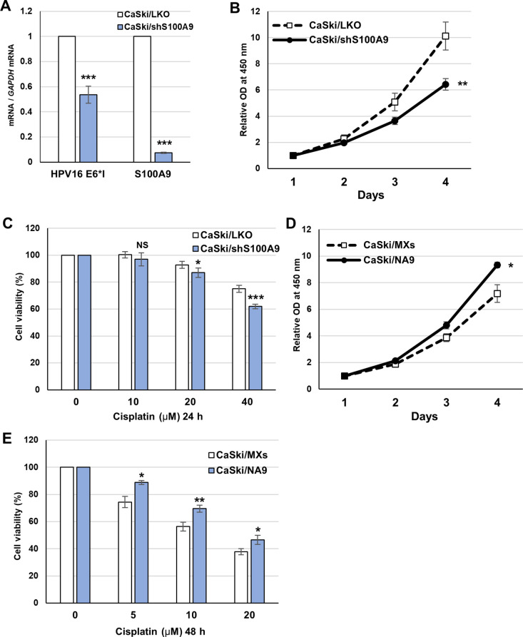
Transcription of the human papillomavirus (HPV) oncogenes, E6 and E7, is regulated by the long control region (LCR) of the viral genome. Although various transcription factors have been reported to bind to the LCR, little is known about the transcriptional cofactors that modulate HPV oncogene expression in association with these transcription factors. Here, we performed in vitro DNA-pulldown purification of nuclear proteins in cervical cancer cells, followed by proteomic analyses to identify transcriptional cofactors that bind to the HPV16 LCR via the transcription factor TEAD1. We detected the proinflammatory cytokine S100A9 that localized to the nucleus of cervical cancer cells and associated with the LCR via direct interaction with TEAD1. Nuclear S100A9 levels and its association with the LCR were increased in cervical cancer cells by treatment with a proinflammatory phorbol ester. Knockdown of S100A9 decreased HPV oncogene expression and reduced the growth of cervical cancer cells and their susceptibility to cisplatin, whereas forced nuclear expression of S100A9 using nuclear localization signals exerted opposite effects. Thus, we conclude that nuclear S100A9 binds to the HPV LCR via TEAD1 and enhances viral oncogene expression by acting as a transcriptional coactivator. IMPORTANCE Human papillomavirus (HPV) infection is the primary cause of cervical cancer, and the viral oncogenes E6 and E7 play crucial roles in carcinogenesis. Although cervical inflammation contributes to the development of cervical cancer, the molecular mechanisms underlying the role of these inflammatory responses in HPV carcinogenesis are not fully understood. Our study shows that S100A9, a proinflammatory cytokine, is induced in the nucleus of cervical cancer cells by inflammatory stimuli, and it enhances HPV oncogene expression by acting as a transcriptional coactivator of TEAD1. These findings provide new molecular insights into the relationship between inflammation and viral carcinogenesis.

Keywords: HPV; S100A9; TEAD1; proinflammatory cytokine; transcriptional coactivator; viral oncogenes.


Conflict of interest statement

The authors declare no conflict of interest.

Figures

Fig 1
Fig 1
Identification of TEAD1-interacting cofactors that bind to the HPV16 LCR. (A) Nucleotide sequences of the WT and Mut LCR probes. Previously identified TEAD-binding sequences (T7–T11) (14, 16) are indicated in blue, and the nucleotide sequences of the Mut LCR probe are denoted in red. The binding motifs for NFI and AP-1 are underlined. (B) A schematic workflow for the identification of TEAD1-interacting cofactors. Biotinylated WT or Mut LCR probes coupled to Dynabeads/M-280 streptavidin were incubated with the nuclear extract of CaSki cells. Bound proteins were affinity purified and analyzed by data-independent acquisition mass spectrometry analyses. (C) The quantitative values of the identified proteins bound to the WT and Mut LCR probes are plotted on the x- and y-axes, respectively. The TEAD family of transcription factors is indicated in blue, and known TEAD-interacting cofactors are indicated in green. The S100A9 protein is indicated in red, and the NFI and AP-1 transcription factors are indicated in black. Other proteins are indicated by gray dots.
Fig 2
Fig 2
Nuclear expression of S100A9 in cervical cancer cells. (A and B) Immunofluorescence staining of S100A9 (red) in CaSki (A) and HeLa (B) cells. Cells were fixed and incubated with an anti-S100A9 monoclonal antibody, followed by an incubation with Alexa Fluor 546-conjugated secondary antibody (upper panels) or with the secondary antibody alone (lower panels). The nuclei were stained with DAPI (blue), and the cells were examined with a confocal microscope. (C) Cytoplasmic (Cp), soluble nuclear (SN), and insoluble nuclear (IN) fractions of CaSki cells treated with 200 ng/mL of 12-O-tetradecanoylphorbol-13-acetate (TPA) or dimethyl sulfoxide (DMSO) alone for 4 h after serum starvation were analyzed by immunoblotting with anti-S100A9, anti-c-Fos, and anti-TEAD1 antibodies, respectively. α-Tubulin and histone H3 (H3) were used as markers for the cytoplasmic and insoluble nuclear fractions, respectively.
Fig 3
Fig 3
S100A9 associates with the HPV LCR via direct binding to TEAD1. (A) Sypro Ruby-stained polyacrylamide gel of purified proteins from HEK293 cells transfected with the expression plasmid for S100A9-HA or FLAG-TEAD1 (arrowhead). (B–D) The WT or Mut LCR probes were coupled to Dynabeads/M-280 streptavidin and incubated with the purified recombinant S100A9-HA (B), FLAG-TEAD1 (C), or the mixture of both proteins (D). Twenty percent of the input volume (Input), the unbound fraction (Unbound), and the total precipitated fraction (Bound) were analyzed by immunoblotting with anti-HA (B, D) or anti-FLAG (C, D) antibodies. (E-G) The nuclear extract isolated from CaSki (E), HaCaT (F), or HCK1T (G) cells was incubated with normal rabbit IgG or anti-S100A9 antibodies. One percent of the input volume (Input) and the total precipitated fractions (IP) were analyzed by immunoblotting with anti-TEAD1, anti-TEAD4, anti-VGLL1, anti-S100A9, and anti-Ku70 antibodies. (H) The mixture of the in vitro-translated S100A9-HA and FLAG-TEAD1 proteins was incubated with normal rabbit IgG, anti-TEAD1, or anti-S100A9 antibodies. Two percent of the input volume (Input) and the total precipitated fractions (IP) were analyzed by immunoblotting with anti-HA and anti-TEAD1 antibodies. (I) The in vitro-translated S100A9-HA protein alone or together with the FLAG-TEAD1 protein was incubated with anti-TEAD1 antibodies. Two percent of the input volume (Input) and the total precipitated fractions (anti-TEAD1 IP) were analyzed by immunoblotting with anti-HA and anti-FLAG antibodies. The asterisk indicates the heavy chains of the anti-TEAD1 antibodies used for immunoprecipitation. (J–L) Cross-linked chromatin prepared from CaSki (J), HeLa (K), or serum-starved CaSki cells treated with TPA or DMSO alone (L) was immunoprecipitated using anti-S100A9 antibodies or normal rabbit IgG, and the recovered DNA was quantified using real-time PCR with primers for the HPV16 (J, L) and HPV18 (K) LCRs. The promoter regions of the complement component 3 ( C3 ) and APOBEC3B genes were amplified as positive and negative controls, respectively. The levels of S100A9 binding to the HPV16 or HPV18 LCR are shown as fold enrichment of the LCR DNA with anti-S100A9 antibodies relative to that with normal IgG. The data are averages from three experiments performed using independent chromatin preparations, with the error bars representing standard deviations. NS, P > 0.05; *, P < 0.05; **, P < 0.01; ***, P < 0.005 (Student’s t test).
Fig 4
Fig 4
S100A9 contributes to HPV oncogene expression. (A–F) CaSki (A, D), HeLa (B, E), and W12 cells (C, F) were transfected with the indicated siRNAs. Two days after transfection, the levels of HPV16 E6*I (A, C) and HPV18 E6*I mRNAs (B) were quantified using RT-qPCR and normalized to the levels of glyceraldehyde-3-phosphate dehydrogenase (GAPDH) mRNA. HPV16 E7 (D, F) and HPV18 E7 (E) proteins were detected by immunoblotting with the anti-HPV16 and anti-HPV18 E7 antibodies, respectively. p53 protein, which is degraded by E6, was detected using anti-p53 antibodies. The effects of the siRNA were verified by immunoblotting with anti-S100A9 and anti-TEAD1 antibodies. For HeLa and W12 cells, endogenous S100A9 was enriched by immunoprecipitation prior to immunoblotting. GAPDH was used as a loading control. (G) HaCaT cells were transfected with indicated siRNAs, and the effects of the siRNA were verified by immunoblotting with anti-S100A9 antibodies. (H) HaCaT cells were transfected with indicated siRNAs. Six hours later, the cells were transfected with the firefly reporter plasmid (pGL3-P97), together with the Renilla luciferase plasmid. Two days after transfection, the firefly luciferase activity was measured and normalized to the Renilla luciferase activity after background subtraction. The data are averages from three independent experiments, with error bars representing standard deviations. *, P < 0.05; **, P < 0.01; ***, P < 0.005 (Student’s t test).
Fig 5
Fig 5
Nuclear S100A9 activates HPV oncogene expression. (A) Cytoplasmic (C) and nuclear (N) fractions of CaSki cells stably expressing S100A9-HA (CaSki/A9), NS100A9-HA (CaSki/NA9), or selection marker alone (CaSki/MXs) were analyzed for expression of S100A9-HA or NS100A9-HA and TEAD1 by immunoblotting with anti-HA and anti-TEAD1 antibodies, respectively. α-Tubulin and lamin B1 were used as cytoplasmic and nuclear markers, respectively. (B) The levels of HPV16 E6*I mRNA in CaSki/MXs, CaSki/A9, or CaSki/NA9 were determined by RT-qPCR with normalization to the level of glyceraldehyde-3-phosphate dehydrogenase (GAPDH) mRNA. (C) CaSki/MXs, CaSki/A9, or CaSki/NA9 cells were analyzed by immunoblotting using anti-HPV16 E7 and anti-p53 antibodies. α-Tubulin was used as a loading control. (D) CaSki cells were transfected with the indicated expression plasmids, together with the HPV16 LCR reporter plasmid (pGL3-P97) and the Renilla luciferase plasmid. Two days after transfection, the firefly luciferase activity was measured with normalization to the Renilla luciferase activity. The levels of luciferase activity are presented as fold increase compared with that obtained from cells transfected with pGL3-P97 with the empty vector (pCMV). (E) Cytoplasmic (C) and nuclear (N) fractions of CaSki cells transiently transfected with the expression vector for S100A9-HA (pCMV-A9), NS100A9-HA (pCMV-NA9), or the empty vector (pCMV) were analyzed for expression of S100A9-HA or NS100A9-HA by immunoblotting with anti-HA antibodies. α-Tubulin and lamin B1 were used as cytoplasmic and nuclear markers, respectively. The data are averages from three independent experiments, with error bars representing standard deviations. NS, P > 0.05; **, P < 0.005; ***, P < 0.001 (Student’s t test).
Fig 6
Fig 6
S100A9 promotes cervical cancer cell growth and reduces their sensitivity to cisplatin. (A) CaSki cells stably expressing short hairpin RNA against S100A9 (CaSki/shS100A9) or selection marker alone (CaSki/LKO) were analyzed for expression of HPV16 E6*I and S100A9 mRNAs by RT-qPCR with normalization to the level of glyceraldehyde-3-phosphate dehydrogenase (GAPDH) mRNA. The RT-qPCR data are averages from three independent experiments, with error bars representing standard deviations. (B) CaSki/shS100A9 or CaSki/LKO cells were examined for cell viability using the Cell Counting Kit-8 (Dojindo) on the indicated days. (C) CaSki/shS100A9 or CaSki/LKO cells were incubated in the culture medium containing the indicated concentrations of cisplatin for 24 h. Cell viability was measured and presented as a percentage of cell viability of cells incubated with DMSO alone (0 µM). (D) CaSki cells stably expressing NS100A9-HA (CaSki/NA9) or selection marker alone (CaSki/MXs) were examined for cell viability as described above. (E) CaSki/NA9 or CaSki/MXs cells were incubated in the culture medium containing the indicated concentration of cisplatin for 48 h. The cell viability data are the average of relative optical density (OD) values (450 nm) obtained from three wells of a 96-well plate, with error bars representing standard deviations. Representative results are shown from three independent experiments with similar results. NS, P > 0.05; *, P < 0.05; **, P < 0.01; ***, P < 0.005 (Student’s t test).

Similar articles

Cited by

References

    1. Doorbar J, Egawa N, Griffin H, Kranjec C, Murakami I. 2015. Human papillomavirus molecular biology and disease association. Rev Med Virol 25 Suppl 1:2–23. doi:10.1002/rmv.1822 - DOI - PMC - PubMed
    1. Longworth MS, Laimins LA. 2004. Pathogenesis of human papillomaviruses in differentiating epithelia. Microbiol Mol Biol Rev 68:362–372. doi:10.1128/MMBR.68.2.362-372.2004 - DOI - PMC - PubMed
    1. Boccardo E, Lepique AP, Villa LL. 2010. The role of inflammation in HPV carcinogenesis. Carcinogenesis 31:1905–1912. doi:10.1093/carcin/bgq176 - DOI - PubMed
    1. Hemmat N, Bannazadeh Baghi H. 2019. Association of human papillomavirus infection and inflammation in cervical cancer. Pathog Dis 77:ftz048. doi:10.1093/femspd/ftz048 - DOI - PubMed
    1. Zhou ZW, Long HZ, Cheng Y, Luo HY, Wen DD, Gao LC. 2021. From microbiome to inflammation: the key drivers of cervical cancer. Front Microbiol 12:767931. doi:10.3389/fmicb.2021.767931 - DOI - PMC - PubMed

Publication types

MeSH terms