Review
. 2023 Oct 1;36(5):318-325.
doi: 10.1097/QCO.0000000000000962. Epub 2023 Aug 14.

Pulmonary cystic echinococcosis

Affiliations
Review

Pulmonary cystic echinococcosis

Tim Frederik Weber et al. Curr Opin Infect Dis. .

Abstract

Purpose of review: The aim of our review is to summarize specific clinical, diagnostic and treatment aspects of pulmonary cystic echinococcosis. The lung is the organ second most affected by cystic echinococcosis with approximately a quarter of cystic echinococcosis cysts. Most cysts are in the liver. Apart from the watch and wait approach for selected inactive cysts [cystic echinococcosis CE4, CE5], the well established WHO cystic echinococcosis cyst classification-based treatment of hepatic cystic echinococcosis cannot be applied to pulmonary cystic echinococcosis cysts. Some standard interventions can even be harmful when applied to pulmonary cystic echinococcosis cysts.

Recent findings: Cystic echinococcosis is one of the neglected tropical diseases (NTDs). Development of new diagnostics and treatment modalities is hampered by low investment into research and is accordingly slow.

Summary: Surgery is the mainstay of treatment for pulmonary cystic echinococcosis cysts. Parenchyma-sparing surgical techniques should be used whenever possible. Albendazole induces decay of the parasitic cyst membrane, opening of cystobronchial fistulas and cyst complications, which can be life threatening. It is strongly recommended to seek advice from expert centres, including differential diagnoses, treatment and a long-term management plan.


Conflict of interest statement

There are no conflicts of interest.

Figures

Box 1
Box 1
no caption available
FIGURE 1
FIGURE 1
Uncomplicated pulmonary cystic echinococcosis and mass effect of pulmonary cystic echinococcosis. (a) Uncomplicated pulmonary cystic echinococcosis. The radiograph shows pulmonary cystic echinococcosis dissemination with multiple well defined masses in both lungs. As there is no evidence of intralesional air, uncomplicated unruptured cystic echinococcosis can be suggested to be present. (b and c) Pulmonary cystic echinococcosis with mass effect on heart and great vessels. T2-weighted MRI (b) shows a large CE3b cyst consisting of solid matrix and daughter cysts in the right hemithorax (asterisk) with severe compression of the heart (arrow). MR angiography (c) depicts severe compression, especially of the distal superior vena cava. Clinically, there was superior vena cava syndrome. MR, magnetic resonance.
FIGURE 2
FIGURE 2
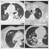
Spectrum of findings in cystobronchial fistula. Depending on the amount of expectorated cyst content (fluid, endocysts, daughter cysts), air-filling and collapse of the lesions and perilesional inflammation, complicated pulmonary cystic echinococcosis lesions will have different imaging appearances. The imaging findings in (a) and (b) are known as the whirl sign (or serpent sign) because of presence of collapsed membranes within the otherwise evacuated cyst cavity. The lesion in (c) may be described as the mass within cavity sign (incarcerated membrane sign) in which the remaining solid parts accumulate in the cavity periphery. Complicated pulmonary cystic echinococcosis may be accompanied and overlaid by perilesional inflammation or atelectasis as seen in (d).
FIGURE 3
FIGURE 3
Secondary cystic echinococcosis. (a and b) Transphrenic expansion of abdominal cystic echinococcosis. The computed tomography (CT) in coronary orientation (a) shows several CE3b lesions (asterices) in the liver (upper lesion) and the peritoneal cavity (lower lesions). The transverse section (b) gives evidence of transphrenic expansion into the right hemithorax with cystobronchial fistulization (air collections, arrow) and rupture into the pleura (open arrow). (c–e) Intrapulmonary and intravascular dissemination of cystic echinococcosis. The radiograph (c) shows a severe dissemination of cystic echinococcosis with a myriad of pulmonary masses. The CT (d) illustrates that the lesions are both intrapulmonary (open arrow in the left lung) and intravascular (arrow in the left pulmonary artery and arrow in the right lower lobe). The T2-weighted MRI (e) proves that there is a CE1 lesion in the left pulmonary artery (arrow) and a CE3b lesion in the left lung (amongst others).
FIGURE 4
FIGURE 4
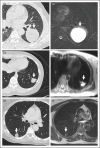
Variety of WHO cystic echinococcosis stages in computed tomography and MRI. (a) Computed tomography (CT) and (b) T2-weighted MRI show an uncomplicated CE1 lesion in the left lower lobe. (c) CT and (d) T2-weighted MRI show a CE2 lesion in the left lower lobe. (e) CT and (f) T2-weighted MRI show a CE4 lesion in the left lower lobe. With T2-weighted MRI, it is possible to depict the lesion content accurately in order to determine the cyst stage. Active lesions (CE1, CE2, CE3b) and transitional lesions (CE3a) have liquid components isointense to cerebrospinal fluid.

Similar articles

Cited by

References

    1. Deplazes P, Rinaldi L, Alvarez Rojas CA, et al. . Global distribution of alveolar and cystic echinococcosis. Adv Parasitol 2017; 95:315–493. - PubMed
    1. Vuitton DA, McManus DP, Rogan MT, et al. . World Association of Echinococcosis. International consensus on terminology to be used in the field of echinococcoses. Parasite 2020; 27:41. - PMC - PubMed
    1. Stojkovic M, Gottstein B, Weber TF, Junghanss T. Cystic, Alveolar and neotropical echinococcosis. In: Farrar J, Garcia PJ, Hotez T, Junghanss T, Kang G, Laloo D, et al., editors. Manson's tropical diseases. 24th ed. July 14, 2023.
    2. Comprehensive and up-to-date chapter on clinical management of cystic echinococcosis.

    1. Brunetti E, Kern P, Vuitton DA. Writing Panel for the WHO-IWGE. Expert consensus for the diagnosis and treatment of cystic and alveolar echinococcosis in humans. Acta Trop 2010; 114:1–16. - PubMed
    1. Stojkovic M, Rosenberger K, Kauczor HU, et al. . Diagnosing and staging of cystic echinococcosis: how do CT and MRI perform in comparison to ultrasound? PLoS Negl Trop Dis 2012; 6:e1880. - PMC - PubMed