. 2023 Oct;146(4):585-610.
doi: 10.1007/s00401-023-02622-9. Epub 2023 Aug 14.

Neuronal tau pathology worsens late-phase white matter degeneration after traumatic brain injury in transgenic mice

Affiliations

Neuronal tau pathology worsens late-phase white matter degeneration after traumatic brain injury in transgenic mice

Fengshan Yu et al. Acta Neuropathol. 2023 Oct.

Abstract

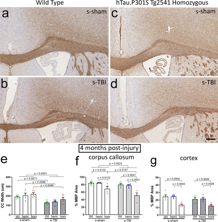
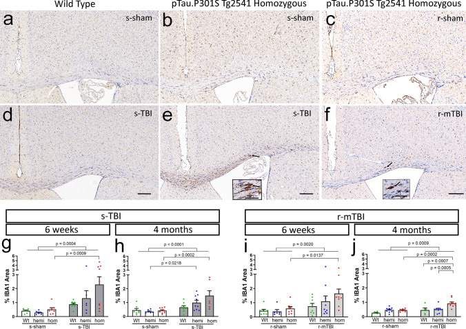
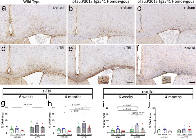
Traumatic brain injury (TBI) causes diffuse axonal injury which can produce chronic white matter pathology and subsequent post-traumatic neurodegeneration with poor patient outcomes. Tau modulates axon cytoskeletal functions and undergoes phosphorylation and mis-localization in neurodegenerative disorders. The effects of tau pathology on neurodegeneration after TBI are unclear. We used mice with neuronal expression of human mutant tau to examine effects of pathological tau on white matter pathology after TBI. Adult male and female hTau.P301S (Tg2541) transgenic and wild-type (Wt) mice received either moderate single TBI (s-TBI) or repetitive mild TBI (r-mTBI; once daily × 5), or sham procedures. Acutely, s-TBI produced more extensive axon damage in the corpus callosum (CC) as compared to r-mTBI. After s-TBI, significant CC thinning was present at 6 weeks and 4 months post-injury in Wt and transgenic mice, with homozygous tau expression producing additional pathology of late demyelination. In contrast, r-mTBI did not produce significant CC thinning except at the chronic time point of 4 months in homozygous mice, which exhibited significant CC atrophy (- 29.7%) with increased microgliosis. Serum neurofilament light quantification detected traumatic axonal injury at 1 day post-TBI in Wt and homozygous mice. At 4 months, high tau and neurofilament in homozygous mice implicated tau in chronic axon pathology. These findings did not have sex differences detected. Conclusions: Neuronal tau pathology differentially exacerbated CC pathology based on injury severity and chronicity. Ongoing CC atrophy from s-TBI became accompanied by late demyelination. Pathological tau significantly worsened CC atrophy during the chronic phase after r-mTBI.

Keywords: Axon damage; Demyelination; Neuroregeneration; Tau; Traumatic brain injury; White matter.


Conflict of interest statement

The authors have no conflicts of interest to disclose. The views, information or content, and conclusions presented do not necessarily represent the official position or policy of, nor should any official endorsement be inferred on the part of, the Uniformed Services University, the Department of Defense, the U.S. Government or the Henry M. Jackson Foundation for the Advancement of Military Medicine, Inc.

Figures

Fig. 1
Fig. 1
Acute injury severity is greater in s-TBI than r-mTBI and is not increased by hTau.P301S genotype at 8 weeks of age, prior to tau-induced neurologic impairment. a The ability of non-injured (naïve) mice to support their weight while hanging from wire cage top bars reveals progressive neurologic impairment with increasing age that is accelerated in homozygotes. This time course in naïve mice was used to select the time points for analysis after TBI. b The study design matches mice across genotype, sex, and TBI models for differential comparisons at phases prior to and after symptom onset. Three distinct approaches examine tau expression for localization, quantification, and translational biomarkers. c Immediately after TBI or sham procedures, a surrogate measure of loss of consciousness is an indicator of more severe injury after s-TBI compared to r-mTBI across genotypes. See also Fig. SI-2 for comparison of sham and injured for each genotype. d At 24 h after a single moderate TBI (s-TBI), acute axon damage in the corpus callosum is increased across genotypes. e At 24 h after the last impact in the repetitive mild TBI (r-mTBI), the acute axon damage is highest in wild-type mice yet less extensive than in s-TBI mice (Fig. SI-3). ce Dots represent individual mice with wild type (Wt) in green, hemizygous (hemi) in blue, and homozygous (hom) in red. Non-significant comparisons are not shown. See Supplemental Information for full statistical analysis of this data along with analysis showing that sex differences were not observed in the righting reflex (Table SI-5), APP immunolabeling (Tables SI-6, SI-7), or hang time results (Table SI-2) even though sex differences were observed among the weights collected for non-injured mice used in the hang time testing (Fig. SI-1; Table SI-3). fh Representative images of β-APP immunolabeling in the corpus callosum (CC) area under the impact site in wild-type mice from the Tg2541 line. Damaged CC axons were identified as β-APP immunolabeled swellings in longitudinal axon profiles (G, yellow arrow enlarged in inset). After s-TBI, damaged axons are also evident in the cingulum (Cg) as β-APP accumulated in transverse axon profiles. Hematoxylin staining of nuclei (blue) was used to exclude β-APP associated with neuron and glial cell bodies. LV lateral ventricle. Scale bars: f–h shown in g = 200 µm, inset = 50 µm
Fig. 2
Fig. 2
Homozygous hTau.P301S genotype increases cortical axon damage at subacute and chronic stages. ad Damaged axons with swellings or endbulbs identified with SMI-34 phosphorylated neurofilament immunolabeling in the medial cerebral cortex under the impact site at 6 weeks (subacute) or 4 months (chronic) stages after TBI or sham procedures. Dots represent individual mice with wild type (Wt) in green, hemizygous (hemi) in blue, and homozygous (hom) in red. Non-significant comparisons are not shown. ae Axon damage is significantly increased in only homozygous mice after s-TBI or the s-sham procedure, which involves anesthesia and scalp incision. c, d Axon damage is increased only in homozygous mice after r-mTBI, but is not increased after the r-sham procedure that involves only anesthesia. ej Representative images of SMI-34 immunolabeling in regions under the impact site in coronal brain sections from homozygous mice at 4 months after TBI or sham procedures. Yellow arrows indicate examples of damaged axons in the medial cortex and corpus callosum (CC) that are enlarged in each inset. Axons cut transversely in the cingulum (Cg) are strongly immunolabeled. Red arrows indicate examples of blood vessels, which are not SMI-34 immunolabeled. Nuclei stained blue with hematoxylin. Scale bars: eg shown in e = 100 µm, hj shown in h = 100 µm, insets shown in h = 50 µm. km Neurologic deficits on the hang time test are not significantly different after r-mTBI as compared to sham procedures in mice of each genotype. See Supplemental Information for full statistical analysis of this data along with analysis showing a sex difference only for the hemizygous mice after r-mTBI (Table SI-4), and no sex differences in the SMI-34 groups (Tables SI-6, SI-7)
Fig. 3
Fig. 3
AT8 detection of phosphorylated tau pathology varies with injury conditions and hTau.P301S genotype at 4 months after TBI or sham procedures. ac Sham wild-type mice do not exhibit AT8 immunolabeling of cortical neurons. In contrast, strong AT8 immunolabeling is found in oligodendrocytes in the corpus callosum (CC). df Wild-type r-mTBI mice exhibit AT8 in the cytoplasm of oligodendrocytes, which typically appear as small round cells that are aligned in rows within the CC and sparsely distributed in cortical regions. gi Wild-type s-TBI mice exhibit clear AT8 immunolabeling in cortical neurons, particularly in superficial layers, along with CC thinning. jl In homozygous sham mice, AT8 immunolabels cortical neurons and callosal oligodendrocytes. mo Homozygous r-mTBI mice exhibit cortical AT8 immunolabeling, along with CC thinning. pr Homozygous s-TBI mice exhibit strong cortical AT8 immunolabeling, along with CC thinning. ar Nuclei stained blue with hematoxylin. Scale bars: left column shown in a = 200 µm; middle column in b = 100 µm, right column in d = 100 µm. Abbreviations: Superior longitudinal fissure (SLF), lateral ventricle (LV), corpus callosum (CC), striatum (STR), indusium griseum (IG)
Fig. 4
Fig. 4
Demonstration of endogenous mouse tau phosphorylation in cortical neurons at 4 months after s-TBI in comparison with human tau expression. a, b After s-TBI in wild-type mice, cortical neurons exhibit clear AT8 immunolabeling of phosphorylated endogenous mouse tau, which is not labeled with the HT7 antibody. c, d After s-TBI in homozygous mice, cortical neurons exhibit strong AT8 immunolabeling and HT7 detection of human tau. ad Nuclei stained blue with hematoxylin. Scale bars for ad shown in B = 200 µm
Fig. 5
Fig. 5
Tau phosphorylation in axons and oligodendrocytes. a, b After s-TBI in homozygous mice, AT8 immunolabels axons and small round oligodendrocytes in the CC. Striatal neurons underlying the CC are not immunolabeled with AT8, but do express the human transgene as detected with the HT7 antibody. c, d In sham wild-type mice, AT8 labels oligodendrocytes in the CC and underlying striatum. A lack of HT7 signal confirms that the oligodendrocyte AT8 signal reflects endogenous mouse tau phosphorylation. e, f After s-TBI in homozygous mice, deeper striatal regions show AT8 immunolabeling of cortical neuron axon bundles and small round oligodendrocytes. The neurons are not immunolabeled with AT8, but do express the human transgene as detected with HT7. g, h In sham wild-type mice, deeper striatal regions show distinct AT8 immunolabeling of oligodendrocytes near axon bundles, in the absence of HT7 immunostaining. ah Nuclei stained blue with hematoxylin. Scale bars: ad shown in c = 100 µm, eh in g = 100 µm
Fig. 6
Fig. 6
Total human tau and phosphorylated tau are increased in brain lysates of tau transgenic mice. a Brain lysates were prepared from the colored cortical regions and white matter tracts, including the corpus callosum (CC) over the lateral ventricles (LV). This superior brain region was dissected from a 2-mm thick coronal slice to capture cortical neuron cell bodies and corresponding axons. b Tau epitopes were quantified in the soluble fraction from brain lysates using Protein Simple Wes capillary electrophoresis immunoassay with actin for normalization of the protein loading amounts. The automated quantification is displayed as electropherograms with the same data also displayed as virtual bands in a blot-like image. cj The HT7 antibody recognizes total human tau to detect the human transgene expression. In contrast, the antibodies to phosphorylated tau epitopes recognize both human and mouse epitopes. Dots represent individual mice with wild type (Wt) in green, hemizygous (hemi) in blue, and homozygous (hom) in red. Non-significant comparisons are not shown. See Supplemental Information for full statistical analysis provided in Table SI-8, which includes analysis showing a general lack of sex differences. cf Comparison across genotypes for s-TBI and s-sham mice at 6 weeks and 4 months post-injury. gj Comparison across genotypes for r-mTBI and r-sham mice at 6 weeks and 4 months post-injury
Fig. 7
Fig. 7
Moderate s-TBI produces corpus callosum atrophy that is not altered by tau genotype. ad Coronal sections through the medial cortex and corpus callosum under the impact site with cell nuclei stained with hematoxylin (blue) and cytoplasm with eosin (pink). Corpus callosum thinning is evident at 4 months post-injury after s-TBI, as compared to sham, in wild-type (a, b) and tau homozygous mice (c, d). Scale bars for ad shown in d = 200 µm. e Corpus callosum width is reduced due to a main effect of injury at the subacute phase. f Corpus callosum width is significantly reduced due to injury in s-TBI versus sham conditions for each genotype at the chronic phase. Tau genotype does not worsen corpus callosum thinning. e, f Dots represent individual mice with wild type (Wt) in green, hemizygous (hemi) in blue, and homozygous (hom) in red. Non-significant comparisons are not shown. See Supplemental Information for full statistical analysis provided in Table SI-6, along with the number and sex of mice for each group
Fig. 8
Fig. 8
Increased pathological tau produces delayed corpus callosum atrophy in chronic r-mTBI. ad Coronal sections through the medial cortex and corpus callosum under the impact site with cell nuclei stained with hematoxylin (blue) and cytoplasm with eosin (pink). Normal adult corpus callosum thickness is seen in wild-type mice, both sham (a) and injured (b), and in sham tau homozygous mice (c) at the 4-month time point. Corpus callosum thinning is evident at 4 months post-injury after r-mTBI only in homozygous tau mice (d). Scale bars for ad shown in d = 200 µm. e Corpus callosum width is not altered by r-mTBI or tau genotype at the subacute phase. f The r-mTBI results in a dramatic reduction of corpus callosum width by the chronic phase only when combined with the homozygous tau genotype, but not in wild-type and hemizygous mice. e, f Dots represent individual mice with wild type (Wt) in green, hemizygous (hemi) in blue, and homozygous (hom) in red. Non-significant comparisons are not shown. See Supplemental Information for full statistical analysis provided in Table SI-7 along with the number and sex of mice for each group
Fig. 9
Fig. 9
Myelin immunolabeling corroborates delayed corpus callosum atrophy with increased pathological tau in chronic r-mTBI. ad Coronal sections through the medial cortex and corpus callosum under the impact site with myelin labeled by immunohistochemistry for myelin basic protein (MBP). Nuclei stained blue with hematoxylin. Normal adult corpus callosum thickness is seen in wild-type mice, both sham (a) and injured (b), and in sham tau homozygous mice (c) at the 4-month time point. Corpus callosum thinning is evident at 4 months post-injury after r-mTBI only in homozygous tau mice (d). Scale bars for ad shown in D = 200 µm. e The r-mTBI results in a dramatic reduction of corpus callosum width by the chronic phase only when combined with the homozygous tau genotype, but not in wild-type and hemizygous mice. f, g The r-mTBI did not cause loss of myelinated areas within the corpus callosum (f) or overlying cortex (g) across genotypes. eg Dots represent individual mice with wild type (Wt) in green, hemizygous (hemi) in blue, and homozygous (hom) in red. Non-significant comparisons are not shown. See Supplemental Information for full statistical analysis provided in Table SI-7, along with the number and sex of mice in each group
Fig. 10
Fig. 10
Myelin immunolabeling shows persistent corpus callosum atrophy after s-TBI, and that increased pathological tau produces late-stage demyelination. ad Coronal sections through the medial cortex and corpus callosum under the impact site with myelin labeled by immunohistochemistry for myelin basic protein (MBP). Nuclei stained blue with hematoxylin. Normal adult corpus callosum thickness is seen in wild-type sham mice (a) with thinning after s-TBI (b). Similarly, in tau homozygous mice, the corpus callosum is notably thicker in sham mice (c) as compared to injured mice at the 4-month time point (d). Scale bars for ad shown in d = 200 µm. e During chronic phase s-TBI, the corpus callosum width is significantly reduced across genotypes. f Increased tau pathology in homozygous mice results in loss of myelinated areas within the corpus callosum that is present after the s-sham procedure and significantly worsened in combination with s-TBI. g Increased tau pathology in homozygous mice results in loss of myelinated areas within the cortex that is similar after s-sham and s-TBI procedures. eg Dots represent individual mice with wild type (Wt) in green, hemizygous (hemi) in blue, and homozygous (hom) in red. Non-significant comparisons are not shown. See Supplemental Information for full statistical analysis provided in Table SI-6, along with the number and sex of mice for each group
Fig. 11
Fig. 11
Both s-TBI and r-mTBI injuries induced a microglial response in the corpus callosum that was most prominent in tau homozygous mice. af Immunohistochemistry for IBA1 to identify microglia in coronal sections under the impact site. Nuclei stained blue with hematoxylin. Images shown are 4 months post-injury or sham procedures. Scale bars inset 50 µm, panel images 200 µm. ac The microglia appeared similar in the cortex and corpus callosum of sham wild-type (a) and tau homozygous mice (b, c). df Both s-TBI (d, e) and r-mTBI (f) appeared to increase IBA1 immunolabeling of microglia in the corpus callosum, with prominent enlargement and elongation after s-TBI in homozygous mice (e). gj Quantification of IBA1 immunolabeling in the corpus callosum shows that the tau homozygous genotype is associated with a significant response to injury at both injury models and at both post-injury time points. Corresponding quantification of IBA1 immunolabeling in the cortex under the impact site is shown in Supplemental Information Fig. SI-5. Dots represent individual mice with wild type (Wt) in green, hemizygous (hemi) in blue, and homozygous (hom) in red. Non-significant comparisons are not shown. See Supplemental Information for full statistical analysis provided in Tables SI-6, SI-7, along with analysis of sex differences which shows that homozygous females have higher IBA1 in the corpus callosum at 6 weeks after s-TBI
Fig. 12
Fig. 12
Both s-TBI and r-mTBI injuries induced reactive astrogliosis in the corpus callosum, which resolved in the chronic phase after r-mTBI. af Immunohistochemistry for GFAP to identify astrocytes in coronal sections under the impact site. Nuclei stained blue with hematoxylin. Images shown are 4 months post-injury or sham procedures. Scale bars inset 50 µm, panel images 200 µm. ac Astrocyte immunolabeling appeared similar in the cortex and corpus callosum of sham wild-type (a) and tau homozygous mice (b, c). df Astrogliosis in the corpus callosum was evident at 4 months after s-TBI (c, e) but localized to a more limited region after r-mTBI (f). gj Quantification of GFAP immunolabeling in the corpus callosum shows a main effect of injury at 6 weeks after s-TBI (g) and r-mTBI (i) that persists to 4 months after s-TBI (h) but resolves after r-mTBI (j). The tau homozygous genotype does not significantly increase astrogliosis in either injury models or at either post-injury time point. Corresponding quantification of GFAP immunolabeling in the cortex under the impact site is shown in Supplemental Information Fig. SI-5. Dots represent individual mice with wild type (Wt) in green, hemizygous (hemi) in blue, and homozygous (hom) in red. Non-significant comparisons are not shown. See Supplemental Information for full statistical analysis provided in Tables SI-6, SI-7, along with analysis that shows a lack of sex differences
Fig. 13
Fig. 13
Clinically used blood biomarker assay shows persistent elevation of human mutant tau while mouse neurofilament light detects axonal injury. al Mouse serum analysis using Simoa® Neurology 4-Plex A to simultaneously detect human total tau and mouse neurofilament light (Nf-L) protein levels. Dots represent individual mice with wild type (Wt) in green, hemizygous (hemi) in blue, and homozygous (hom) in red. Non-significant comparisons are not shown. Full details of statistical analysis in Table SI-6. a In the acute phase, tau protein levels were elevated after s-TBI in serum from homozygous Tg2541 mice, as compared to matched sham mice or to s-TBI wild-type mice. b, c At subacute and chronic time points, human tau protein levels were consistently elevated in s-sham and s-TBI homozygous mice. df Human tau protein levels were consistently elevated in r-sham and r-mTBI mice. In contrast to s-TBI mice, the r-mTBI mice did not show an effect of injury in the acute phase. g, j In the acute phase, mouse Nf-L protein levels in serum showed a significant injury effect, which was most pronounced after s-TBI in wild-type mice. hi, kl Nf-L showed a significant increase in sham and injured homozygous mice that was similar in s-TBI and r-mTBI cohorts. The subacute elevation of Nf-L in homozygous mice further increased at the chronic stage. See Supplemental Information for full statistical analysis provided in Table SI-9, which includes analysis showing a general lack of sex differences

References

    1. Adamek GD, Shipley MT, Sanders MS. The indusium griseum in the mouse: architecture, Timm’s histochemistry and some afferent connections. Brain Res Bull. 1984;12:657–668. doi: 10.1016/0361-9230(84)90147-3. - DOI - PubMed
    1. Allen B, Ingram E, Takao M, Smith MJ, Jakes R, Virdee K, Yoshida H, Holzer M, Craxton M, Emson PC, et al. Abundant tau filaments and nonapoptotic neurodegeneration in transgenic mice expressing human P301S tau protein. J Neurosci. 2002;22:9340–9351. doi: 10.1523/JNEUROSCI.22-21-09340.2002. - DOI - PMC - PubMed
    1. Alosco ML, Stein TD, Tripodis Y, Chua AS, Kowall NW, Huber BR, Goldstein LE, Cantu RC, Katz DI, Palmisano JN, et al. Association of white matter rarefaction, arteriolosclerosis, and tau with dementia in chronic traumatic encephalopathy. JAMA Neurol. 2019;76:1298–1308. doi: 10.1001/jamaneurol.2019.2244. - DOI - PMC - PubMed
    1. Andorfer C, Kress Y, Espinoza M, de Silva R, Tucker KL, Barde YA, Duff K, Davies P. Hyperphosphorylation and aggregation of tau in mice expressing normal human tau isoforms. J Neurochem. 2003;86:582–590. doi: 10.1046/j.1471-4159.2003.01879.x. - DOI - PubMed
    1. Bachstetter AD, Morganti JM, Bodnar CN, Webster SJ, Higgins EK, Roberts KN, Snider H, Meier SE, Nation GK, Goulding DS, et al. The effects of mild closed head injuries on tauopathy and cognitive deficits in rodents: primary results in wild type and rTg4510 mice, and a systematic review. Exp Neurol. 2020;326:113180. doi: 10.1016/j.expneurol.2020.113180. - DOI - PMC - PubMed

Publication types