. 2023 Aug 14;80(9):247.
doi: 10.1007/s00018-023-04898-2.

LSM14B controls oocyte mRNA storage and stability to ensure female fertility

Affiliations

LSM14B controls oocyte mRNA storage and stability to ensure female fertility

Li-Ying Shan et al. Cell Mol Life Sci. .

Abstract

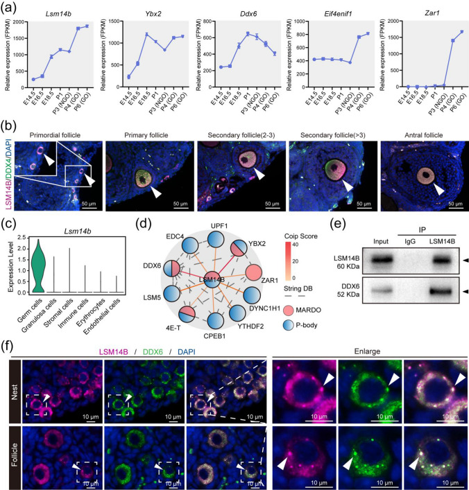
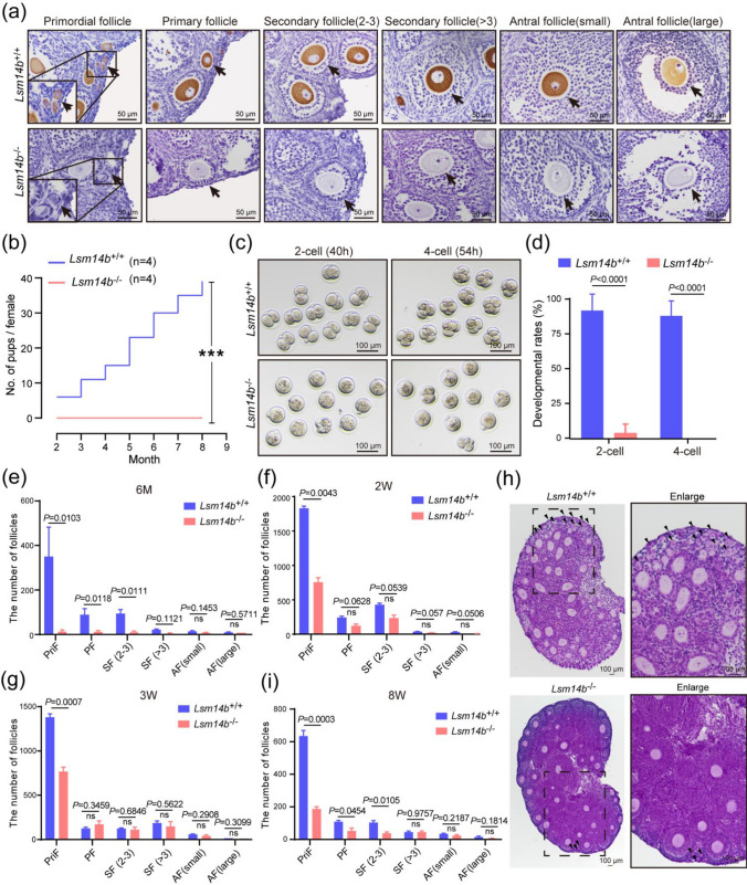
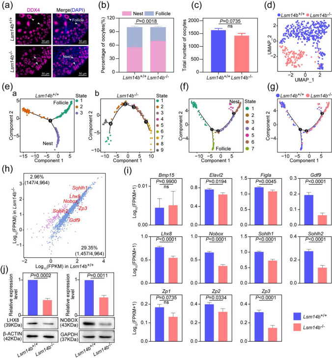
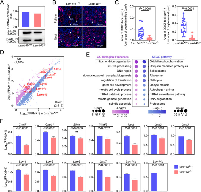
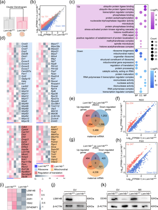
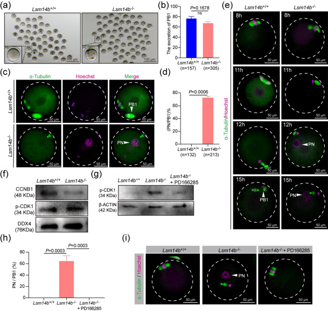
Controlled mRNA storage and stability is essential for oocyte meiosis and early embryonic development. However, how to regulate mRNA storage and stability in mammalian oogenesis remains elusive. Here we showed that LSM14B, a component of membraneless compartments including P-body-like granules and mitochondria-associated ribonucleoprotein domain (MARDO) in germ cell, is indispensable for female fertility. To reveal loss of LSM14B disrupted primordial follicle assembly and caused mRNA reduction in non-growing oocytes, which was concomitant with the impaired assembly of P-body-like granules. 10× Genomics single-cell RNA-sequencing and immunostaining were performed. Meanwhile, we conducted RNA-seq analysis of GV-stage oocytes and found that Lsm14b deficiency not only impaired the maternal mRNA accumulation but also disrupted the translation in fully grown oocytes, which was closely associated with dissolution of MARDO components. Moreover, Lsm14b-deficient oocytes reassembled a pronucleus containing decondensed chromatin after extrusion of the first polar body, through compromising the activation of maturation promoting factor, while the defects were restored via WEE1/2 inhibitor. Together, our findings reveal that Lsm14b plays a pivotal role in mammalian oogenesis by specifically controlling of oocyte mRNA storage and stability.

Keywords: Lsm14b; Oocytes; P-body-like granules; Primordial follicle assembly; mRNA stability.


Conflict of interest statement

The authors declare no conflict of interest.

Figures

Fig. 1
Fig. 1
LSM14B exhibits oocyte-specific expression and as an integral constituent of both P-body-like granules and MARDO. a Gene expression dynamics of MARDO in oocyte at embryonic day 14.5 (E14.5), E16.5, E18.5, postnatal day 1 (P1), P3, P4, and P6. The averaged FPKM values of each of the indicated genes are shown. b Immunostaining of LSM14B (magenta) and germ cell specific markers DDX4 (green) in different classes of follicles. Cell nuclei were counterstained with 4’,6-diamidino-2-phenylindole (DAPI, blue). Scale bar, 50 μm. c Violin diagram showed that Lsm14b was specifically expressed in ovarian germ cells. d Co-IP followed by mass spectrometry analysis was carried to identify the interactions between LSM14B and protein associated with the membraneless compartment, which contains pink-indicated MARDO and blue-indicated P-body. e Co-IP followed by Western blot was performed to prove the interaction between LSM14B and DDX6. f Representative image of immunostaining of oocytes in nest and follicle with LSM14B (magenta) and DDX6 (green) in P3 ovaries. Nuclei were counterstained with DAPI (blue). Scale bar, 10 μm
Fig. 2
Fig. 2
Knockout of Lsm14b compromises female fertility concomitant with a decline in primordial follicle reserve. a Representative immunohistochemistry of LSM14B protein (dark brown) of different classes of follicles in the WT and Lsm14b KO mice. b Cumulative numbers of pups per female showing fertility of WT and Lsm14B KO female mice. Data are the mean ± SEM (n = 4 females for each genotype). c Representative images of 2-cell embryos and 4-cell embryos formed by ovulated oocytes after in vitro fertilization of WT mice and Lsm14b KO mice. d The proportion of 2-cell embryos and 4-cell embryos in WT mice and Lsm14b KO mice. Data are the mean ± SEM (n = 3 biologically independent repeats). e Quantification of the number of different classes of follicles (primordial follicle: PriF; primary follicle: PF; secondary follicle: SF; antral follicle: AF) in ovaries of 6 months WT mice and Lsm14b KO mice. Data are the mean ± SEM (n = 3 and 5 females for WT and Lsm14b KO for 6 months mice, respectively). ns: non-significant. f–g Quantification of the number of different classes of follicles in ovaries of WT mice and Lsm14b KO mice at 2 and 3 weeks, respectively. Data are the mean ± SEM (n = 2 females for each genotype for 2 weeks mice, n = 3 females for each genotype for 3 weeks mice). ns: non-significant. h Representative H&E image of different classes of follicles in 3 weeks ovaries. The arrows represent primordial follicle. Scale bar, 100 μm. i Quantification of the number of different classes of follicles in ovaries of 8 weeks WT mice and Lsm14b KO mice. Data are the mean ± SEM (n = 3 females for each genotype for 8 weeks mice). ns: non-significant
Fig. 3
Fig. 3
Loss of LSM14B impaired the assembly of primordial follicle assembly. a Representative image of nests and follicles staining of ovaries sections from P3 mice. Germ cells are labeled with MVH (magenta), and nucleus was counterstained with DAPI (blue). Scale bar, 50 μm. Data are the mean ± SEM (n = 5 for each genotype). b The percentage of germ cells within nests and follicles in WT and Lsm14b KO ovaries. c The count of germ cells in WT and Lsm14b KO ovaries. Data are the mean ± SEM (n = 3 for each genotype). ns: non-significant. d Clustering of germ cell population with UMAP, colored based on sample groups. e (a) Single-cell pseudotime developmental trajectory of WT germ cells, (b) Single-cell pseudotime developmental trajectory in Lsm14b KO germ cells, which are colored according to cell development state. f The pseudotime trajectory of germ cell populations colored by seven states. g The pseudotime trajectory of germ cell population colored by two sample groups. h The scatter plot shows how genes whose expression levels increased more than threefold from E16.5 to P3 differ between WT and Lsm14b KO P3 oocytes. i Relative expression levels of genes involved in primordial follicle assembly in ovaries. j Detection of LHX8, NOBOX and GAPDH proteins of P3 ovaries by Western blot. GAPDH (for LHX8 and NOBOX control expression)
Fig. 4
Fig. 4
Disrupted assembly of P-body-like granules in Lsm14b KO oocytes. a Detection of DDX6 and β-ACTIN proteins expression level of P3 ovaries by Western blot. b Immunostaining of DDX6 (magenta) in nest and follicle of P3 ovaries. The arrows represent the foci of DDX6. Cell nuclei were counter stained with DAPI (blue). Scale bar, 50 μm. c Quantification of nest and follicle containing DDX6 foci. Data are the mean ± SEM (n = 4 for each genotype). d Scatter plot comparing transcripts between WT and Lsm14b KO from P3 ovaries. Transcripts decreased or increased in the Lsm14b KO sample, highlighted in blue or purple, respectively. e Gene Ontology and KEGG enrichment analysis of down-regulated genes list. f Relative expression levels of P-body-like granules related genes in P3 ovaries
Fig. 5
Fig. 5
LSM14B deletion impaired the maternal mRNA accumulation and translation in GV oocyte. a Sample clustering of the transcriptome of RNA-seq data from GV oocytes. Gray color indicates WT oocytes, and pink color indicates Lsm14b KO oocytes. b Scatter plot comparing transcripts between WT and Lsm14b KO from GV oocytes. Transcripts decreased or increased in the Lsm14b KO oocytes, highlighted in blue or red, respectively. c Gene Ontology enrichment analysis of differential transcripts. d The heatmap shows the expression levels of “Ribosome,” “Mitochondrial,” and “Regulation of translational” related transcripts in WT and Lsm14b KO oocytes. e The Venn plot showing the overlap of maternal mRNA with differential genes of WT and Lsm14b KO NGO, where differential transcripts were acquired by scRNA-seq. f Scatterplot of transcripts between maternal transcripts in up-regulated and down-regulated in Fig. 5E. Down-regulated is displayed in blue; up-regulated is displayed in red. g The Venn plot showing the overlap of maternal mRNA with differential genes of WT and Lsm14b KO FGO, where differential transcripts were acquired by RNA-seq. h Scatterplot of transcripts between maternal mRNA in up-regulated and down-regulated in Fig. 5G. Down-regulated is displayed in blue; up-regulated is displayed in red. i The heatmap shows the expression of MARDO-associated proteins in WT and Lsm14b KO GV oocytes. j Detection of LSM14B and β-ACTIN proteins of GV oocytes by Western blot. k Detection of DDX6 and β-ACTIN proteins of GV and MII oocytes by Western blot
Fig. 6
Fig. 6
Knockout of LSM14B interferes with oocyte meiotic progression. a Representative image of WT and Lsm14b KO oocytes showed PB1 emission 15 h after hCG injection. Scale bar, 100 μm. b Percentage of PB1 released in WT and Lsm14b KO oocytes in vivo. Data are the mean ± SEM (n = 157 and 305 in the WT and Lsm14b KO oocytes, respectively). ns: non-significant. c Confocal microscopy results of WT and Lsm14b KO ovulated oocytes. Arrows indicate PB1 or PN. Spindle is labeled with α-Tubulin (green). Chromosome is labeled with Hoechst (magenta). Scale bar, 50 μm. d Percentage of PN/PB1 in WT and Lsm14b KO ovulated oocytes. Data are the mean ± SEM (n = 132 and 213 in the WT and Lsm14b KO oocytes, respectively). ns: non-significant. e Immunofluorescence showing spindle and chromosome in WT and Lsm14b KO oocytes after injecting hCG in different times. Scale bar, 50 μm. f Western blot results showing the levels of CCNB1 and p-CDK1 between WT and Lsm14b KO oocytes at MII stages. 200 oocytes are loaded in each lane. g Western blot results showed the levels of p-CDK1 and β-ACTIN in WT and Lsm14b KO oocytes by treatment with WEE kinase inhibitor, PD16685. 180 oocytes are loaded in each lane. h Percentage of PN/PB1 in WT, Lsm14b KO, and Lsm14b KO + PD166285 oocytes. (n = 3 biologically independent repeats). i Confocal microscopy results showing PN formation in WT, Lsm14b KO, and Lsm14b KO + PD166285 oocytes. Scale bar, 50 μm

References

    1. Wang JJ, Ge W, Zhai QY, Liu JC, Sun XW, Liu WX, et al. Single-cell transcriptome landscape of ovarian cells during primordial follicle assembly in mice. PLoS Biol. 2020;18(12):e3001025. doi: 10.1371/journal.pbio.3001025. - DOI - PMC - PubMed
    1. He Y, Chen Q, Dai J, Cui Y, Zhang C, Wen X, et al. Single-cell RNA-Seq reveals a highly coordinated transcriptional program in mouse germ cells during primordial follicle formation. Aging Cell. 2021;20(7):e13424. doi: 10.1111/acel.13424. - DOI - PMC - PubMed
    1. Tingen C, Kim A, Woodruff TK. The primordial pool of follicles and nest breakdown in mammalian ovaries. Mol Hum Reprod. 2009;15(12):795–803. doi: 10.1093/molehr/gap073. - DOI - PMC - PubMed
    1. Gu C, Liu S, Wu Q, Zhang L, Guo F. Integrative single-cell analysis of transcriptome, DNA methylome and chromatin accessibility in mouse oocytes. Cell Res. 2019;29(2):110–123. doi: 10.1038/s41422-018-0125-4. - DOI - PMC - PubMed
    1. Gosden RG. Oogenesis as a foundation for embryogenesis. Mol Cell Endocrinol. 2002;186(2):149–153. doi: 10.1016/s0303-7207(01)00683-9. - DOI - PubMed