Review
. 2023 Oct;21(5):503-518.
doi: 10.1007/s11914-023-00815-5. Epub 2023 Aug 14.

Role of the Peripheral Nervous System in Skeletal Development and Regeneration: Controversies and Clinical Implications

Affiliations
Review

Role of the Peripheral Nervous System in Skeletal Development and Regeneration: Controversies and Clinical Implications

Mohamed G Hassan et al. Curr Osteoporos Rep. 2023 Oct.

Abstract

Purpose of review: This review examines the diverse functional relationships that exist between the peripheral nervous system (PNS) and bone, including key advances over the past century that inform our efforts to translate these discoveries for skeletal repair.

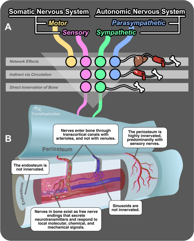
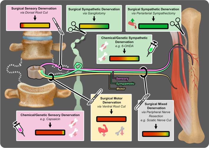
Recent findings: The innervation of the bone during development, homeostasis, and regeneration is highly patterned. Consistent with this, there have been nearly 100 studies over the past century that have used denervation approaches to isolate the effects of the different branches of the PNS on the bone. Overall, a common theme of balance emerges whereby an orchestration of both local and systemic neural functions must align to promote optimal skeletal repair while limiting negative consequences such as pain. An improved understanding of the functional bidirectional pathways linking the PNS and bone has important implications for skeletal development and regeneration. Clinical advances over the next century will necessitate a rigorous identification of the mechanisms underlying these effects that is cautious not to oversimplify the in vivo condition in diverse states of health and disease.

Keywords: Bone regeneration; Denervation; Fracture healing; NGF/TrkA; Peripheral nerve; Skeletal development.


Conflict of interest statement

The authors declare no competing interests.

Figures

Fig. 1
Fig. 1
Innervation and regulation of the bone by the peripheral nervous system (PNS). (A) Three pathways of regulation of the bone by the PNS: (1) network effects, where all four divisions of the PNS indirectly influence the bone via their actions on diverse organ systems (represented by a liver icon) and downstream circulating factors (blood vessel icon); (2) indirect effects via circulation, where the sensory, parasympathetic, and sympathetic divisions of the PNS influence the bone by secreting neurotransmitters into the bloodstream; and (3) direct innervation of the bone, where only the sensory and sympathetic divisions directly release signals into the local bone microenvironment. Solid lines represent direct innervation, and dotted lines represent subsequent indirect effects mediated by circulating factors. (B) Direct innervation of bone structures by the sensory and sympathetic divisions of the PNS. The colored lines originating from (A) continue into this figure as nerve fibers from the sensory (pink) and sympathetic (green) divisions. This figure provides a deconstructed view of a bone, with the periosteum being pulled away from the surface and the bone sliced to expose the marrow and vessels within. Multiple call-out boxes elucidate the interactions of these nerve fibers with various bone structures. The colored circles near the nerve endings represent neurotransmitters secreted by these nerve fibers in response to local molecular, chemical (e.g., H +), and mechanical signals
Fig. 2
Fig. 2
Effects of peripheral denervation on bone development, homeostasis, and repair. This figure illustrates the key methods and outcomes of 65 denervation experiments from 1900–2023, as detailed in Table 1. The schematic features a section of the spinal cord with sensory (pink), sympathetic (green), and motor (yellow) nerve fibers extending to a long bone, with accompanying muscle and vasculature. Seven boxes, each color-coded to the relevant nerve type, detail specific types of interventions and their collective outcomes. Icons within the boxes represent the method of denervation: a scalpel for surgical cuts, forceps for the removal of nervous tissue, and syringes for chemical/genetic denervation. The outcomes of these interventions are summarized by colored gradient bars (green for positive effects on the bone, yellow for neutral, and red for negative), reflecting the proportion of experimental studies yielding the indicated results. Additional icons highlight specific outcomes: artery with an upward arrow indicates increased blood flow, cracked bone signifies poor fracture healing, bone with a downward arrow represents bone loss, and muscle with a downward arrow indicates muscle loss

Similar articles

Cited by

References

    1. Wan Q-Q, Qin W-P, Ma Y-X, Shen M-J, Li J, Zhang Z-B, et al. Crosstalk between bone and nerves within bone. Adv Sci (Weinh) 2021;8:2003390. doi: 10.1002/advs.202003390. - DOI - PMC - PubMed
    1. Maryanovich M, Takeishi S, Frenette PS. Neural regulation of bone and bone marrow. Cold Spring Harb Perspect Med. 2018;8(9):a031344. 10.1101/cshperspect.a031344. - PMC - PubMed
    1. Kacena MA, White FA. No pain, no gain: will migraine therapies increase bone loss and impair fracture healing? EBioMedicine. 2020;60:103025. doi: 10.1016/j.ebiom.2020.103025. - DOI - PMC - PubMed
    1. Lorenz MR, Brazill JM, Beeve AT, Shen I, Scheller EL. A neuroskeletal atlas: spatial mapping and contextualization of axon subtypes innervating the long bones of C3H and B6 mice. J Bone Miner Res. 2021;36:1012–1025. doi: 10.1002/jbmr.4273. - DOI - PMC - PubMed
    1. Castañeda-Corral G, Jimenez-Andrade JM, Bloom AP, Taylor RN, Mantyh WG, Kaczmarska MJ, et al. The majority of myelinated and unmyelinated sensory nerve fibers that innervate bone express the tropomyosin receptor kinase A. Neuroscience. 2011;178:196–207. doi: 10.1016/j.neuroscience.2011.01.039. - DOI - PMC - PubMed

Publication types