. 2023 Aug 14;19(8):e1011386.
doi: 10.1371/journal.pcbi.1011386. eCollection 2023 Aug.

In-silico and in-vitro morphometric analysis of intestinal organoids

Affiliations

In-silico and in-vitro morphometric analysis of intestinal organoids

Sandra Montes-Olivas et al. PLoS Comput Biol. .

Abstract

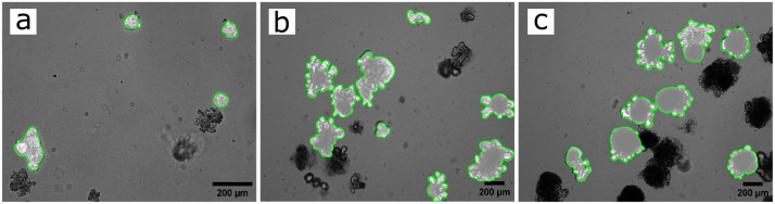
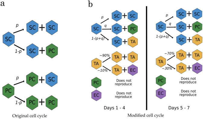
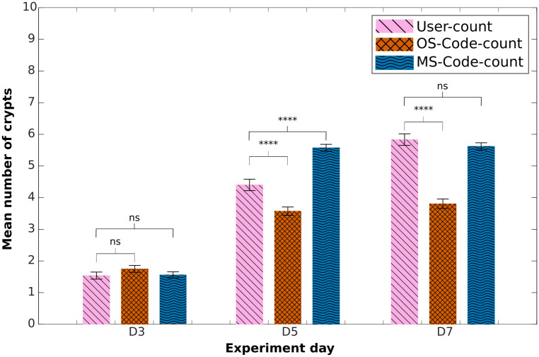
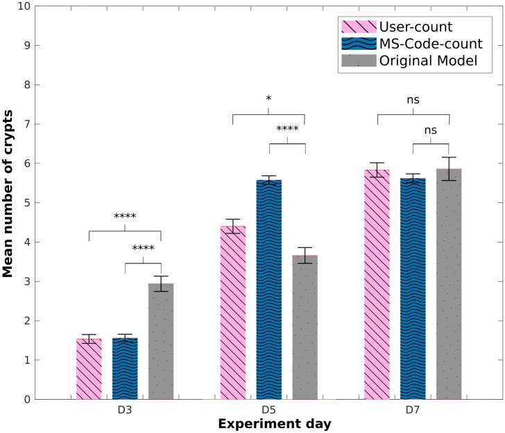
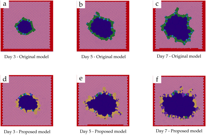
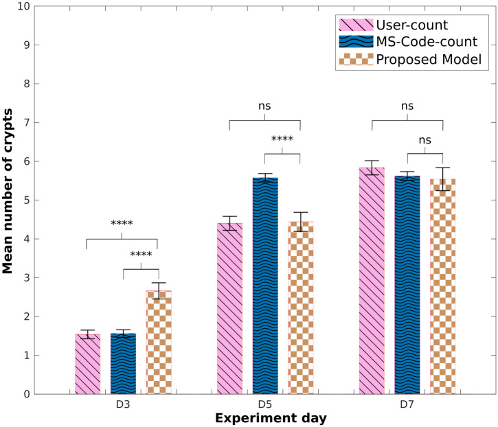
Organoids offer a powerful model to study cellular self-organisation, the growth of specific tissue morphologies in-vitro, and to assess potential medical therapies. However, the intrinsic mechanisms of these systems are not entirely understood yet, which can result in variability of organoids due to differences in culture conditions and basement membrane extracts used. Improving the standardisation of organoid cultures is essential for their implementation in clinical protocols. Developing tools to assess and predict the behaviour of these systems may produce a more robust and standardised biological model to perform accurate clinical studies. Here, we developed an algorithm to automate crypt-like structure counting on intestinal organoids in both in-vitro and in-silico images. In addition, we modified an existing two-dimensional agent-based mathematical model of intestinal organoids to better describe the system physiology, and evaluated its ability to replicate budding structures compared to new experimental data we generated. The crypt-counting algorithm proved useful in approximating the average number of budding structures found in our in-vitro intestinal organoid culture images on days 3 and 7 after seeding. Our changes to the in-silico model maintain the potential to produce simulations that replicate the number of budding structures found on days 5 and 7 of in-vitro data. The present study aims to aid in quantifying key morphological structures and provide a method to compare both in-vitro and in-silico experiments. Our results could be extended later to 3D in-silico models.


Conflict of interest statement

The authors have declared that no competing interests exist.

Figures

Fig 1
Fig 1. Timeline images of in-vitro intestinal organoid culture.
Samples of in-vitro intestinal organoid culture stacked images with segmented organoid boundaries highlighted in green. The images were obtained at days 3 (a), 5 (b) and 7 (c) after seeding, using 10x (a) and 5x (b and c) objective. Scale bar: 200 μm.
Fig 2
Fig 2. Cell cycle diagrams.
(a) Original cell-cycle model used by Langlands et al. and Almet et al. [25, 30], (b) ‘proposed model’ modification that requires a production probability for each cell type derived from stem cells. Both models maintain the stochasticity of the system, as the selection of cell type is dependent on a random number generated at each time-step. Cell types: stem cell (SC); Paneth cell (PC); transit amplifying cell (TA); general differentiated enteroid cell (EC).
Fig 3
Fig 3. Mean number of crypts in experimental images.
Comparison of mean number of crypts obtained using the counting-crypts code applied to the OrganoSeg- or manually-segmented images (‘OS-Code-count’ and ‘MS-Code-count’, respectively). ‘User-count’ refers to manually counted crypts. P-values from two-tailed unpaired t-test computed over the data sets shown, *p<0.05, **p<0.01, ***p<0.001, ****p<0.0001.
Fig 4
Fig 4. Mean number of crypts: Original in-silico model.
Comparison of mean number of crypts obtained using the counting-crypts code applied to manually segmented in-vitro data (‘MS-Code-count’), in-silico organoids obtained from the ‘original model’ and the ‘user-counted’ crypts. P-values from two-tailed unpaired t-test computed over the data sets shown, *p<0.05, **p<0.01, ***p<0.001, ****p<0.0001.
Fig 5
Fig 5. Timeline of in-silico organoids.
Samples of simulation results at days 3 (a,d), 5 (b,e) and 7 (c,f). The ‘original model’ results are shown from a to c; and the ‘proposed model’ results are presented from d to f. Colour code: Stem cells (blue), transit amplifying cells (yellow), Paneth cells (green), general differentiated enteroid cells (purple), Matrigel (pink), lumen (dark blue) and simulation boundary (red).
Fig 6
Fig 6. Mean number of crypts: Modified in-silico model.
Comparison of mean number of crypts obtained using the counting-crypts code applied to the in-silico organoids simulated with the ‘proposed model’, and to manually segmented in-vitro data (‘MS-Code-count’); ‘user-counted’ crypt numbers are also shown. P-values from two-tailed unpaired t-test computed over the data sets shown, *p<0.05, **p<0.01, ***p<0.001, ****p<0.0001.

Similar articles

Cited by

References

    1. Wang Q, Dong X, Lu J, Hu T, Pei G. Constitutive activity of a G protein-coupled receptor, DRD1, contributes to human cerebral organoid formation. Stem Cells. 2020;38(5):653–665. doi: 10.1002/stem.3156 - DOI - PMC - PubMed
    1. Li Y, Muffat J, Omer A, Bosch I, Lancaster MA, Sur M, et al.. Induction of Expansion and Folding in Human Cerebral Organoids. Cell Stem Cell. 2017;20(3):385–396.e3. doi: 10.1016/j.stem.2016.11.017 - DOI - PMC - PubMed
    1. Berger E, Magliaro C, Paczia N, Monzel AS, Antony P, Linster CL, et al.. Millifluidic culture improves human midbrain organoid vitality and differentiation. Lab on a Chip. 2018;18(20):3172–3183. doi: 10.1039/C8LC00206A - DOI - PubMed
    1. Sato T, Stange DE, Ferrante M, Vries RGJ, Van Es JH, Van den Brink S, et al.. Long-term expansion of epithelial organoids from human colon, adenoma, adenocarcinoma, and Barrett’s epithelium. Gastroenterology. 2011;141(5):1762–1772. doi: 10.1053/j.gastro.2011.07.050 - DOI - PubMed
    1. Low JT, Ho GY, Scott M, Tan CW, Whitehead L, Barber K, et al.. Transplantable programmed death ligand 1 expressing gastroids from gastric cancer prone Nfkb1−/− mice. Cell Death and Disease. 2021;12(12):1091. doi: 10.1038/s41419-021-04376-2 - DOI - PMC - PubMed

Publication types