. 2023 Aug 22;120(34):e2308807120.
doi: 10.1073/pnas.2308807120. Epub 2023 Aug 14.

Triple-negative breast tumors are dependent on mutant p53 for growth and survival

Affiliations

Triple-negative breast tumors are dependent on mutant p53 for growth and survival

Denada Dibra et al. Proc Natl Acad Sci U S A. .

Abstract

The TP53 tumor suppressor gene is mutated early in the majority of patients with triple-negative breast cancer (TNBC). The most frequent TP53 alterations are missense mutations that contribute to tumor aggressiveness. We developed an autochthonous somatic K14-Cre driven TNBC mouse model with p53R172H and p53R245W mutations in which mutant p53 can be toggled on and off genetically while leaving the tumor microenvironment intact and wild-type for p53. These mice develop TNBCs with a median latency of 1 y. Deletion of mutant p53R172H or p53R245W in vivo in these tumors blunts their tumor growth and significantly extends survival of mice. Downstream analyses revealed that deletion of mutant Trp53 activated the cyclic GMP-AMP Synthase-Stimulator of Interferon Genes pathway but did not cause apoptosis implicating other mechanisms of tumor regression. Furthermore, we determined that only tumors with stable mutant p53 are dependent on mutant p53 for growth.

Keywords: breast cancer; mouse models; mutant p53 stability; tumor cell vulnerabilities.


Conflict of interest statement

G.L. is on the scientific advisory board for PMV, receives honoraria from various universities for her lectures, and owns stock in PMV.

Figures
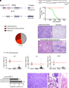
Fig. 1.
Fig. 1.
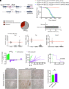
Mutant p53 R172H drives development of TNBCs. (A) A schematic representation of the Trp53wm-R172H allele, which normally expresses wild-type p53 (15). Cre recombinase removes the wild-type cDNA sequences and converts the allele to one expressing the p53R172H mutant. Red triangles indicate loxP sites; “A” indicates the Trp53 native polyadenylation signal; asterisks indicate the CGC to CAC alteration at codon 172 in exon 5, which generates the p53R172H mutant (15). (B) Kaplan–Meier breast-specific survival curves for P172CC mice (N = 47), with mutant p53R172H expression specifically in epithelial cells; and control P172C mice lacking Cre (N = 10). (C) Pathological subtypes of mammary tumors from P172CC mice: adenocarcinoma, 47%; sarcoma, 31%; and sarcomatoid adenocarcinoma 22%. (D) Representative hematoxylin and eosin–stained sections of mammary tumors from mice with different pathological subtypes. (Scale bars, 50 µm.) (E) RT-qPCR analysis for Esr1, Pgr, and Erbb2 in 59 breast tumors from 47 P172CC mice. Mouse ovaries and spleens from normal mice, and breast tumors from MMTV-Erbb2 mice were used as positive and negative controls, respectively (15). Each dot represents one sample; the red dashed line represents median of positive controls. (F) Comparison of metastases incidence of mammary tumors from the P172CC cohort based on pathological subtypes. (G) Hematoxylin and Eosin–stained sections of a primary adenocarcinoma (Left) and metastatic lesions in the lung (Right). (Scale bars, 500 µm.) Black arrowheads point to metastatic lesions in the lung.
Fig. 2.
Fig. 2.
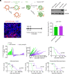
Somatic TNBCs with p53R172H depend on mutant p53 for maintenance. (A) A schematic of the experimental process of turning on and off mutant p53. (B) A murine cell line with mutant p53 was infected with AAV-Control or AAV-Δmut-p53 (1 × 10⁵ genome copies/cell), and p53 levels were measured by western blotting 72 h after infection. (C) Detection of tdTomato in a frozen section of a P172CC breast tumor injected with AAV-EF1-tdTomato-WPRE-hGH (which induces the expression of tdTomato in infected cells), 72 h after infection. (Scale bar, 100 µm.) (D) Treatment regimen; arrows indicate days of AAV injections. (E) Tumor volumes of breast tumors from P172CC mice prior to treatment with AAV-Control or AAV-Δmut-p53. (F) Waterfall plot of percent changes in tumor volume in P172CC mice 3 d after the first AAV injection. *nonresponders after treatment with AAV-Δmut-p53. (G) Breast tumor growth (adenocarcinomas, sarcomas, and adenocarcinoma with sarcomatoid features) in individual P172CC mice after three injections with AAV-Control or AAV-Δmut-p53. Arrows denote injection times. (H) Kaplan–Meier survival curves of all P172CC mice with breast cancer treated with AAV-Control (n = 9) or AAV-Δmut-p53 (n = 18; P < 0.001). (I) Kaplan–Meier survival curves of P172CC mice separated by pathological subtypes. Arrows and arrowheads indicate mice killed due to second tumor formation. Data are mean ± SEM. Significant differences between groups was evaluated by Student’s t test. Survival curve comparisons were made using a log-rank Mantel–Cox test.
Fig. 3.
Fig. 3.
Tumors are dependent on stable mutant p53 for growth. (A) Tumor growth rates for breast cancers treated with AAV-Control or AAV-Δmut-p53. (B) Kaplan–Meier survival curves of P172CC mice with breast cancer treated with AAV-Control (N = 9) or AAV-Δmut-p53 (N = 18; P < 0.001) with (16) or without (2) stable p53. (C) Immunofluorescence imaging of mutant p53 (red) and DAPI (blue) in breast tumors from four mice treated with AAV-Control or AAV-Δmut-p53. Outlined regions indicate tumor cell loss. (Scale bar, 100 µm.) ****P < 0.0001. Survival curve comparisons were made using a log-rank Mantel–Cox test.
Fig. 4.
Fig. 4.
Mutant p53 dependency in vivo is likely not due to apoptosis. (A) GSEA enrichment plots indicating that deletion of mutant Trp53 in breast cancers from P172CC mice induces enrichment in gene sets associated with metabolism, and immune activation processes, including NADH metabolic processes and Interferon α/β. NES, normalized enrichment score; FDR, False discovery rate q-value. (B) GSEA pathways from datasets GO and Reactome, ranked by NES (black columns) and FDR (orange line) q-values from tumors treated with AAV-Control or AAV-Δmut-p53. Dotted orange line, FDR q-values significance set at <0.05. #, apoptotic pathways are nonsignificant (FDR q-values ranging between 0.55 to 0.93 indicated by the orange line). (C) Normalized read counts from tumors treated with AAV-Control or AAV-Δmut-p53 for Bbc3 (Puma), Bid, Bax, and Bcl2l11 (Bim) genes whose protein products induce apoptosis from the RNA-seq dataset. Ns, not significant.
Fig. 5.
Fig. 5.
Mutant p53R245W drives development of TNBCs that depend on mutant p53 for maintenance in vivo. (A) A schematic representation of the Trp53wm-R245W allele, which normally expresses wild-type p53 (15). Cre recombinase removes the wild-type cDNA sequences and converts the allele to one expressing p53R245W mutant. Red triangles indicate loxP sites; “A” indicates the Trp53 native polyadenylation signal; asterisks indicate the GCC to TGG mutations at codon 245 in exon 7, which generate the p53R245W mutant. (B) Kaplan–Meier breast-specific survival curves for Trp53wm-172/fl, K14-Cre, Rosa26LSL CAS9-P2A-EGFP/+ (P172CC) mice (N = 47), and Trp53wm-245/fl, K14-Cre, Rosa26LSL CAS9-P2A-EGFP/+ (P245CC) mice (N = 23), with mutant p53 expression specifically in epithelial cells; and control Trp53wm-172/fl, Rosa26LSL CAS9-P2A-EGFP/+ (P172C) mice lacking Cre (N = 10). (C) Pathological subtypes of mammary tumors from P245CC mice: adenocarcinomas 65%, sarcomas 30%, sarcomatoid adenocarcinomas 5%. (D) Comparison of metastasis incidence of mammary tumors based on pathological subtypes. (E) RT-qPCR analysis for Esr1, Pgr, and Erbb2 in breast tumors from P245CC mice. Mouse ovaries and spleens from normal mice, and breast tumors from MMTV-Erbb2 mice were used as positive and negative controls, respectively (15). Each dot represents one sample; the red dashed line represents median of positive controls. (F) Waterfall plot of percent changes in tumor volume in P245CC mice 3 d after the first AAV injection. (G) Breast tumor growth of individual P245CC mice after injections with AAV-Control or AAV-Δmut-p53. (H) Kaplan–Meier survival curves of P245CC mice with breast cancer treated with AAV-Control (N = 2) or AAV-Δmut-p53 (N = 8; P < 0.0009). (I) Immunohistochemical staining for cleaved caspase 3 (CC3) in tumors from P245CC injected with AAV-Control (n = 5) or AAV-Δmut-p53 (n = 8) 6 d after two injections. [Scale bars, 500 µm (Left).] CC3 staining was quantified in three fields of view (FOV) per sample (Right).
Fig. 6.
Fig. 6.
Activation of the cGAS–STING pathway post mutant Trp53 deletion in vivo. (A) GSEA enrichment plots indicating that deletion of mutant Trp53 in breast cancers from P172CC mice induces enrichment in gene sets associated cytosolic sensing of pathogen associated DNA and Rig I like receptor signaling. NES, normalized enrichment score; FDR, False discovery rate q-value. (B) Normalized read counts from tumors treated with AAV-Control (N = 3) or AAV-Δmut-p53 (N = 6) for Cxcl10 and Ifit10. (C) Representative immunofluorescence imaging of pSTING-Ser366 (red) and DAPI (blue) of breast P172CC (Left) or P245CC (Right) tumors treated with AAV-Control or AAV-Δmut-p53. (Scale bars, 50 µm.)

Similar articles

Cited by

References

    1. Ying H., et al. , Oncogenic Kras maintains pancreatic tumors through regulation of anabolic glucose metabolism. Cell 149, 656–670 (2012). - PMC - PubMed
    1. Koboldt D. C., et al. , Comprehensive molecular portraits of human breast tumours. Nature 490, 61–70 (2012). - PMC - PubMed
    1. Freed-Pastor W. A., Prives C., Mutant p53: One name, many proteins. Genes Dev. 26, 1268–1286 (2012). - PMC - PubMed
    1. Aschauer L., Muller P. A., Novel targets and interaction partners of mutant p53 Gain-Of-Function. Biochem. Soc. Trans. 44, 460–466 (2016). - PubMed
    1. Kim M. P., Lozano G., Mutant p53 partners in crime. Cell Death Differ. 25, 161–168 (2018). - PMC - PubMed

Publication types

Substances