. 2023 Aug 22;120(34):e2220269120.
doi: 10.1073/pnas.2220269120. Epub 2023 Aug 14.

Ceramide as an endothelial cell surface receptor and a lung-specific lipid vascular target for circulating ligands

Affiliations

Ceramide as an endothelial cell surface receptor and a lung-specific lipid vascular target for circulating ligands

Daniela I Staquicini et al. Proc Natl Acad Sci U S A. .

Abstract

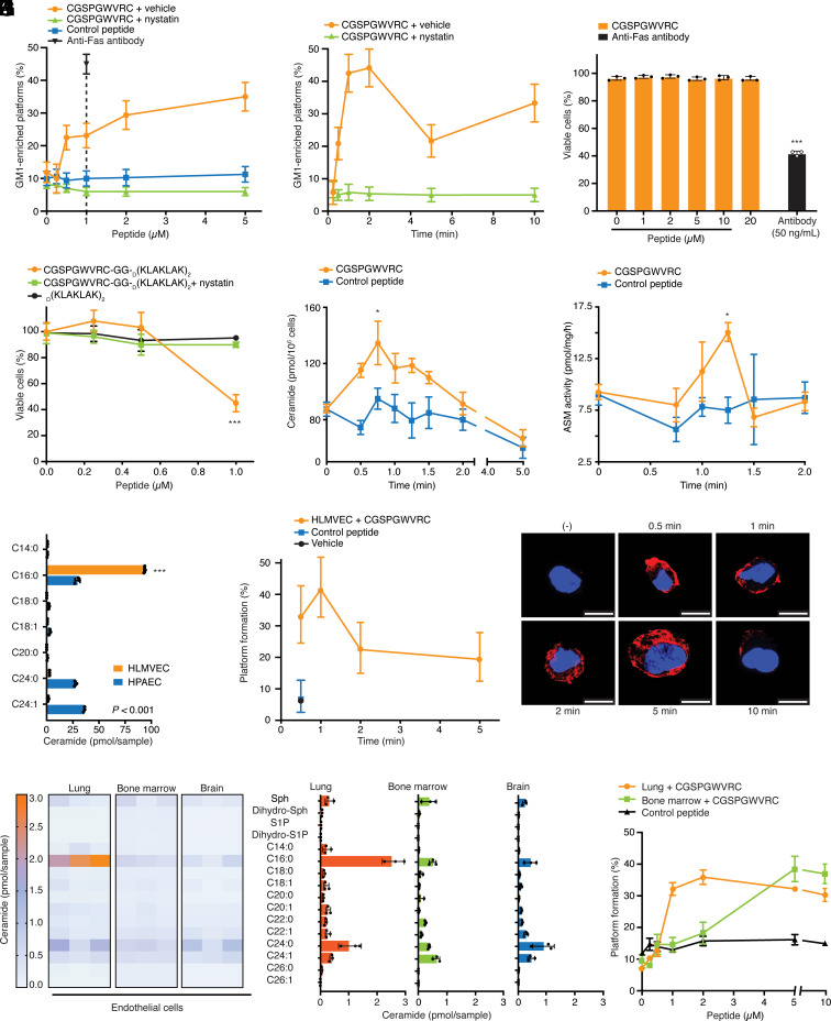
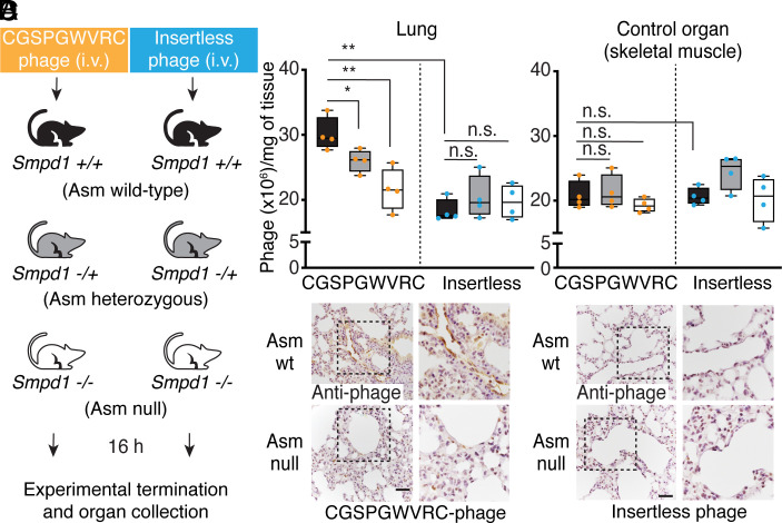
The vascular endothelium from individual organs is functionally specialized, and it displays a unique set of accessible molecular targets. These serve as endothelial cell receptors to affinity ligands. To date, all identified vascular receptors have been proteins. Here, we show that an endothelial lung-homing peptide (CGSPGWVRC) interacts with C16-ceramide, a bioactive sphingolipid that mediates several biological functions. Upon binding to cell surfaces, CGSPGWVRC triggers ceramide-rich platform formation, activates acid sphingomyelinase and ceramide production, without the associated downstream apoptotic signaling. We also show that the lung selectivity of CGSPGWVRC homing peptide is dependent on ceramide production in vivo. Finally, we demonstrate two potential applications for this lipid vascular targeting system: i) as a bioinorganic hydrogel for pulmonary imaging and ii) as a ligand-directed lung immunization tool against COVID-19. Thus, C16-ceramide is a unique example of a lipid-based receptor system in the lung vascular endothelium targeted in vivo by circulating ligands such as CGSPGWVRC.

Keywords: acid sphingomyelinase; ceramide; endothelial cells; lung; phage display.


Conflict of interest statement

J.A.R., R.N.K., R.P., and W.A. are inventors on US patents #8,562,993, #9,592,238 and #11,207,329, which describe the anti-ceramide monoclonal antibody 2A2 used in this study; as such, they will be entitled to standard royalties if commercialization occurs. J.G.G., R.P., and W.A. are founders and equity stockholders of PhageNova Bio. R.P. and W.A. receive research support from PhageNova Bio. R.P. is a paid consultant for PhageNova Bio and serves as its Chief Scientific Officer. D.I.S., F.H.F.T., T.L.S., R.P., and W.A. are inventors on international patent application PCT/US2021/040392, which describes immunization technology adapted for COVID-19. PhageNova Bio has licensed this intellectual property portfolio and D.I.S., F.H.F.T., T.L.S., R.P., and W.A. will be entitled to standard royalties if commercialization occurs. R.P. and W.A. are founders, stockholders, and scientific advisors for MBrace Therapeutics, a company developing antibodies for oncology applications. R.P. and W.A. receive research support from MBrace Therapeutics. R.P. and W.A. are paid consultants for MBrace Therapeutics. R.N.K. is founder and stockholder of Ceramedix Holding LLC. R.N.K. is an inventor on US patent #10,413,533 and pending international patent applications describing methods to activate the ceramide/sphingomyelin pathway; he will be entitled to standard royalties if commercialization occurs. J.A.R. and R.N.K. are inventors on US patent #10,450,385 and pending US and international patent applications describing anti-ceramide antibodies; as such, they will be entitled to standard royalties if commercialization occurs. R.N.K. is an inventor on US patents #7,195,775 and #7,850,984 describing methods of use of antagonists to acid sphingomyelinase and US patent #10,052,387 describing methods of use of exogenous ceramide for therapy. Y.A.H. and L.M.O. are inventors on US patent #8,093,393. Y.A.H. is also inventor on US patents #8,592,419 and #8,697,379, all of which describe ceramide-related compounds; as such, they will be entitled to standard royalties if commercialization occurs. Each of the corresponding institutions currently manages these arrangements according to their established institutional conflict-of-interest policies. None of these conflicts affected the experimental design, interpretation, or reporting of the results. Other authors declare that they have no competing interests.

Figures

Fig. 1.
Fig. 1.
Ceramide is a lipid target for the CGSPGWVRC peptide. (AC) LUVs fluorescence (6-FAM) leakage assay in the presence of increasing molar concentrations of the targeted CGSPGWVRC peptide or an unrelated negative control peptide. (D) Targeted CGSPGWVRC-phage particles bind preferentially to immobilized ceramide species. Data are means ± SD. ***P < 0.001; two-way ANOVA. (E) A site-directed point mutation of the tryptophan residue to alanine residue abolishes the mutant CGSPGAVRC phage binding to ceramide species. Nontargeted (insertless) phage particles were used as negative controls. (F) The monoclonal anti-ceramide 2A2 antibody inhibits the binding of targeted CGSPGWVRC-phage to C16-ceramide. Insertless phage particles and BSA were used as negative controls. Data are means ± SD. ***P < 0.001; two-tailed unpaired Student’s t test.
Fig. 2.
Fig. 2.
Soluble CGSPGWVRC induces platform formation and ceramide generation through ASM activity. (A and B) CGSPGWVRC induces raft coalescence into platforms in Jurkat T-cells. An anti-Fas antibody and an unrelated control peptide were used as positive and negative controls, respectively. Disruption of ordered lipid rafts by nystatin prevents CGSPGWVRC-induced platform formation independently of the peptide molar concentration or the time of peptide stimulation. GM1, a lipid intrinsic to rafts and a marker of platform formation, was stained using FITC-conjugated cholera toxin B-subunit. (C) Cell viability with increasing molar concentrations of CGSPGWVRC or anti-Fas antibody. Data are means ± SD. ***P < 0.001; two-tailed unpaired Student’s t test. (D) Viability of endothelial cells is reduced with equimolar concentrations of the CGSPGWVRC-GG-D(KLAKLAK)2, but the pro-apoptotic effect is abrogated in the presence of nystatin; D(KLAKLAK)2 served as a negative control. Data are means ± SD. ***P < 0.001; two-tailed unpaired Student’s t test. (E) Kinetics of ceramide generation and ASM activity measured in cells stimulated with CGSPGWVRC (F). Data are means ± SD. *P < 0.05; two-tailed unpaired Student’s t test. (G) Quantification and comparison of the lipid abundance in human lung microvascular endothelial cells (HLMVEC) versus human pulmonary artery endothelial cells (HPAEC). Data are means ± SD. ***P < 0.001; two-tailed unpaired Student’s t test. (H) CGSPGWVRC induces platform formation in HLMVEC. (I) Representative images of ceramide-enriched platforms in HLMVEC stimulated with CGSPGWVRC peptide. The data are representative of at least two independent experiments. (Scale bar, 10 μm.) (J) Quantification of lipid abundance in immortalized mouse lung-, bone marrow-, and brain-derived endothelial cells by LC-MS/MS. The scale represents ceramide (pmol/sample) and the relative quantification is shown (K). (L) CGSPGWVRC induces platform formation in the lung- and bone-marrow-derived endothelial cells in a dose-dependent manner. Lung-derived endothelial cells incubated with an unrelated peptide were used as controls. Data are means ± SEM; two-way ANOVA.
Fig. 3.
Fig. 3.
Targeted CGSPGWVRC-phage homing to the lung is dependent on the Smpd1 gene expression in vivo. (A) Experimental scheme of side-by-side i.v. administration in vivo of targeted CGSPGWVRC-phage or control insertless phage in Smpd1+/+ (Asm wt), Smpd1−/+ (Asm heterozygous), or Smpd1−/− (Asm null) mice. Phage particles were allowed to circulate for 16 h, after which time the experiment was terminated, and lung and control organs or tissues (shown is skeletal muscle) were collected. (B and C) Targeted CGSPGWVRC-phage and control insertless phage homing to the lungs or to a control organ (n = 4 mice per each group) as assessed by qPCR. Data are shown as box-and-whisker plots. *P < 0.05, **P < 0.01; two-way ANOVA; n.s. stands for nonstatistically significant. (D) IHC analysis with an anti-phage antibody in lung tissues. No difference in phage homing in the lung and control organ among the groups of mice given the control insertless phage; only background staining was observed for the negative control insertless phage. (Scale bar, 100 μm.) Data are representative of three independent experiments.
Fig. 4.
Fig. 4.
Molecular imaging of the lungs by a targeted CGSPGWVRC-phage AuNP hydrogel. (A and B) Confocal images of targeted CGSPGWVRC-phage AuNP hydrogel or negative control insertless phage AuNP hydrogel homing to the lungs upon i.v. administration. Blood vessels were co-stained with an anti-CD31 antibody (red, phage; green, CD31). (Scale bar, 100 μm.) (CE) Scanning electron microscopy of targeted CGSPGWVRC-phage AuNP hydrogel in the lung vascular endothelium. (FH) Negative control insertless phage AuNP hydrogel. Images represent 10,000-fold magnification (C and F), 20,000-fold magnification (D and G), or 60,000-fold magnification (E and H). (I) NIR surface-enhanced Raman spectroscopy analysis of the lungs from mice receiving either targeted CGSPGWVRC-phage AuNP hydrogel or negative control insertless phage AuNP hydrogel i.v. Heat-maps specific to peaks at 1,325 cm−1 and 1,263 cm−1 demonstrate increased localization of the targeted CGSPGWVRC-phage AuNP hydrogel in the lung relative to the lower signal of the negative control AuNP hydrogel. (J) Micro-CT images of the lungs 2 min after i.v. administration of CGSPGWVRC-phage AuNP hydrogel (Left) relative to the negative control phage AuNP hydrogel (Right). (K) Three-dimensional rendering of the micro-CT data pseudocolored for visualization. Blue represents air, yellow represents bone, and red represents Au.
Fig. 5.
Fig. 5.
Immunogenicity of the SARS-CoV-2 S protein epitope on targeted CGSPGWVRC-dual-display phage particles. (A) Representation of dual-display, single-display or control insertless phage particles. (B) Five-week-old BALB/c mice were immunized via i.v. or s.c. with each of the phage constructs. Animals received a boost injection 3 wk after the first immunization. S protein-specific IgG antibodies’ titers were evaluated with ELISA by using a recombinant full-length S protein. Data shown as box-and-whisker plots. ***P < 0.001, two-way ANOVA. (C) Pharmacokinetic model simulation of dual-display phage particles’ accumulation in the lungs of Smpd1 wild-type, heterozygous, or null mice following i.v. administration. (D) Immune response model simulation of SARS-CoV-2 epitope deposition in the lungs after administration of dual-display phage particles via i.v. or s.c. routes. (E) Simulation of the antigen-presenting cells activation in mice administered with dual- and single-display phage particles. (F) Fit of the anti-S protein antibody titers recapitulate the experimentally observed data.

References

    1. Pasqualini R., Ruoslahti E., Organ targeting in vivo using phage display peptide libraries. Nature 380, 364–366 (1996). - PubMed
    1. Rajotte D., et al. , Molecular heterogeneity of the vascular endothelium revealed by in vivo phage display. J. Clin. Invest. 102, 430–437 (1998). - PMC - PubMed
    1. Giordano R. J., et al. , Targeted induction of lung endothelial cell apoptosis causes emphysema-like changes in the mouse. J. Biol. Chem. 283, 29447–29460 (2008). - PMC - PubMed
    1. Arap W., et al. , Steps toward mapping the human vasculature by phage display. Nat. Med. 8, 121–127 (2002). - PubMed
    1. Arap W., Pasqualini R., Ruoslahti E., Cancer treatment by targeted drug delivery to tumor vasculature in a mouse model. Science 279, 377–380 (1998). - PubMed

Publication types