. 2023 Dec;1869(8):166846.
doi: 10.1016/j.bbadis.2023.166846. Epub 2023 Aug 12.

Iron promotes glycolysis to drive colon tumorigenesis

Affiliations

Iron promotes glycolysis to drive colon tumorigenesis

Zhaoli Liu et al. Biochim Biophys Acta Mol Basis Dis. 2023 Dec.

Abstract

Colorectal cancer (CRC) is the third most common cancer and is also the third leading cause of cancer-related death in the USA. Understanding the mechanisms of growth and progression of CRC is essential to improve treatment. Macronutrients such as glucose are energy source for a cell. Many tumor cells exhibit increased aerobic glycolysis. Increased tissue micronutrient iron levels in both mice and humans are also associated with increased colon tumorigenesis. However, if iron drives colon carcinogenesis via affecting glucose metabolism is still not clear. Here we found the intracellular glucose levels in tumor colonoids were significantly increased after iron treatment. 13C-labeled glucose flux analysis indicated that the levels of several labeled glycolytic products were significantly increased, whereas several tricarboxylic acid cycle intermediates were significantly decreased in colonoids after iron treatment. Mechanistic studies showed that iron upregulated the expression of glucose transporter 1 (GLUT1) and mediated an inhibition of the pyruvate dehydrogenase (PDH) complex function via directly binding with tankyrase and/or pyruvate dehydrogenase kinase (PDHK) 3. Pharmacological inhibition of GLUT1 or PDHK reactivated PDH complex function and reduced high iron diet-enhanced tumor formation. In conclusion, excess iron promotes glycolysis and colon tumor growth at least partly through the inhibition of the PDH complex function.

Keywords: GLUT1; Glucose; Iron; PDH; PDHK3; Tankyrase.


Conflict of interest statement

Declaration of competing interest C.A.L. has received consulting fees from Astellas Pharmaceuticals and Odyssey Therapeutics and is an inventor on patents pertaining to Kras regulated metabolic pathways, redox control pathways in pancreatic cancer, and targeting the GOT1-pathway as a therapeutic approach (US Patent No: 2015126580-A1, 05/07/2015; US Patent No: 20190136238, 05/09/2019; International Patent No: WO2013177426-A2, 04/23/2015).

Figures
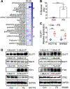
Figure 1.
Figure 1.. Iron increases glucose accumulation in colon tumor cells.
A. Heatmap of metabolites change and B. relative quantification of D-Glucose after FS treatment in tumor colonoids revealed by untargeted metabolomics analysis. C. qPCR analysis of GLUT1 expression change after 100 μM FS overnight treatment in HCT116 and SW480 cells. D. Western blot analysis after 100 μM FS overnight treatment in HCT116 and SW480 cells. *p<0.05, ***p<0.001, Student’s t-test.
Figure 2.
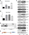
Figure 2.. Iron increases glycolysis in tumor colonoids.
A. 13C-labeled glucose flux assay shows changes in metabolites of the glycolysis pathway and TCA cycle from FS treated tumor colonoids. B. Schematic representation of the inhibitory effects of PDHK1 on PDHE1α, which catalyzes pyruvate to form acetyl-CoA. Red up arrows (↑) indicate increased metabolites or enzymes, blue down arrows (↓) indicate decreased metabolites. C. Western blot analysis after overnight FS treatment in tumor colonoids. *p<0.05, Student’s t-test.
Figure 3.
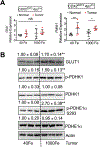
Figure 3.. The protein levels of GLUT1, p-PDHK1 and p-PDHE1α Ser293 are increased in mouse colon tumors.
A. qPCR analysis and B. Western blot analysis in CDX2ERT2 ApcF/+ mice. *p<0.05, **p<0.01, NS, not significant. Two-way ANOVA followed by Sidak’s multiple comparisons test or Student’s t-test.
Figure 4.
Figure 4.. Iron increases PDHE1α Ser293 phosphorylation via directly binding with PDHK3 that is abundant in colon tumors.
Gene expression of PDHK1-4 in A. normal colon and B. colon tumors from human protein atlas database. C. Western blot analysis after overnight FS treatment in SW480 cells. D. Immunoblotting analysis of proteins pulled down (PD) by Fe2+ or empty beads in HEK293T cells. E. Diagram shows that iron increases PDHE1α Ser293 phosphorylation via directly binding with PDHK3. *p<0.05, **p<0.01 and ***p<0.001, Student’s t-test.
Figure 5.
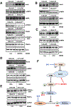
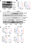
Figure 5.. GLUT1 inhibitor BAY876 and pan PDHK inhibitor DCA reactivate iron inhibited PDH complex function.
Western blot analysis of SW480 cell lysates after treatment with A. GLUT1 inhibitor 5μM BAY876, B. 100μM FS, or C. 100μM iron chelation deferoxamine (DFO). D. Western blot analysis in CDX2ERT2 ApcF/+ mice. E. Western blot analysis of SW480 cell lysates after treatment with 10mM PDHK1 inhibitor DCA. F. Diagram shows that PDHK inhibitor DCA reactivate iron inhibited AMPK-PDH signaling. *p<0.05, **p<0.01, ***p<0.001 vs Con, # p<0.05 vs FS. Student’s t-test.
Figure 6.
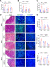
Figure 6.. Pharmacological inhibition of GLUT1 or PDHK reduces iron-driven colon tumorigenesis.
A. Total tumor number, B. tumor number at size >3mm, C. tumor burden, D. histological analysis and E. quantification of colon tumors in CDX2ERT2 ApcF/+ mice treated with 100mg/kg TAM for 3 days, 40Fe (n=31) or 1000Fe diet (n=24), vehicle (n=22), GLUT1 inhibitor BAY (n=19) or PDHK1 inhibitor DCA (n=14), DSS for 7 days and regular water for a month. *p<0.05, **p<0.01, ***p<0.001, ****p<0.0001. Two-way ANOVA followed by Sidak’s multiple comparisons test.
Figure 7.
Figure 7.. On target efficacy validation of GLUT1 and PDHK inhibitors in vivo.
A. Western blot analysis and B. quantification of AMPK phosphorylation as a biomarker in tumors from CDX2ERT2 ApcF/+ mice treated with 1000Fe combined with PBS or GLUT1 inhibitor BAY876. C. Western blot analysis and D. quantification of tumors from CDX2ERT2 ApcF/+ mice treated with 40Fe or 1000Fe combined with PBS or BAY876.

References

    1. Siegel RL, Miller KD, Fuchs HE, Jemal A, 2021. Cancer Statistics, 2021. CA: A Cancer Journal for Clinicians 71(1): 7–33, Doi: 10.3322/caac.21654. - DOI - PubMed
    1. Nelson RL, 2001. Iron and colorectal cancer risk: Human studies. Nutrition Reviews 59(5): 140–8, Doi: 10.1111/j.1753-4887.2001.tb07002.x. - DOI - PubMed
    1. Xue X, Ramakrishnan SK, Weisz K, Triner D, Xie L, Attili D, et al., 2016. Iron Uptake via DMT1 Integrates Cell Cycle with JAK-STAT3 Signaling to Promote Colorectal Tumorigenesis. Cell Metabolism 24(3): 447–61, Doi: 10.1016/j.cmet.2016.07.015. - DOI - PMC - PubMed
    1. Xue X, Taylor M, Anderson E, Hao C, Qu A, Greenson JK, et al., 2012. Hypoxia-inducible factor-2α activation promotes colorectal cancer progression by dysregulating iron homeostasis. Cancer Research 72(9): 2285–93, Doi: 10.1158/0008-5472.CAN-11-3836. - DOI - PMC - PubMed
    1. Radulescu S, Brookes MJ, Salgueiro P, Ridgway RA, McGhee E, Anderson K, et al., 2012. Luminal Iron Levels Govern Intestinal Tumorigenesis after Apc Loss In Vivo. Cell Reports 2(2): 270–82, Doi: 10.1016/j.celrep.2012.07.003. - DOI - PubMed

Publication types

LinkOut - more resources