. 2023 Nov;82(11):1415-1428.
doi: 10.1136/ard-2023-224479. Epub 2023 Aug 14.

New biologic (Ab-IPL-IL-17) for IL-17-mediated diseases: identification of the bioactive sequence (nIL-17) for IL-17A/F function

Affiliations

New biologic (Ab-IPL-IL-17) for IL-17-mediated diseases: identification of the bioactive sequence (nIL-17) for IL-17A/F function

Anella Saviano et al. Ann Rheum Dis. 2023 Nov.

Abstract

Objectives: Interleukin (IL) 17s cytokines are key drivers of inflammation that are functionally dysregulated in several human immune-mediated inflammatory diseases (IMIDs), such as rheumatoid arthritis (RA), psoriasis and inflammatory bowel disease (IBD). Targeting these cytokines has some therapeutic benefits, but issues associated with low therapeutic efficacy and immunogenicity for subgroups of patients or IMIDs reduce their clinical use. Therefore, there is an urgent need to improve the coverage and efficacy of antibodies targeting IL-17A and/or IL-17F and IL-17A/F heterodimer.

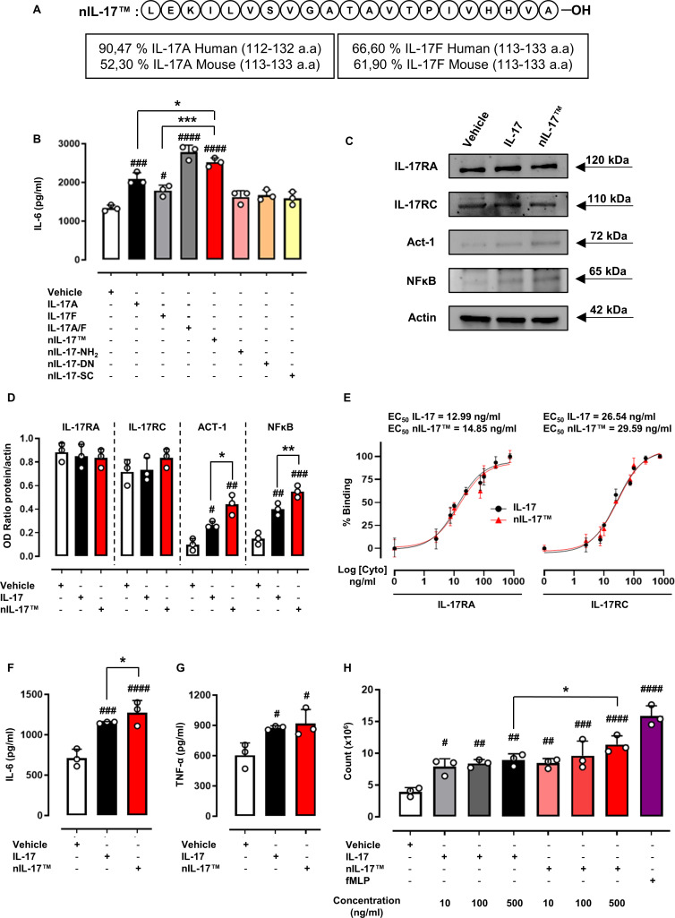
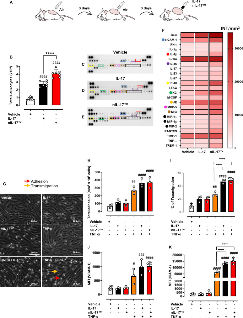
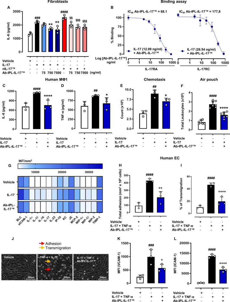
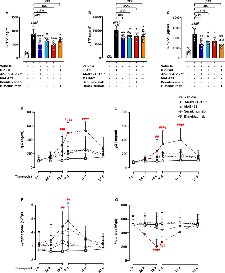
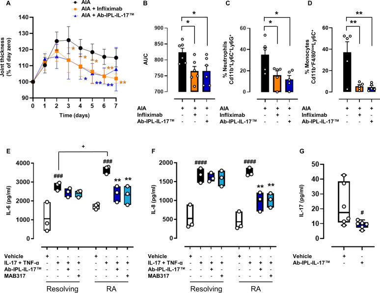
Methods and results: Here, we initially identified a bioactive 20 amino acid IL-17A/F-derived peptide (nIL-17) that mimics the pro-inflammatory actions of the full-length proteins. Subsequently, we generated a novel anti-IL-17 neutralising monoclonal antibody (Ab-IPL-IL-17) capable of effectively reversing the pro-inflammatory, pro-migratory actions of both nIL-17 and IL-17A/F. Importantly, we demonstrated that Ab-IPL-IL-17 has less off-target effects than the current gold-standard biologic, secukinumab. Finally, we compared the therapeutic efficacy of Ab-IPL-IL-17 with reference anti-IL-17 antibodies in preclinical murine models and samples from patients with RA and IBD. We found that Ab-IPL-IL-17 could effectively reduce clinical signs of arthritis and neutralise elevated IL-17 levels in IBD patient serum.

Conclusions: Collectively, our preclinical and in vitro clinical evidence indicates high efficacy and therapeutic potency of Ab-IPL-IL-17, supporting the rationale for large-scale clinical evaluation of Ab-IPL-IL-17 in patients with IMIDs.

Keywords: arthritis, rheumatoid; autoimmune diseases; autoimmunity; inflammation.


Conflict of interest statement

Competing interests: This article has been conducted and written in the absence of any commercial or financial relationships that could be construed as a potential conflict of interest. The authors ASaviano, FR, FMerlino, RB, PG, MB, AJI and FMaione hold patents for the diagnostic and therapeutic use of nIL-17 and Ab-IPL-IL-17 (IT patent no.: 102022000016722) in autoimmune disease, chronic inflammatory disease and other diseases in which IL-17 producing cells contribute to pathogenesis.

Figures

Figure 1
Figure 1
Biological characterisation of a novel IL-17-derived peptide (nIL-17). (A) Amino acids sequence of nIL-17 was obtained after a study of primary structures of both mouse/human IL-17A and IL-17F. (B) To assess the biological activity of nIL-17 peptide, IL-6 production was evaluated in NIH-3T3 cell supernatants following 24 hours of incubation in the presence of either IL-17A protein (50 ng/mL), IL-17F protein (50 ng/mL), IL-17A/F heterodimer (50 ng/mL), nIL-17 (50 ng/mL), nIL-17 (50 ng/mL) with terminal NH2 sequence, denatured (–DN) form or ‘scrambled’ (–SC) sequence (both at 50 ng/mL). (C)–(D) Whole cell lysates from NIH-3T3 cells stimulated with IL-17 or nIL-17 (50 ng/mL) were analysed, by western blot, for IL-17RA (~120 kDa), IL-17RC (~110 kDa), Act-1 (~72 kDa), NFκB (~65 kDa) and actin (~42 kDa) expression. Representative western blot images are shown from three pooled experiments with similar results. (E) To evaluate the binding interaction of nIL-17 with IL-17RA and IL-17RC, biotinylated IL-17 and nIL-17 (0–750 ng/mL) were co-incubated for 30 min with IL-17RA-Fc or IL-17RC-Fc prior to fluorescence being measured. (B)–(E) Data are presented as mean±SD of n=3 independent experiments. (F)–(G) Macrophages, derived from primary human CD14+ monocytes, were stimulated with LPS and IFN-γ (M1 stimuli) for 16 hours. Following differentiation, M1 macrophages were treated with IL-17 vehicle, IL-17 or nIL-17 (100 ng/mL) for 24 hours. Supernatants from all experimental conditions were assayed by ELISA for (F) IL-6 and (G) TNF-α. (H) Transwell chemotaxis assay was employed to determine the chemotactic activity of nIL-17. M199 media (final volume: 700 µL) was added to the bottom well of a Transwell-24 permeable support with 3.0 µm pores with IL-17 (10–500 ng/mL), nIL-17 (10–500 ng/mL) or fMLP (10−6 M as positive control). Neutrophils were added to the top chamber, which had a confluent stimulated (TNF-α and IFN-γ) HDBEC monolayer. (H) After 2 hours of incubation at 37°C, neutrophils were collected from the bottom of the wells and quantified using flow cytometry. (F)–(H) Data are presented as mean±SD of n=3 healthy donors. Statistical analysis was performed using the one-way analysis of variance test followed by Bonferroni. #p≤0.05, ##p≤0.01, ###p≤0.001, ####p≤0.0001 vs vehicle group; *p≤0.05, **p≤0.01, ***p≤0.001 vs IL-17s group. fMLP, formyl-methionyl-leucyl-phenylalanine; HDBEC, human dermal blood endothelial cell; IFN-γ, interferon gamma; IL-17, interleukin 17; LPS, lipopolysaccharide; NFκB, nuclear factor kappa B; TNF-α, tumour necrosis factor α.
Figure 2
Figure 2
nIL-17 promotes leucocyte recruitment in vivo and migration in vitro. To evaluate the pro-inflammatory activity of nIL-17, we used a subchronic model of inflammation, the dorsal air pouch. (A) Mice were treated with IL-17 vehicle (0.5% CMC), IL-17 (1 µg/pouch) or nIL-17 (1 µg/pouch). (B) Total CD45+ leucocyte numbers were quantified by flow cytometry. (B) Data are presented as means±SD of n=7 mice per group. (C)–(E) Inflammatory supernatants obtained from pouch cavities were assayed using a Proteome Profiler Cytokine Array. (F) Densitometric analysis is presented as a heat map with dots indicating the most significant modulated cyto-chemokines mediators. (F) Data are presented as means±SD.D. of positive spots from three independent experiments run each with n=7 mice per group pooled. (G)–(I) To determine the impact of nIL-17 on leucocyte adhesion and transmigration on HDBEC, a static migration assay was used. HDBECs were treated with IL-17 vehicle (HCl 4 mM PBS), IL-17 (100 ng/mL) or nIL-17 (100 ng/mL), alone or in combination with TNF-α (100 U/mL) for 24 hours. (G) Representative images of the static adhesion assay are shown (200 µm magnification). PBMCs were added for 20 min on stimulated HDBEC, followed by washing to remove all non-adherent cells. Phase bright PBMCs were considered (H) adherent (red arrow), whereas phase-dark were quantified as (I) transmigrated (% of adherent cells) (orange arrow). (J)–(K) VCAM-1 and ICAM-1expression on HDBECs was quantified by flow cytometry. (H)–(K) Data are presented as means±SD of n=3 independent healthy donors. Statistical analysis was conducted by one or two-way analysis of variance test followed by Bonferroni’s correction for multiple comparisons. #p≤0.05, ##p≤0.01, ###p≤0.001, ####p≤0.0001 vs vehicle group; ****p≤0.0001 vs IL-17 group; +++p≤0.001 vs TNF-α group. CMC, carboxymethyl cellulose; HDBECs, human dermal blood endothelial cells; ICAM-1, intercellular adhesion molecule-1; IL-17, interleukin 17; PBMCs, peripheral blood mononuclear cells; PBS, phosphate-buffered saline; TNF-α, tumour necrosis factor α; VCAM-1, vascular cell adhesion molecule-1.
Figure 3
Figure 3
Biological characterisation of a novel IL-17 neutralising antibody (Ab-IPL-IL-17). (A) To assess the biological activity of Ab-IPL-IL-17, IL-6 production was evaluated in NIH-3T3 cell supernatants following 24 hours treatment with IL-17 (50 ng/mL) or nIL-17 (50 ng/mL) alone or in combination with Ab-IPL-IL-17 (75–750 ng/mL). (B) To analyse the neutralisation effect of Ab-IPL-IL-17 on IL-17/IL-17Rs interactions, biotinylated IL-17 (EC50 concentrations) and Ab-IPL-IL-17 (0–750 ng/mL) complex was co-incubated for 30 min with IL-17RA-Fc or IL-17RC-Fc prior to fluorescence being measured. (A)–(B) Data are presented as mean±SD of n=3 independent experiments. (C)–(D) Macrophages, derived from primary human CD14+ monocytes, were stimulated with LPS and IFN-γ (M1-stimuli) over 16 hours. Following differentiation, cells were treated with IL-17 vehicle, IL-17 (100 ng/mL) alone or in combination with Ab-IPL-IL-17 (10 µg/mL) for 24 hours. Supernatants from all experimental conditions were assayed by ELISA for (C) IL-6 and (D) TNF-α. (E) For the transwell chemotaxis assay, neutrophils were added to the top chamber, which had a confluent stimulated (TNF-α and IFN-γ) HDBEC monolayer. (E) Chemotactic migration to IL-17 (500 ng/mL) alone or in combination with Ab-IPL-IL-17 (10 µg/mL) was quantified using flow cytometry. (C)–(E) Data are presented as means±SD of n=3 independent healthy donors. (F) For in vivo experiment, mice were treated with IL-17 vehicle (0.5% CMC), IL-17 (1 µg/pouch) alone or in co-administration with Ab-IPL-IL-17 (10 µg/mL), and thereafter total CD45+ leucocyte numbers were quantified by flow cytometry. (F) Data are presented as means±SD of n=7 mice per group. (G) Inflammatory supernatants obtained from the pouch cavities were assayed using a Proteome Profiler Cytokine Array. Densitometric analyses are presented as a heat map indicating the most significant modulated cyto-chemokines mediators. (G) Data are presented as means±SD of positive spots of three separate independent experiments run each with n=7 mice per group pooled. (H)–(J) HDBECs were treated with IL-17 vehicle (HCl 4 mM PBS), IL-17 (100 ng/mL) plus TNF-α (100 U/mL) alone or in combination with Ab-IPL-IL-17 (10 µg/mL) for 24 hours. Phase bright PBMCs were considered (H) adherent (red arrow), whereas phase-dark were quantified as (I) transmigrated (% of adherent cells) (orange arrow). (J) Representative images of the static adhesion assay are shown (200 µm magnification). (K)–(L) VCAM-1 and ICAM-1 expression on HDBECs was quantified by flow cytometry. (H)–(L) Data are presented as means±SD of n=3 independent healthy donors. Statistical analysis was conducted by one or two-way analysis of variance test followed by Bonferroni’s for multiple comparisons. ##p≤0.01, ###p≤0.001, ####p≤0.0001 vs vehicle group; *p≤0.05, **p≤0.01, ****p≤0.0001 vs IL-17 group; §§p≤0.01, §§§p≤0.001, §§§§p≤0.0001 vs nIL-17 group; +p≤0.05, ++p≤0.01, ++++p≤0.0001 vs IL-17+TNF-α group. CMC, carboxymethyl cellulose; HDBECs, human dermal blood endothelial cells; ICAM-1, intercellular adhesion molecule-1; IFN-γ, interferon gamma; IL-17, interleukin-17; LPS, lipopolysaccharide; PBMCs, peripheral blood mononuclear cells; PBS, phosphate-buffered saline; TNF-α, tumour necrosis factor α; VCAM-1, vascular cell adhesion molecule-1.
Figure 4
Figure 4
Ab-IPL-IL-17 displays a protective profile in murine preclinical models of immune-mediated inflammatory diseases. To assess the neutralising activity of Ab-IPL-IL-17, CD-1 mice were injected i.p. with 100 µg/mouse of Ab-IPL-IL-17, MAB421, secukinumab or bimekizumab as positive controls. After 30 min, an i.p. injection of 10 µg/mouse of IL-17A, IL-17F or IL-17A/F heterodimer was administered. After 2hours, blood was collected by intracardiac puncture and serum levels of (A) IL-17A, (B) IL-17F or (C) IL-17A/F were quantified by ELISA. (D)–(G) For the evaluation of immunogenic effects, CD-1 mice were injected i.p. with 100 µg of IgG1 isotype antibody (vehicle) or IL-17 neutralising antibodies (secukinumab, bimekizumab, MAB421 or Ab-IPL-IL-17). In the selected time-point (2 hours, 24 hours, 72 hours, 7 days, 14 days and 21 days), (D) total IgG, (E) IgG1, (F) lymphocytes and (G) platelets levels were determined by ELISA and haematological blood count test, respectively. Data are presented as mean±SD for n=5 mice per group. Statistical analysis was conducted by one or two-way analysis of variance test followed by Bonferroni’s for multiple comparisons. *p≤0.05, **p≤0.01, ***p≤0.001 vs IL-17 group; #p≤0.05, ##p≤0.01, ###p≤0.001, ####p≤0.0001 vs vehicle group (in red refers to secukinumab and in light blue refers to MAB421, respectively). IgG, immunoglobuin G; IL-17, interleukin 17; i.p., intraperitoneal.
Figure 5
Figure 5
Ab-IPL-IL-17 displays a protective profile in human preclinical models of IMIDs. Monoarthritic mice (AIA group) were therapeutically administered Ab-IPL-IL-17 or infliximab (anti-TNF-α) on day 1 and day 3. Joint inflammation was scored daily and expressed as (A) percentage of baseline joint thickness or (B) AUC. Flow cytometry analysis was employed to determine in situ neutrophil and monocyte levels. At 7-day time-point, ankle joints were digested, and total cells were gated, followed by single cells, before the identifications of CD45. CD45+ cells were plotted to identify the % of CD11b+/LY6C+/LY6G+ as neutrophils (C) and CD11b+/F4/80low/LY6C+ as monocytes (D). Data are presented as mean±SD for n=5–6 mice per group. Statistical analysis was conducted by one or two-way analysis of variance test followed by Dunnett post-test. *p≤0.05, **p≤0.01 vs AIA group. (E)–(F) Fibroblasts from patients with resolving arthritis or RA were treated with IL-17 (10 ng/mL) and TNF-α (100 U/mL) alone or in combination with MAB317 or Ab-IPL-IL-17 (10 µg/mL). Secretion of (E) IL-6 or (F) IL-8 were measured by ELISA. Data are median±IQRs (min. 25%, max. 75%) for n=3 independent donors. (E)–(F) Statistical analysis was conducted by one-way ANOVA followed by Bonferroni’s for multiple comparisons. ###p≤0.001, ####p≤0.0001 vs own vehicle group; **p≤0.01 vs own IL-17+TNF-α; +p≤ 0.05 vs resolving IL-17+TNF-α. (G) Human whole blood from patients with IBD was treated with or without Ab-IPL-IL-17 (10 µg/mL) for 4 hours, after which serum IL-17 levels were assessed by ELISA assay. Data are median±IQRs (min. 25%, max. 75%) for n=6 independent donors. Statistical analysis was conducted by one-way analysis of variance test followed by Bonferroni’s for multiple comparisons. #p≤0.05 vs vehicle group. AIA, antigen-induced arthritis; AUC, area under the curve; IBD, inflammatory bowel disease; IL-17, interleukin 17; IMIDs, immune-mediated inflammatory diseases; RA, rheumatoid arthritis; TNF-α, tumour necrosis factor α.

References

    1. Gaffen SL, Jain R, Garg AV, et al. . The IL-23-IL-17 immune axis: from mechanisms to therapeutic testing. Nat Rev Immunol 2014;14:585–600. 10.1038/nri3707 - DOI - PMC - PubMed
    1. Ishigame H, Kakuta S, Nagai T, et al. . Differential roles of Interleukin-17A and -17F in host defense against mucoepithelial bacterial infection and allergic responses. Immunity 2009;30:108–19. 10.1016/j.immuni.2008.11.009 - DOI - PubMed
    1. Miossec P, Kolls JK. Targeting IL-17 and TH17 cells in chronic inflammation. Nat Rev Drug Discov 2012;11:763–76. 10.1038/nrd3794 - DOI - PubMed
    1. Maione F. Commentary: IL-17 in chronic inflammation: from discovery to targeting. Front Pharmacol 2016;7:250. 10.3389/fphar.2016.00250 - DOI - PMC - PubMed
    1. Mills KHG. IL-17 and IL-17-producing cells in protection versus pathology. Nat Rev Immunol 2023;23:38–54. 10.1038/s41577-022-00746-9 - DOI - PMC - PubMed

Publication types