. 2023 Aug 14;13(1):13192.
doi: 10.1038/s41598-023-38797-0.

Iridescent biofilms of Cellulophaga lytica are tunable platforms for scalable, ordered materials

Affiliations

Iridescent biofilms of Cellulophaga lytica are tunable platforms for scalable, ordered materials

Claretta J Sullivan et al. Sci Rep. .

Abstract

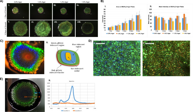
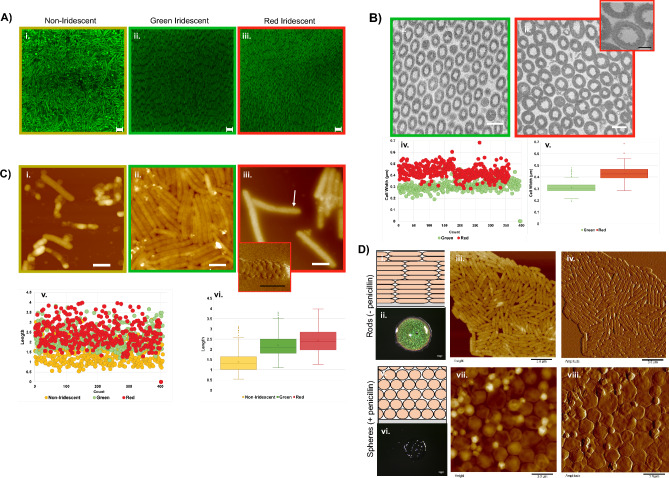
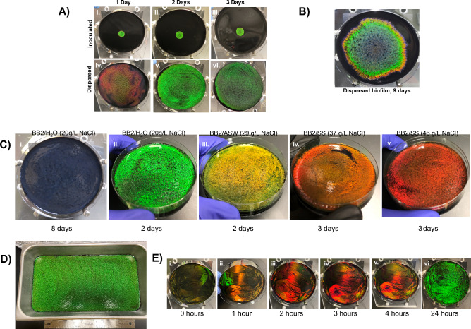
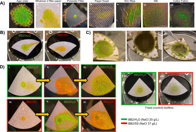
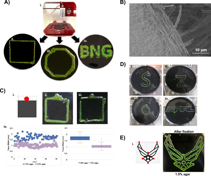
Nature offers many examples of materials which exhibit exceptional properties due to hierarchical assembly of their constituents. In well-studied multi-cellular systems, such as the morpho butterfly, a visible indication of having ordered submicron features is given by the display of structural color. Detailed investigations of nature's designs have yielded mechanistic insights and led to the development of biomimetic materials at laboratory scales. However, the manufacturing of hierarchical assemblies at industrial scales remains difficult. Biomanufacturing aims to leverage the autonomy of biological systems to produce materials at lower cost and with fewer carbon emissions. Earlier reports documented that some bacteria, particularly those with gliding motility, self-assemble into biofilms with polycrystalline structures and exhibit glittery, iridescent colors. The current study demonstrates the potential of using one of these bacteria, Cellulophaga lytica, as a platform for the large scale biomanufacturing of ordered materials. Specific approaches for controlling C. lytica biofilm optical, spatial and temporal properties are reported. Complementary microscopy-based studies reveal that biofilm color variations are attributed to changes in morphology induced by cellular responses to the local environment. Incorporation of C. lytica biofilms into materials is also demonstrated, thereby facilitating their handling and downstream processing, as would be needed during manufacturing processes. Finally, the utility of C. lytica as a self-printing, photonic ink is established by this study. In summary, autonomous surface assembly of C. lytica under ambient conditions and across multiple length scales circumvent challenges that currently hinder production of ordered materials in industrial settings.


Conflict of interest statement

The authors declare no competing interests.

Figures

Figure 1
Figure 1
Given particular growth conditions, C. lytica 7489 makes intensely iridescent biofilms though previously reported as lacking color. (A) Representative C. lytica 7489 biofilms were grown at 27°C on BB2/H2O agar plates containing 0.8%, 1.0%, 1.2% or 1.5% agar. Biofilms were imaged each day for 5 days. Agar concentration impacts color saturation and expansion of DSM 7489 C. lytica biofilms. Scale bar = 1 mm. (B) Areas (i) and mean intensity (ii) measurements of biofilms in A were recorded daily using ImageJ. Data are presented as averages for each timepoint with standard deviation shown as ±error (n = 10 for each timepoint). (C) C. lytica 7489 is capable of generating a range of intense colors as shown in this photo of a biofilm acquired from an oblique viewing angle (i) and schematic showing the concentric coloration (ii). (D) Optical images showing that regions of the biofilm appearing green (i) and red (ii) macroscopically are mosaics of pointillistic colors. (bar = 1 mm) (E) A representative hyperspectral data cube reveals region-specific variations in signal intensity in mature biofilms. (i) The biofilm was grown in a 10 cm petri dish on nutrient agar containing black ink. Note that the detector is normal to the surface of the biofilm. Its position is the reason for the reduction in reflection intensity compared to the biofilm in (C-i). Outer regions of the biofilm generate an especially sharp peak centered near 550 nm suggesting constructive and coherent reflection through the biofilm. (ii, location 1) In contrast, reflections in the center region remain near baseline. (ii, location 2)
Figure 2
Figure 2
Microscopy of biofilm cells. (A) Confocal images of biofilms stained with SYTO9 showing that cellular organization differs between non-iridescent (i) and iridescent regions (ii, iii) (bar = 2.0 µm). (B) Transmission Electron Microscopy (TEM) cross section images of green (i) and red regions (ii). (bar = 0.5 µm) Inset (iii) showing small protrusions surrounding the cell walls (bar = 200 nm). Width measurements (iv, v) also differ by region. (C) Atomic Force Microscopy (AFM) height images of non-iridescent (i), green iridescent (ii), and red iridescent (iii) regions showing that distinct cellular morphologies are associated with each region (bar = 1.0 µm). Inset (iv) is an AFM amplitude image of the region indicated by the arrow (bar = 0.5 µm). Length measurements from specified biofilm regions (v, vi). (D) 2-day old biofilms grown in ambient conditions on BB2/H2O agar (optical scale bars = 1 mm; AFM bars = 3 µm). Schematic showing typical arrangement of cells in iridescent biofilms (i). Optical image of iridescent biofilm. (ii) AFM height (iii) and amplitude (iv) images of cells from iridescent biofilm showing typical rod shape morphology. Schematic showing predicted arrangement of cells in biofilms grown when sublethal penicillin is added (v). Optical image of biofilm showing that sub-lethal penicillin disrupts structural coloration. (vi) AFM height (vii) and amplitude (viii) images of cells confirm conversion to spheres due to penicillin treatment.
Figure 3
Figure 3
Monochrome biofilms. (A) A comparison of inoculated (i–iii) versus dispersed (iv–vi) biofilms at ambient temperature. Dispersed biofilms peak in monochrome color and fill the plate at 2 days (v). (B) Whereas, inoculated biofilms develop banded coloration and require significantly more time to cover the surface. (C) Photos showing that dispersing C. lytica cells leads to monochrome biofilms of various colors presumably by simultaneously exposing all the cells to homogeneous growth conditions (nutrients, metabolites, etc.). The range of colors can be accessed by extending the growth period (i) or changing media salinity (ii – v) using sea water simulants. Effective NaCl in recipe given in parenthesis. Plates were grown in ambient conditions for the indicated time. (D) A large green biofilm was generated in 3 days in ambient conditions by dispersing a proportional inoculum on the surface of a 41 x 23 cm pan. (E) Sequential application and dispersal of 50-fold concentrated aliquots of culture reduces monochrome biofilm formation to 24 h or less, suggesting that the cells immediately begin to organize and that cell density will be an important consideration for industrial manufacturing applications. Plates were incubated at 27 °C between cell applications. Additional trials shown in Supplemental figure 4E,F.
Figure 4
Figure 4
Ambient growth of iridescent biofilms on porous substrates including paper facilitates handling for characterization and downstream processing. (A) C. lytica biofilms were grown in ambient conditions on a variety of porous substrates. (B) Whatman 2 filter paper placed atop nutrient agar is one of several porous substrates that allow C. lytica to form iridescent colonies. As on agar plates, living paper-associated biofilms (PABs) are green after 3 days of growth atop BB2/H2O agar (A-i) and red-shifted when salinity increases as on BB2/SS (A-ii). (C) PABs retain their iridescence after removal from agar and fixation with glutaraldehyde (B-i). Drying the fixed PABs with nitrogen causes them to lose their iridescence (B-ii). However, the structural color is restored upon rehydration (B-iii). (D) Living PABs retain their ability to respond to environmental cues. PABs from BB2/H2O agar reflect mostly green until they are moved to BB2/SS plates where reflections are shifted red (C-i and C-ii, respectively). Similarly, PABs originating on BB2/SS agar plates are red but change to green when placed on BB2/H2O (C-iv and C-v, respectively). In both cases, biofilms are able to revert back to original color when returned to the original media condition (C-iii and C-vi). Fixed biofilms do not show this dynamic behavior (C-vii and C-viii).
Figure 5
Figure 5
C. lytica can be used as an iridescent bioink. (A) 3D printed designs containing C. lytica were generated on agar using an Allevi 3 Bioprinter setup (A-i to A-iv). As previously shown for dispersed biofilms, increased salinity red shifts the C. lytica ink’s reflection. (Supplemental fig. 9) (B) SEM image of edge of 3D printed biofilm showing ordered cells of the printed biofilm. (C, D) C. lytica trace the edges of a paper template (e.g. schematic in C-i). The red circle on the template design indicates the site of inoculation. Cells behave like a self-printing bioink to write various patterns in ambient conditions. (B) Increasing the agar concentration from 1.0% (C-ii) to 1.5% (C-iii) reduces the width of the tracing as revealed in Keyence measurements (C-iv). This result suggests that gliding motility is modulated in a way that confines the cells closer to the template on the higher concentration agar. (D) Additional tracings show BACTracing can be used with shapes of varying complexity, angles and connections. Agar concentration can be used to confine the traces when the distance between features is small as is the case in intricate patterns such as the Air Force Symbol (supplemental Fig. 6c). (E) Template of a complex pattern (i) and its BACTraced counterpart (ii) after fixation showing that the iridescent pattern can be preserved.

Similar articles

References

    1. Sun JY, Bhushan B, Tong J. Structural coloration in nature. RSC Adv. 2013;3:14862–14889. doi: 10.1039/c3ra41096j. - DOI
    1. Tadepalli S, Slocik JM, Gupta MK, Naik RR, Singamaneni S. Bio-optics and bio-inspired optical materials. Chem. Rev. 2017;117:12705–12763. doi: 10.1021/acs.chemrev.7b00153. - DOI - PubMed
    1. Zhang W, et al. Butterfly effects: Novel functional materials inspired from the wings scales. Phys. Chem. Chem. Phys. 2014;16:19767–19780. doi: 10.1039/c4cp01513d. - DOI - PubMed
    1. Zhou H, et al. Bio-inspired photonic materials: Prototypes and structural effect designs for applications in solar energy manipulation. Adv. Funct. Mater. 2018 doi: 10.1002/adfm.201705309. - DOI - PubMed
    1. Chung K, et al. Flexible, angle-independent, structural color reflectors inspired by morpho butterfly wings. Adv. Mater. 2012;24:2375–2379. doi: 10.1002/adma.201200521. - DOI - PubMed

Publication types

Supplementary concepts

LinkOut - more resources