. 2023 Aug 14;14(1):4895.
doi: 10.1038/s41467-023-40635-w.

Toll-like receptor 4 and macrophage scavenger receptor 1 crosstalk regulates phagocytosis of a fungal pathogen

Affiliations

Toll-like receptor 4 and macrophage scavenger receptor 1 crosstalk regulates phagocytosis of a fungal pathogen

Chinaemerem U Onyishi et al. Nat Commun. .

Abstract

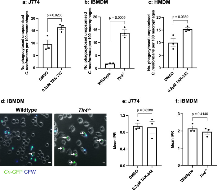
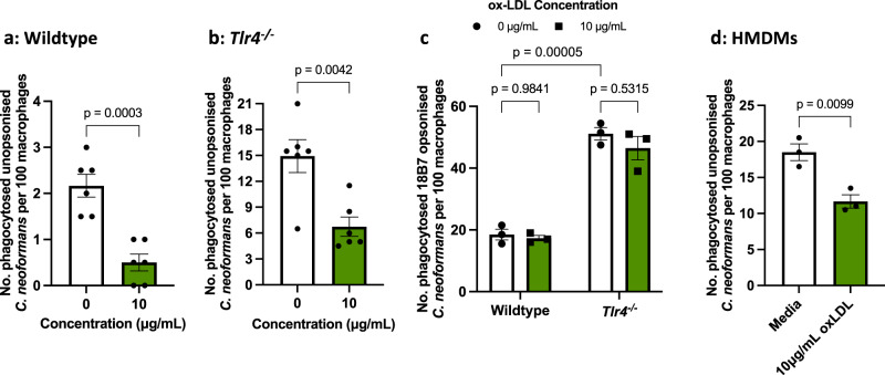
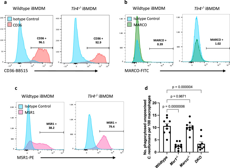
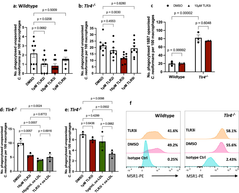
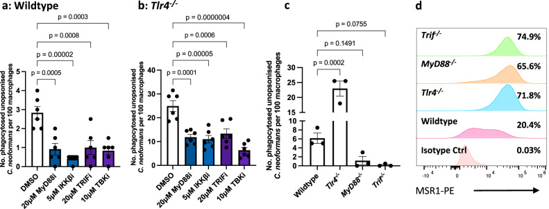
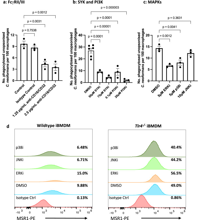
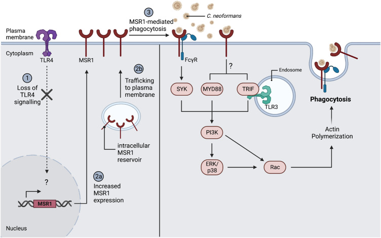
The opportunistic fungal pathogen Cryptococcus neoformans causes lethal infections in immunocompromised patients. Macrophages are central to the host response to cryptococci; however, it is unclear how C. neoformans is recognised and phagocytosed by macrophages. Here we investigate the role of TLR4 in the non-opsonic phagocytosis of C. neoformans. We find that loss of TLR4 function unexpectedly increases phagocytosis of non-opsonised cryptococci by murine and human macrophages. The increased phagocytosis observed in Tlr4-/- cells was dampened by pre-treatment of macrophages with oxidised-LDL, a known ligand of scavenger receptors. The scavenger receptor, macrophage scavenger receptor 1 (MSR1) (also known as SR-A1 or CD204) was upregulated in Tlr4-/- macrophages. Genetic ablation of MSR1 resulted in a 75% decrease in phagocytosis of non-opsonised cryptococci, strongly suggesting that it is a key non-opsonic receptor for this pathogen. We go on to show that MSR1-mediated uptake likely involves the formation of a multimolecular signalling complex involving FcγR leading to SYK, PI3K, p38 and ERK1/2 activation to drive actin remodelling and phagocytosis. Altogether, our data indicate a hitherto unidentified role for TLR4/MSR1 crosstalk in the non-opsonic phagocytosis of C. neoformans.


Conflict of interest statement

The authors declare no competing interests.

Figures

Fig. 1
Fig. 1. Both chemical inhibition and genetic loss of TLR4 results in an increase in the phagocytosis of C. neoformans in both murine and human macrophages.
a J774A.1 macrophages or (c) human monocyte derived macrophages (HMDMs) were treated with DMSO (control) or 0.2 μM TAK-242, a TLR4 specific inhibitor, for 1 h before infection with non-opsonised C. neoformans. b Immortalised bone marrow derived macrophages (iBMDM) from wildtype and Tlr4−/− macrophages were infected with non-opsonised C. neoformans. Phagocytosis was quantified as the number of individual internalised cryptococci within 100 macrophages. Figures are representative of at least three independent experiments. HMDM data represents two independent experiments with separate donors. d Representative image showing the phagocytosis of GFP-labelled C. neoformans (Cn-GFP) by wildtype and Tlr4−/− iBMDM. Calcofluor White (CFW) was used to stain extracellular fungi. White arrows show phagocytosed fungi. Scale bar = 10 μm. The intracellular proliferation (IPR) of C. neoformans was measured in (e) J774A.1 macrophages and (f) wildtype and Tlr4−/− iBMDMs using timelapse imaging. Images were captured every 5 mins for 18 h. The number of internalised fungi per 100 macrophages at the ‘first frame’ (T0) and ‘last frame’ (T10) was quantified and IPR was determined using the equation: IPR = T10/T0. Data is representative of two independent experiments. All data shown as mean ± SEM; n = 3 per condition; statistical significance was evaluated using an unpaired two-sided t-test; P-values are shown above each graph. Source data are provided as a Source Data file.
Fig. 2
Fig. 2. The increased phagocytosis observed in Tlr4−/− macrophages is partially driven by scavenger receptors.
a Wildtype iBMDMs (n = 6 per condition), b Tlr4−/− iBMDMs (n = 6 per condition), and d human monocyte derived macrophages (HMDMs) (n = 3 per condition) were treated with oxidised low-density lipoprotein (ox-LDL), a general scavenger receptor ligand, for 30 mins prior to infection with non-opsonised C. neoformans. iBMDM data is pooled from two independent experiments. HMDM data represents two independent experiments with separate donors. c Wildtype and Tlr4−/− iBMDMs (n = 3 per condition) were treated with 10 μg/mL ox-LDL for 30 mins, then infected with C. neoformans opsonised with the anti-capsular 18B7 antibody. The number of internalised fungi per 100 macrophages was quantified from fluorescent microscopy images. Data represents two independent experiments. All data is shown as mean ± SEM; statistical significance was evaluated using an unpaired two-sided t-test (a, b, d); or two-way ANOVA followed by Tukey’s post-hoc test (c). P-values are shown above each graph. Source data are provided as a Source Data file.
Fig. 3
Fig. 3. The scavenger receptor, MSR1, mediates the nonopsonic uptake of C. neoformans.
Baseline surface expression of a CD36 stained with anti-mouse CD36-BB515 antibody; b Macrophage Receptor with Collagenous structure (MARCO) stained with anti-mouse MARCO-Fluorescein antibody; and c Macrophage Scavenger Receptor 1 (MSR1), also known as CD204, stained with anti-mouse CD204-PE antibody on wildtype and Tlr4−/− macrophages. Receptor expression was measured using flow cytometry. Data is representative of three independent experiments which gave similar results. Numbers above gates refer to the percentage of CD36-, MARCO- and MSR1-positive cells. d MPI cells, a non-transformed GM-CSF-dependent murine macrophage cell line, isolated from wildtype, Msr1−/−, Marco−/− and MSR1/MARCO double knockout (DKO) mice, were infected with non-opsonised C. neoformans. The data shown is pooled from three independent experiments (n = 9). Data is mean ± SEM; statistical significance was evaluated using a one-way ANOVA; P-values are shown above the graph. Source data are provided as a Source Data file.
Fig. 4
Fig. 4. The increased phagocytosis observed in Tlr4−/− macrophages is dependent on TLR3 signalling.
a Wildtype and b Tlr4−/− iBMDMs were treated with chemical inhibitors of TLR2, TLR3 and TLR9 for 1 h, then infected with non-opsonised C. neoformans. Data is pooled from three independent experiments (n = 9 per condition). c Wildtype and Tlr4−/− iBMDMs were treated with a TLR3 inhibitor then infected with C. neoformans opsonised with the anti-capsular 18B7 antibody (n = 3 per condition). d, e Tlr4−/− macrophages were pre-treated with optimal and suboptimal concentrations of TLR3 inhibitor and ox-LDL, individually and in combination (n = 3 per condition). Phagocytosis was quantified as the number of internalised cryptococci within 100 macrophages. Data is representative of two independent experiments and is shown as mean ± SEM; statistical significance was evaluated using (a, b, d, e) one-way ANOVA or (c) two-way ANOVA followed by Tukey’s post-hoc test. P-values are shown above each graph. f Wildtype and Tlr4−/− iBMDMs were treated with 0.025% DMSO (control) or 10 μM TLR3 inhibitor for 1 h. MSR1 (also known as CD204) expression was measured using anti-mouse CD204-PE monoclonal antibody. PE-labelled rat IgG2a κ was used as an isotype control. Samples were run through a flow cytometer and analysed in the FlowJo software. Percentages refer to the percentage of MSR1-positive cells. Data is representative of two independent experiments. Source data are provided as a Source Data file.
Fig. 5
Fig. 5. MyD88 and TRIF are required for nonopsonic uptake of C. neoformans without affecting MSR1 expression.
a Wildtype and b Tlr4−/− macrophages were treated with inhibitors of MyD88, IKKβ (a kinase downstream of MyD88 that is necessary for NF-κB activation), TRIF, and TBK1 (a kinase downstream of TRIF that phosphorylates and activates IRF3) (n = 6 per condition). Following pre-treatment with the various inhibitors, cells were infected with non-opsonised C. neoformans. Data are pooled from two independent experiments. c Immortalised BMDMs from wildtype, Tlr4−/−, MyD88−/− and Trif−/− macrophages were infected with non-opsonised C. neoformans (n = 3). Data is representative of three independent experiments. Phagocytosis was quantified as the number of internalised cryptococcus within 100 macrophages. Data is mean ± SEM and analysed using one-way ANOVA followed by Tukey’s post-hoc test. P-values are shown above each graph. d Baseline surface expression of Macrophage Scavenger Receptor 1 (MSR1) (also known as CD204) was measured in wildtype, Tlr4−/−, MyD88−/− and Trif−/− macrophages using anti-mouse CD204-PE antibody. PE-labelled rat IgG2a κ was used as an isotype control. Receptor expression was measured using flow cytometry and analysed using the FlowJo software. Data is representative of two independent experiments. Percentages refer to the percentage of MSR1-positive cells. Source data are provided as a Source Data file.
Fig. 6
Fig. 6. Increased uptake in Tlr4−/− macrophages is dependent on FcγRs, SYK, PI3K, ERK1/2 and p38, but not JNK.
Tlr4−/− macrophages were pre-treated with inhibitors of a FcγRs (n = 3 per condition), b SYK and PI3K (control, n = 6; treatment, n = 3), and c Mitogen Activate Protein Kinases (MAPKs) (n = 3 per condition) for 1 h, then infected with non-opsonised C. neoformans. The number of internalised fungi per 100 macrophages was quantified from images from a fluorescence microscope. Figures are representative of three independent experiments. Data shown as mean ± SEM; a one-way ANOVA with Tukey’s post-hoc test was performed to evaluate statistical significance. P-values are shown above each graph. d Wildtype and Tlr4−/− iBMDMs were exposed to MAPK inhibitors for 24 h, then cell surface expression of MSR1 was detected with using anti-mouse CD204-PE antibody. PE-labelled rat IgG2a κ was used as an isotype control. Receptor expression was measured using flow cytometry and analysed using the FlowJo software. Data is representative of two independent experiments. Percentages refer to the percentage of MSR1-positive cells. Source data are provided as a Source Data file.
Fig. 7
Fig. 7. Proposed model of MSR1-meidated non-opsonic phagocytosis.
TLR4 modulates the surface expression of MSR1 through a yet-to-be-identified mechanism, such that loss of TLR4 signalling drives increased surface expression of MSR1. This may be through modulation of MSR1 gene expression or modulation of intracellular MSR1 reservoir trafficking to the plasma membrane. MSR1 is then responsible for the direct binding and internalisation of C. neoformans. MSR1-mediated uptake may rely on the formation of a signalling complex with FcγRII/III. The ITAM domain of FcγRs enables SYK and PI3K activation which then activates Rac GTPases that drive actin polymerisation and phagocytosis. At the same time, TLR3, MyD88 and TRIF may also serve as coreceptors or adaptor proteins leading to the activation of ERK1/2 and p38 which will also drive actin remodelling and pathogen uptake. Figure created with BioRender.com.

References

    1. May RC, Stone NRH, Wiesner DL, Bicanic T, Nielsen K. Cryptococcus: from environmental saprophyte to global pathogen. Nat. Rev. Microbiol. 2016;14:106–117. - PMC - PubMed
    1. Maziarz EK, Perfect JR. Cryptococcosis. Infect. Dis. Clin. North Am. 2016;30:179–206. - PMC - PubMed
    1. Rajasingham R, et al. Global burden of disease of HIV-associated cryptococcal meningitis: an updated analysis. Lancet Infect. Dis. 2017;17:873–881. - PMC - PubMed
    1. Osterholzer JJ, et al. Role of dendritic cells and alveolar macrophages in regulating early host defense against pulmonary infection with Cryptococcus neoformans. Infect. Immun. 2009;77:3749–3758. - PMC - PubMed
    1. Lim J, et al. Characterizing the mechanisms of nonopsonic uptake of cryptococci by macrophages. J. Immunol. 2018;200:3539–3546. - PMC - PubMed

Publication types