. 2023 Aug 14;13(1):13183.
doi: 10.1038/s41598-023-40531-9.

Adipose-derived mesenchymal stem cell therapy for reverse bleomycin-induced experimental pulmonary fibrosis

Affiliations

Adipose-derived mesenchymal stem cell therapy for reverse bleomycin-induced experimental pulmonary fibrosis

Xiansheng Zhao et al. Sci Rep. .

Abstract

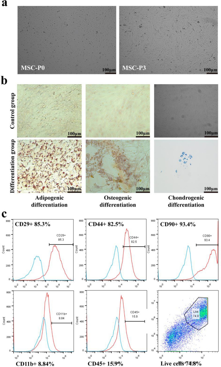
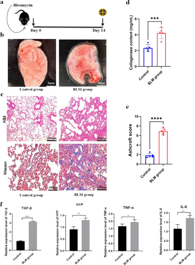
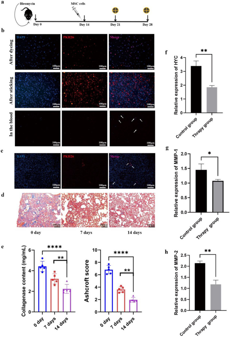
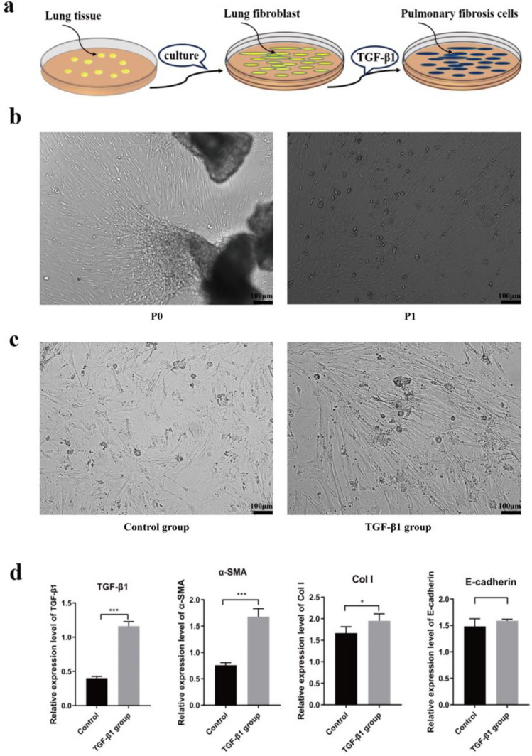
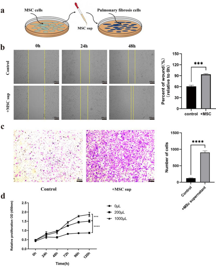
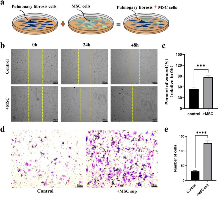
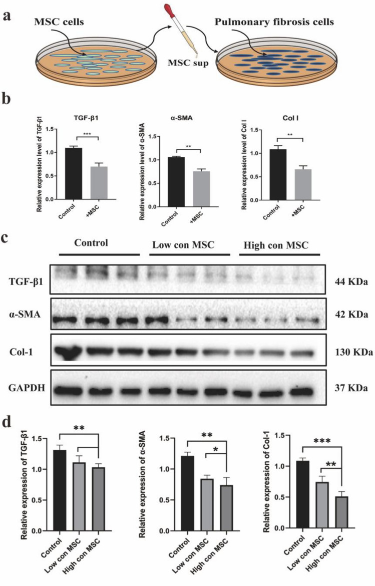
Idiopathic pulmonary fibrosis (IPF) is a chronic, progressive respiratory disease. Arguably, the complex interplay between immune cell subsets, coupled with an incomplete understanding of disease pathophysiology, has hindered the development of successful therapies. Despite efforts to understand its pathophysiology and develop effective treatments, IPF remains a fatal disease, necessitating the exploration of new treatment options. Mesenchymal stromal/stem cell (MSC) therapy has shown promise in experimental models of IPF, but further investigation is needed to understand its therapeutic effect. This study aimed to assess the therapeutic effect of adipose-derived mesenchymal stem cells in a bleomycin-induced pulmonary fibrosis model. First, MSC cells were obtained from mice and characterized using flow cytometry and cell differentiation culture methods. Then adult C57BL/6 mice were exposed to endotracheal instillation of bleomycin and concurrently treated with MSCs for reversal models on day 14. Experimental groups were evaluated on days 14, 21, or 28. Additionally, lung fibroblasts challenged with TGF-β1 were treated with MSCs supernatant or MSCs to explore the mechanisms underlying of pulmonary fibrosis reversal. Mesenchymal stem cells were successfully isolated from mouse adipose tissue and characterized based on their differentiation ability and cell phenotype. The presence of MSCs or their supernatant stimulated the proliferation and migration of lung fibrotic cells. MSCs supernatant reduced lung collagen deposition, improved the Ashcroft score and reduced the gene and protein expression of lung fibrosis-related substances. Bleomycin-challenged mice exhibited severe septal thickening and prominent fibrosis, which was effectively reversed by MSCs treatment. MSC supernatant could suppress the TGF-β1/Smad signaling pathway and supernatant promotes fibroblast autophagy. In summary, this study demonstrates that MSCs supernatant treatment is as effective as MSCs in revert the core features of bleomycin-induced pulmonary fibrosis. The current study has demonstrated that MSCs supernatant alleviates the BLM-induced pulmonary fibrosis in vivo. In vitro experiments further reveal that MSC supernatant could suppress the TGF-β1/Smad signaling pathway to inhibit the TGF-β1-induced fibroblast activation, and promotes fibroblast autophagy by Regulating p62 expression. These findings contribute to the growing body of evidence supporting the therapeutic application of MSCs in cell therapy medicine for IPF.


Conflict of interest statement

The authors declare no competing interests.

Figures

Figure 1
Figure 1
Cultivation, differentiation, and identification of MSCs. (a) Morphology of MSCs at different culture passages. The first-generation MSCs are generally triangular or dot-shaped, while the third-generation MSCs displayed a spindle-like or elongated shape. Images were taken at × 40 magnification. (b) MSCs subjected to differention and stained accordingly. Successfully differentiation into adipocytes (stained red with Oil Red), osteoblasts (stained with Alizarin Red), and chondrocytes (stained with Alcian Blue). Scale bar = 100 μm. Images were taken at × 100 magnification. (c) Flow cytometric analysis of MSCs after three passages of culture. The expression of CD45 and CD11b antigen/cell markers was found to be negative, while CD29, CD44, CD90, and other antibodies exhibited positive expression, Statistical analysis was performed using the Student’s t-test (two-tailed).
Figure 2
Figure 2
Bleomycin-induced pulmonary fibrosis mouse model. (a) Eight-week-old mice (C57BL/6 strain) received endotracheal bleomycin (3 U/kg) or 0.9% normal saline (control). on day 0. Mice were sacrificed on day 14. The cross symbol represents animal harvest. (b) Macroscopic observation of mouse lung tissue was conducted to assess the effects of the interventions. (c) Lung sections were stained with Hematoxylin–eosin and Masson’s trichrome. Images were taken at × 100 magnification. Bleomycin group showed architectural destruction, alveolar septal thickening, and fibrotic changes. Scale bar: 100/200 μm. (d) Collagen deposition was assessed by Sircol assay and represented as milligrams per milliliter (mg/mL) of left lung homogenate. (e) The extent of lung fibrosis was measured on day 14 by using the Ashcroft score. (f) qRT-PCR analysis of bleomycin induces changes in the expression of pulmonary fibrosis-related genes. The mice were sacrificed humanely, and lung tissues were collected to assess the mRNA levels of fibrosis-related genes. Expression of TGF-β, HYP, TNF-α, and IL-6 levels (n = 6 mice per group). Data are representative of 5 independent experiments, mean ± SD. n = 5 per experimental group; each symbol represents 1 mouse. *P < 0.05; **P < 0.01, ***P < 0.001, ****P < 0.0001, 1-way ANOVA followed by Fisher’s LSD post hoc analysis.
Figure 3
Figure 3
MSC therapy reverts bleomycin-induced pulmonary fibrosis. (a) Fourteen days after the administration of bleomycin, MSCs were administered, and mice were sacrificed on day 21 or 28 for further analysis. The cross symbol represents the time of animal harvest. (b) MSCs were labeled with fluorescent dyes, DAPI for the nucleus, and PKH26 for the cell membrane. After the mice were sacrificed, the presence of fluorescently labeled MSCs in their peripheral blood was detected. (c) Lung slices are used for fluorescence microscopy. The image was taken at × 100 magnification. Fluorescently labeled MSCs can be observed after cell therapy. (d) Lung sections were stained with Mason's trichrome. The image was taken at × 100 magnification. Collagen deposition decreased in a time dependent fashion following MSC therapy. Scale bar: 100 μm. (e) Collagen deposition was assessed by Sircol assay and the result were represented as mg/mL of left lung homogenate. The degree of lung fibrosis was measured using the Ashcroft score at days 0, 7 and 14 after treatment. (fh) qRT-PCR analysis of gene expression of related cytokines in lung tissue after MSC cell therapy. The mice were sacrificed humanely, and lung tissues were collected to assess the mRNA levels of fibrosis-related genes. Expression of HYP, MMP-1, and MMP-2 levels (n = 6 mice per group). Data are representative of 5 independent experiments, mean ± SD. n = 5 per experimental group; each symbol represents 1 mouse. *P < 0.05; **P < 0.01, ***P < 0.001, ****P < 0.0001, 1-way ANOVA followed by Fisher’s Dunnett post hoc analysis.
Figure 4
Figure 4
The cellular model of pulmonary fibrosis induced by TGF-β1. (a) Pulmonary fibroblasts were isolated from lung tissue using an adherent culture method, and subsequently, TGF-β1 was employed to induce their transformation into pulmonary fibrotic cells. (b) Pulmonary fibroblasts were purified from lung tissue and cultured for cell passage. Images were taken at × 40 magnification. Scale bar: 100 μm. (c) Following treatment with TGF-β1 (10 mg/L), lung fibroblasts underwent induction and acquired a fibrotic phenotype. Images of the induced cells were taken at × 40 magnification. Scale bar: 100 μm. (d) qRT-PCR analysis of gene expression supports the formation of a cellular model of pulmonary fibrosis following induction by TGF-β1. Expression of TGF-β1, α-SMA, Col I and E-cadherin levels (n = 3 per group). Data are representative of 5 independent experiments, mean ± SD. n = 5 per experimental group. *P < 0.05; **P < 0.01, ***P < 0.001, 1-way ANOVA followed by Fisher’s LSD post hoc analysis.
Figure 5
Figure 5
MSC supernatant promotes the migration and proliferation of fibrotic lung cells in vitro. (a) MSC supernatant was harvested and added to the culture medium of lung fibrosis cells. (b) The migration level of lung fibrotic cells was evaluated through a wound healing experiment following the addition of MSC supernatant. The results of this experiment provided insights into the migratory capacity of the cells upon exposure to MSC supernatant. (c) The transwell migration assay method detects the migration level of lung fibrotic cells after adding MSC supernatant. (d) The CCK-8 viability assay of the lung fibrotic cells added to the MSC supernatant. All results are represented as mean ± SD. n = 5 per experimental group. ***P < 0.001, ****P < 0.0001, 2-way ANOVA followed by Fisher’s Dunnett post hoc analysis.
Figure 6
Figure 6
MSCs promotes the migration and proliferation of lung fibrotic cells in vitro. (ac) Promotion of Migration and Proliferation of Lung Fibrotic Cells by MSCs In Vitro. (a) Co-culture of MSCs and lung fibrotic cells. MSCs were harvested and added to the culture medium of lung fibrotic cells. (b,c) Wound healing assay. (d,e) Transwell invasion assay. All results are represented as mean ± SD. n = 5 per experimental group. ***P < 0.001, ****P < 0.0001,1-way ANOVA followed by Fisher’s LSD post hoc analysis.
Figure 7
Figure 7
MSCs supernatant reduces the expression of fibrosis-related genes and protein levels of pulmonary fibrosis cells. (a) The MSC supernatant was harvested and added to the culture medium of lung fibrosis cells. (b) qRT-PCR analysis showed that the application of MSC supernatant resulted in the reversel of pulmonary fibrotic cells formation induced by TGF-β1. The expression levels of TGF-β1, α-SMA, and collagen type I were assessed (n = 3 per group). (c,d) Western blot analysis was performed to evaluate the relative protein levels at different doses of MSC supernatant. The expression levels of TGF-β1, α-SMA, and Col I levels (n = 3 per group). Data are representative of 5 independent experiments, mean ± SD. n = 5 per experimental group. *P < 0.05; **P < 0.01, ***P < 0.001, 1-way ANOVA followed by Fisher’s LSD post hoc analysis. Original blots are presented in Supplementary Fig. S3.

References

    1. Brooks PJ, Samuel LT, Levin JM, et al. Mortality after hip resurfacing versus total hip arthroplasty in young patients: A single surgeon experience. Ann. Transl. Med. 2019;7(4):77. - PMC - PubMed
    1. George PM, Wells AU, Jenkins RG. Pulmonary fibrosis and COVID-19: The potential role for antifibrotic therapy. Lancet Respir. Med. 2020;8(8):807–815. - PMC - PubMed
    1. Akram A. Overwhelming COVID-19 sepsis in a patient with idiopathic pulmonary fibrosis. Cureus. 2020;12(7):e9320. - PMC - PubMed
    1. Mishra M, Sindhwani G. Antifibrotics for COVID-19 related lung fibrosis: Agents with benefits? Adv. Respir. Med. 2021;89(2):231–233. - PubMed
    1. Shao D, Liu X, Wu J, et al. Identification of the active compounds and functional mechanisms of Jinshui Huanxian formula in pulmonary fibrosis by integrating serum pharmacochemistry with network pharmacology. Phytomedicine. 2022;102:154177. - PubMed

Publication types