. 2023 Sep;55(9):1542-1554.
doi: 10.1038/s41588-023-01471-2. Epub 2023 Aug 14.

In vivo screening characterizes chromatin factor functions during normal and malignant hematopoiesis

Affiliations

In vivo screening characterizes chromatin factor functions during normal and malignant hematopoiesis

David Lara-Astiaso et al. Nat Genet. 2023 Sep.

Abstract

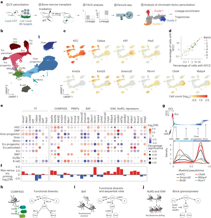
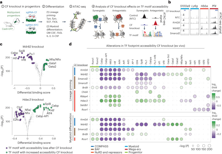
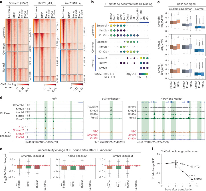
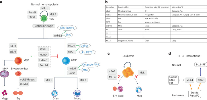
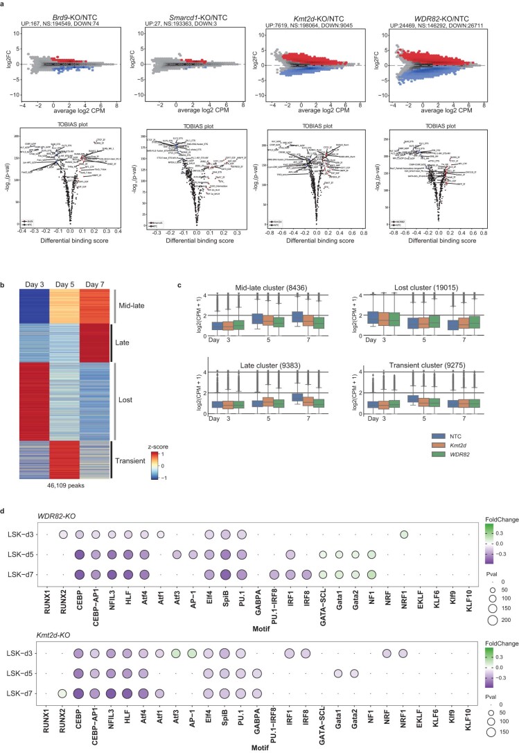
Cellular differentiation requires extensive alterations in chromatin structure and function, which is elicited by the coordinated action of chromatin and transcription factors. By contrast with transcription factors, the roles of chromatin factors in differentiation have not been systematically characterized. Here, we combine bulk ex vivo and single-cell in vivo CRISPR screens to characterize the role of chromatin factor families in hematopoiesis. We uncover marked lineage specificities for 142 chromatin factors, revealing functional diversity among related chromatin factors (i.e. barrier-to-autointegration factor subcomplexes) as well as shared roles for unrelated repressive complexes that restrain excessive myeloid differentiation. Using epigenetic profiling, we identify functional interactions between lineage-determining transcription factors and several chromatin factors that explain their lineage dependencies. Studying chromatin factor functions in leukemia, we show that leukemia cells engage homeostatic chromatin factor functions to block differentiation, generating specific chromatin factor-transcription factor interactions that might be therapeutically targeted. Together, our work elucidates the lineage-determining properties of chromatin factors across normal and malignant hematopoiesis.


Conflict of interest statement

T.G. and J.P.T.-K. are employees receiving compensation from Relation Therapeutics. J.P.T.-K. is a founder of Relation Therapeutics. D.L.-A. is a consultant of Relation Therapeutics. The other authors declare no competing interests.

Figures

Fig. 1
Fig. 1. Functional screens identify lineage specificities for chromatin factors in hematopoiesis.
a, Schema of the experimental approach used, describing the murine progenitor populations used, their isolation and transduction with the CRISPR library, subsequent differentiation, flow sorting of specific differentiation readouts and read-count based analysis of function. CF, chromatin factor; TF, transcription factor. b, Differentiation systems and FACS-based readouts: (1) self-renewal versus differentiation; (2) lineage priming: mega-erythroid versus myeloid; (3) myeloid differentiation: mature myeloid versus non-myeloid; and (4) terminal myeloid maturation versus immature. c, Averaged lineage scores of ‘differentiation versus self-renewal’ (y axis) versus ‘lineage priming’ (x axis) for 554 genes. Significant hits (n = 93 genes with 50% or more significant guide RNAs (gRNAs) in either comparison) are shown with a red cross. NTC sgRNAs are shown with a blue cross. Data for non-Cas9 cells are shown in the background using a yellow-blue density. d, Lineage scores for chromatin factors grouped on the basis of complex membership. The dot color represents the lineage score, the dot size the percentage of significant guides (Supplementary Table 3). HDAC, histone deacetylase; PRMT, protein arginine methyltransferase. e, Exemplar immunophenotypic validations for chromatin factors in the lineage priming (top) and myeloid differentiation (bottom) systems. SSC-A, side scatter area.
Fig. 2
Fig. 2. Perturb-seq highlights disparate lineage dependencies for chromatin complexes during hematopoiesis.
a, Schematic drawing of the in vivo Perturb-seq: LSK progenitors were sorted from Cas9-GFP mice infected with the chromatin factor knockout library and transplanted into irradiated recipient mice; bone marrow was collected 14 days after transplant, sorted for an immature phenotype (lineage and Lin+c-Kit+) and the Perturb-seq and downstream analyses were performed. b, Uniform manifold approximation and projection (UMAP) of the single-cell transcriptomes. Clusters are annotated using external reference maps. The analysis integrates seven different biological replicates. CLP, common lymphoid progenitor; Eo/Ba, esosinophil-basophil progenitor; Ery, erythroblast; G1, G1 phase; Gran, granulocyte; IMP, immature myeloid progenitor; MEP, mega-erythroid progenitor; MKP, megakaryocyte progenitor; Mono, monocyte; S, S phase. c, UMAP showing the distribution of unperturbed cells (NTC sgRNAs) and specific perturbations. d, Scatterplot showing comparisons between experimental batches. Each dot represents the abundance of two NTC sgRNAs in a given population. Pearson correlation between NTC and sgRNAs per experimental batch = 0.962 with P = 1.20 × 10−67. e, Enrichment and depletion of chromatin factor knockouts across hematopoietic populations (Supplementary Table 4). Dot color and size relate to the log2 odds ratio (OR) and the percentage of significant enrichments. f, Effect of specific chromatin factor knockouts on myeloid versus erythroid priming. Positive values (red) show enhanced myeloid priming. Negative values (blue) indicate reduced myeloid priming. g, Trajectory analysis of specific chromatin factor knockouts along myeloid differentiation. Cells are ordered from HSCs to mature granulocytes using pseudotime. DC1, diffusion component 1; DC2, diffusion component 2. hj, Graphic representation of the roles of key chromatin regulatory complexes. Source data
Fig. 3
Fig. 3. Chromatin factors regulate the accessibility of lineage-determining transcription factors.
a, Schema of the experiment; LSK progenitors were collected from Cas9-GFP mice, transduced with a single chromatin factor or NTC control gRNA, placed into differentiation medium for 7 days and ATAC-seq was performed and analyzed to infer the interacting synergistic and antagonistic transcription factors. b, Chromatin accessibility for representative myeloid and mega-erythroid loci after disruption of specific chromatin factors. The accessibility profiles correspond to two merged replicate experiments. The S100a9, Ly6g, Hbba and Pf4 loci coordinates are chr3:90685583–90703276, chr15:75134355–75144437, chr7:103869996–103874051 and chr5:90771031–90773155, respectively. The y axis ranges for all knockouts in the same regions are 0.02–1.5, 0.02–1, 0.02–2 and 0.02–4, respectively. c, Volcano plot showing differentially bound transcription factor motifs (estimated by TOBIAS) in Wdr82 and Hdac3 knockouts under lineage priming conditions. Transcription factor motifs demonstrating gained and lost accessibility in each chromatin factor knockouts (compared to NTCs) are shown in green and purple, respectively. n = 2 biologically independent experiments. d, Heatmap summarizing the effect of ten chromatin factor knockouts on transcription factor motif footprints estimated by TOBIAS. n = 2 biologically independent experiments. Dot color and size relate to the log2 fold change and the −log10(Padj) value, respectively. Source data
Fig. 4
Fig. 4. Disruption of ncBAF leads to a pre-leukemic accumulation of myeloid progenitors with diminished Cebp-AP-1 activity.
a, Schema of the in vivo perturbation of cBAF (Smarcd2 sgRNA) and ncBAF (Brd9 sgRNA); in vivo transplantation of LSK cells transduced with a library of Smarcd1, Smarcd2, Brd9 and NTC guides, into irradiated recipient mice and lineage-negative cells were collected and sorted at either 14 or 28 days for Perturb-seq as in Fig. 2. b, Proportions of cells with a specific sgRNA at 14 and 28 days after transplant. c, UMAP showing the distribution of Brd9 knockout and control (NTC sgRNA) cells at 28 days after transplant. d, Schema of the in vivo ATAC-seq experiment; Brd9 knockout LSK were generated and transplanted as in a and collected at day 28 for the ATAC-seq analysis. e, The ATAC-seq signal at myeloid progenitor (top) and differentiated (bottom) loci. Coordinates: Hoxa7, Hoxa9, Hoxa10: chr6:52214971–52236669, chr11:18912448–19036437; Meis1: chr6:88186822–8820729; Gata2: chr11:87788022–87796472; Mpo: chr3:90651905–90703284; S100a7, S100a8, S100a9: chr14:56098019–56107286; Ctsg: chr14:56098019–56107286. f, Volcano plot showing differentially bound transcription factor motifs (estimated by TOBIAS) between control (NTC) and Brd9 knockout GMPs. n = 2 biologically independent experiments. g, Genome browser tracks showing ATAC-seq, Cebpa ChIP–seq and Cebpe ChIP–seq in wild-type (WT) myeloid progenitors (GMPs). Loci coordinates are the same as in Fig. 4e. h,i, Quantification of accessibility changes between Brd9 knockout and control (NTC) GMPs. h, MA plot showing loci overlapping with Cebpa binding (red). i, Box plots showing accessibility loss (statistically tested using a two-sided Kolmogorov–Smirnov test, n = 2) at Cebpa (n = 11,316, statistic = 0.86, P = 1 × 10−323) and Cebpe sites (n = 10,409, statistic = 0.85, P = 1 × 10−323). The box plot displays the median as the center line of the box, with the box representing the distribution’s 25th (minima) and 75th (maxima) percentiles. The whiskers extend up to 1.5 times the interquartile range (IQR) (Q3–Q1) from the minima and maxima. j, Schema of the ChIP–seq analysis. k, Heatmaps showing specific binding of Smarcb1 (cBAF) and Brd9 (ncBAF) in myeloid progenitors (GMPs) and bone marrow monocytes. Two merged independent ChIP–seq experiments were used. l, Transcription factor motif enrichment measured by HOMER at the cBAF and ncBAF sites between progenitor (GMP) and mature (monocytes) myeloid cells. The axes represent the transcription factor motif odds-ratio (OR). All colored transcription factor motifs have Padj < 0.001. The analysis was performed with two independent experiments per population.
Fig. 5
Fig. 5. Npm1c and Flt3-ITD leukemia abrogates normal chromatin factor function to maintain leukemic fitness through enforcing differentiation blockade.
a, UMAP projection of single-cell transcriptomes from Npm1c and Flt3-ITD primary leukemia. The color-coded clusters correspond to cells with specific signatures: leukemic stem cells (LSC, green), granulocyte-like (Gra.1 and Gra.2, Gran-perturbed, blue), erythroid-like (Ery.1 and Ery.2, red) and basophil-like (Baso-like, yellow). The analysis integrates datasets from six different Perturb-seq experiments. b, mRNA-derived and CITE-seq-derived expression of lineage makers in the differentiated leukemia subpopulations. c,d, Clonogenic (c) and proliferation (d) assays for differentiated leukemia subpopulations, isolated according to the strategy in Extended Data Fig. 9e. Colonies were counted after 7 days of culture in methylcellulose. Proliferation and clonogenic values were obtained from n = 4 biologically independent experiments. ****P < 0.0001 (two-way analysis of variance (ANOVA)). The error bars are the s.e.m., the midpoints show the mean. e, Enrichment analyses of specific chromatin factor knockouts across differentiated leukemia subpopulations. Dot color and size relate to the log2(OR) and the percentage of significant enrichments versus NTCs, respectively. The analysis is based on measurements for two sgRNAs per chromatin factor target. All values are shown in Supplementary Table 6. Gra.P1 and Gra.P2, granulocyte-like. f, Perturbed growth curves for leukemic chromatin factor knockouts. The assay measures the change in the proportion of blue fluorescent protein (BFP) BFP sgRNA-expressing cells over time, n = 4 biologically independent experiments. All ***P < 0.001, except for Smarcd1 versus NTC (day 9) where ***P = 0.0009 (two-way ANOVA). The error bars are the s.e.m., the midpoints show the mean. g, FACS analysis of mega-erythroid (CD55) and myeloid (CD11b, Gr1) surface differentiation markers in leukemia cells depleted for cBAF (Smarcb1 and Smarcd2 knockout) and MLL (Kmt2a knockout) components. h, FACS analysis of myeloid surface differentiation markers (CD11b, Gr1) in leukemia cells treated with increasing doses of Men1 inhibitor (revumenib). Raw data can be found in Supplementary Data 1. Source data
Fig. 6
Fig. 6. Chromatin factors enforce differentiation blockade in AML through corrupted transcription factor interactions.
a, Heatmaps showing ChIP–seq signal for cBAF (Smarcb1), MLL (Kmt2a) and MLL4 (Kmt2d) in leukemia (Npm1c and Flt3-ITD), in vivo myeloid progenitors (GMPs), in vivo monocytes and ex vivo-derived primary monocytes. n = 2 independent ChIP–seq experiments per factor. b, Motif enrichment analysis of cBAF, MLL and MLL4 binding patterns specific for leukemia (leukemic), common between leukemia and myeloid progenitors (common), and specific for normal myeloid cells (normal). c, Box plot showing the Stat5a, Runx1 and Runx2 binding signal (ChIP–seq) at the leukemic, common and normal loci defined in Fig. 6a (n = 2). The number of loci comprising the leukemic, common and normal categories are 2,019, 581 and 2,361 for Smarcb1; 2,427, 2,346 and 5,789 for Kmt2a; and 3,144, 810 and 4,129 for Kmt2d. d, Genome browser tracks showing the ChIP–seq signal for Smarcb1, Kmt2a, Kmt2d, Stat5a and Runx2 in leukemia, and ATAC-seq for control and chromatin factor-depleted leukemia cells. The chosen loci are leukemic-specific. n = 2 independent experiments. The green highlighted regions shown identify chromatin factor–transcription factor binding and altered accessibility on chromatin factor knockout. e, Box plots showing changes in chromatin accessibility at leukemic loci bound by Stat5a, Runx1 and Runx2 on depleting specific chromatin factors. cBAF, n = 2 independent experiments. Smarcd2 knockout: n = 1,385, 1,050, 1,711; statistic = 0.73, 0.75, 0.76; P = 5 × 10−15, 0, 0. Kmt2a knockout: n = 1,288, 695, 1,427; statistic = 0.78, 0.86, 0.78; P = 0, 9 × 1016, 0. Kmt2d knockout: n = 1,482, 990, 1,913; statistic = 0.70, 0.70, 0.69; P = 2 × 10−15, 0, 0. The decay in accessibility was tested statistically using a two-sided Kolmogorov–Smirnov test. f, Growth curves for Cas9-leukemic cells expressing NTC and anti-Stat5a sgRNAs, n = 3 independent experiments. ***P < 0.001 (two-way ANOVA). The error bars are the s.e.m., the midpoints show the mean. The box plots in c and e display the median and the distribution’s 25th (minima) and 75th (maxima) percentiles. The whiskers extend up to 1.5 times the IQR (Q3–Q1) from the minima and maxima. Source data
Fig. 7
Fig. 7. Summary model of chromatin factor function and chromatin factor–transcription factor interactions in normal and malignant hematopoiesis.
a, Roadmap of chromatin factor requirements for major hematopoietic cell fate decisions, identifying individual chromatin factors required for specific lineages and the transcription factor families they interact with to orchestrate these decisions. b, Table explaining the roles of specific chromatin factor–transcription factor complexes. c, Examples of how chromatin factor function is hijacked in leukemia, where cBAF-, MLL4- and MLL1-containing complexes block rather than facilitate hematopoietic differentiation. d, Examples of ‘transcription factor switches’ that mechanistically underpin the different functions of chromatin factors in normal and malignant hematopoiesis.
Extended Data Fig. 1
Extended Data Fig. 1. Characterization of CRISPR Screen systems.
(a) Comparison of expression profiles of the different readout populations from our screens to lineage specific signatures from 3 different studies, named along the bottom of the graph. Comparisons are based on enrichment analyses between the screen signatures and the reference signatures. Dot colour and size relate to log2 odds ratio and -log10 adjusted p-value, respectively. CLP – Common Lymphoid Progenitor, MkP – Megakaryocyte Progenitor, IMP – Immature Myeloid Progenitor, Ery1-4 – Erythroblasts, Neu1-4 – Neutrophils, Mono1-3 – Monocytes. P-values were calculated using the Fisher’s exact test. (b) Comparison between the expression profiles of FACS-sorted populations from our ex vivo systems and a single-cell map of normal haematopoiesis. Bulk transcriptomic signatures derived from FACS-sorted populations were projected on the single-cell map from Izzo and colleagues. (c) Example distribution of the CF lineage scores calculated from the Cas9 (green border) and Non-Cas9 (grey) populations. (d) Replicate analysis for 200 CFs screened in a second experiment under Self-renewal (top) and Lineage Priming conditions (bottom). (left) heatmaps showing correlation (Spearman) between two replicates, (right) scatter plots showing correlation (Spearman) between replicates. P-values are based on the algorithm AS 89 using the function cor.test in R. (e) Lineage scores for all hits. The color of each dot represents the aggregated lineage score. The size represents the number of significant guides, as per key to the right. All values are shown in Supplementary Table 3.
Extended Data Fig. 2
Extended Data Fig. 2. Validation of the effects of individual CF-KOs.
(a-b) Heatmap showing changes in representative populations for each CF-KO compared to a Non-Targeting Control (Fold-change in population abundances versus NTC) under lineage-priming (a) and Myeloid differentiation and terminal Myeloid maturation (b). The Myeloid master regulator Pu.1 (Spi1) was included as a positive control. Gates and values for the selected populations are derived from Supplementary Fig. 2c, d. (c-d) Exemplar FACS plots showing validation results for individual CF-KOs under lineage priming conditions (c) and Terminal Myeloid Differentiation (d). These validations were performed in different batches. Each batch included a Non-Targeting Control condition. All results were compared with the NTC included in each batch.
Extended Data Fig. 3
Extended Data Fig. 3. Characterization of the in vivo Perturb-seq system.
(a) Comparison of expression profiles from our in vivo single-cell clusters and external cell-type signatures from Izzo et al, 2020. (b) Number of cells per cell type. (c) UMAP projection of the Lineage- and Lineage+ ckit+ fractions. Color scale represents the number of cells in each area. (d) Number of cells with a sgRNAs targeting specific CFs. CF-KOs for which less than 50 cells (in red) were detected were removed from subsequent analysis. (e) Visualization of TF- and CF-KO patterns derived from in vivo Perturb-seq of Chromatin Regulatory Complexes during lineage specification. The distribution of NTCs is shown as background in grey in all plots. Cells are aggregated and the color of each area represents the density of cells in each area.
Extended Data Fig. 4
Extended Data Fig. 4. Extended in vivo Perturb-seq analysis of Chromatin Regulatory Complexes during lineage specification.
(a) Enrichment analyses of CF-KOs across 11 cellular states spanning the main hematopoietic lineages, all values are shown in Supplementary Table 4. Dot color and size relate to the log2 odds ratio and the percent of significant enrichments versus NTCs, respectively. The analysis is based on measurements of two aggregated sgRNAs per CF target. (b) CF-KO effects on early Myeloid versus Erythroid lineage branching, positive values (red) indicate CF-KOs leading to increased Myeloid outputs. (c) CF-KO effects on Myeloid versus Erythroid total outputs, positive values (red) indicate CF-KOs leading to increased Myeloid outputs. (d) CF-KO effects on Granulocyte versus Monocyte total outputs, positive values (red) indicate CF-KOs leading to increased monocytic outputs. (e) CF-KO effects on viability/survival of CF-KOs after 14 days post-transplant. Negative values (blue) indicate that cells with specific CF-KOs have growth/engraftment disadvantages a when compared to the Control (NTC harboring) cells. Positive values (red) indicate that cells with CF-KOs have growth advantages when compared to the Control (NTC harboring) cells.
Extended Data Fig. 5
Extended Data Fig. 5. Extended in vivo Perturb-seq analysis of Chromatin Regulatory Complexes during lineage specification.
(a) Heatmap summarizing trajectory analysis for CF-KOs along Myeloid and Erythroid branches ordered from HSCs to mature lineage using pseudotime. Colors are given by signed negative log10 p-values (for p<0.01) generated by a t-test between targeting and non-targeting control populations such that negative values correspond to reduced differentiation capability and positive values correspond to increased differentiation capability. (b-e) Analysis of aberrant cellular states generated after specific CF-KOs. (b) UMAP showing localization of 53 subclusters across the hematopoietic landscape. (c) Plot showing the abundance of CF-sgRNAs with respect to Control-sgRNAs across the 53 hematopoietic subclusters. Clusters deviating from the diagonal are rare or absent in the unperturbed scenario. (d) Enrichment of specific CF-KO cells in three representative aberrant subclusters: Erythroid-perturbed (cluster 26) and Granulocytic-Perturbed (clusters 45 and 51). (e) Marker genes of aberrant clusters. (f) Functions specific of the Erythroid-perturbed cluster (26). P-values were calculated by random sampling as implemented in the fgsea R package. (g) Barplots showing the expression levels of selected Chromatin and Transcription factors. The bars represent the normalized read counts taken from an RNA-seq dataset.
Extended Data Fig. 6
Extended Data Fig. 6. Extended analysis of transcriptomic effects of CF-KOs.
(a) Analysis of the effect of Chromatin factor disruption (CF-KOs) on lineage specific expression patterns, comprising markers and transcription factors specific for progenitor, Myeloid, Erythroid, Megakaryocytic, Basophil and B-cell lineages. The color of each dot represents the log2 fold change (compared to NTCs), the size represents the –log10 adjusted p-value, as per key to the bottom right. P-values were calculated using negative binomial mixed models from the nebula R package. All values are shown in Supplementary Table 5. (b) Gene set enrichment analysis (GSEA) of differentially expressed genes in knockouts of factors belonging to repressive complexes. The color of each dot represents normalized enrichment score, the size represents the –log10 adjusted p-value. P-values were calculated by random sampling as implemented in the fgsea R package.
Extended Data Fig. 7
Extended Data Fig. 7. Extended analysis of effects of CF-KOs on chromatin accessibility and TF footprints.
(a) MA plots demonstrating differential accessibility analysis between selected CF-KOs and Control (NTC). Up- and down-regulated genomic loci are indicated in red and blue, respectively. (lower panel) Volcano Plots showing the differentially bound TF motifs (estimated by TOBIAS) between the same CF-KOs and Control (NTC). Gained and lost footprints are indicated in red and blue, respectively, n = 2 independent experiments. (b) Time-series analysis of chromatin accessibility dynamics under ex vivo priming conditions at day 3, 5 and 7, n=2 independent experiments. (c) Effect of Kmt2d- and Wdr82-KOs on the differential accessible patterns derived from the time-series analysis. n=8436, 19015, 9383, and 9275 for all conditions and days in the mid-late, lost, late, and transient clusters, respectively (n=2). Boxplots display the median and the distribution’s 25th (minima) and 75th (maxima) percentiles. The whiskers extend up to 1.5 times the interquartile range (Q3-Q1) from the minima and maxima. (d) Time-series analysis of differentially bound TF motifs (estimated by TOBIAS) under lineage priming conditions for Kmt2d- and Wdr82-KOs.
Extended Data Fig. 8
Extended Data Fig. 8. In vivo binding patterns of BAF and COMPASS complexes.
(a) CF binding at representative loci in Myeloid (GMP) and Erythroid (MEP) progenitors. Genomic Coordinates: Cebpa chr7:35,114,878-35,131,210; Cebpe chr14:54,702,383-54,717,520; Elane chr10:79,871,207-79,893,610 ; Gfi1b chr2:28,585,038-28,624,000; Hbb chr7:103,845,151-103,886,745; Car2 chr3:14,855,264-14,912,573. (b) Heatmaps showing lineage-specific binding patterns for each CF. (c) Heatmap showing joint analysis of Smarcb1/cBAF and Brd9/ncBAF binding in Myeloid progenitors (GMPs) and in mature Myeloid cells (monocytes). This analysis shows that Smarcb1/cBAF has widespread binding at early Myeloid stages while Brd9/ncBAF exhibits more presence in mature Myeloid cells (Monocytes). Strong overlap between cBAF and ncBAF complexes seems limited to few regions. (d) Representative binding tracks of Smarcb1 and Brd9 binding in GMP and Monocytes at progenitor loci (upper panel) and mature Myeloid loci (lower panel). (e) TF motif co-occurrence in lineage specific binding patterns of Smarcb1 (cBAF), Brd9 (ncBAF), Kmt2a (MLL) and Kmt2d (MLL4). TF motifs (discovered with HOMER) are sorted by their odds ratios (y-axis) in Kmt2a- Kmt2d- Brd9- and Smarcb1- lineage specific peaks: GMP, Mye (GMP & Monocytes), MEP, Ery (MEP & Erythrocytes) and B-cells. The color scale reflects the –log10 p-adjusted values for each TF motif.
Extended Data Fig. 9
Extended Data Fig. 9. Extended analysis of Chromatin Factor roles in Npm1c/Flt3-ITD Leukemia.
(a) UMAP showing original projection of Npm1c/Flt3-ITD single-cell transcriptomes. (b) UMAP projection of Npm1c/Flt3-ITD single-cell transcriptomes projected over the Hematopoietic in vivo map derived from bone marrow at 14-day post-transplant. (c) Scaled CITE-seq signal for 9 surface markers in leukemic cells. (d) Expression analysis of markers over the different leukemic clusters. According to their mRNA and Surface marker patterns these are classified into: Leukemic Stem Cells (LSC), GMP-like, Monocyte-like, Granulocyte-like, Basophil-like, Megakaryocyte-like and Erythroid-like. Clusters in red are absent in the unperturbed (NTC) cells. (e) Exemplar sorting strategy of leukemic subpopulations showing traits of differentiation into Granulocyte (Gran) or mixed Erythroid-Basophil populations. (f) Enrichment analyses of all CF-KOs across leukemia subpopulations. Disruption of factors highlighted in red induce differentiation pathways in leukemia. All values are shown in Supplementary Tables 6 and 7. (g). Plot showing the abundance of specific CF-sgRNAs with respect to Control-sgRNAs across the leukemic subclusters. Subclusters deviating from the diagonal are rare or absent in the unperturbed scenario. (h) Growth curves of Prmt1- and Prmt5-KO cells, n=3 biologically independent experiments. The cells expressing each sgRNA harbor a BFP reporter and, the assay measures the change in the proportion of BFP expressing cells over time. ***P<0.001 (Two Way ANOVA). Error bars are SEM.
Extended Data Fig. 10
Extended Data Fig. 10. Extended analysis of cBAF, MLL and MLL4 binding and TF-partnership in Npm1c/Flt3-ITD Leukemia.
(a) Correlation (spearman) analysis of CF binding patterns in Myeloid progenitors, Mature myeloid and leukemic populations. (b-d) Enrichment analysis of Smarcb1-, Kmt2a- and Kmt2d- bound loci specific of leukemia cells. Bar graphs show enriched terms across input gene lists, sorted by p-values. Targets connected to leukemia specific peaks (nearest TSS) were run in Metascape. Functions with particularly high relevance for Npm1c/Flt3-ITD leukemia are highlighted in bold. (e) Genome-browser snapshots showing binding of cBAF/Smarcb1, MLL/Kmt2a and MLL4/Kmt2d on leukemic specific loci. (f) MA plots showing accessibility changes after acute depletion of Smarcd2-KO (cBAF), Kmt2a-KO (MLL) and Kmt2d-KO (MLL4) with respect to Control cells harbouring NTC sgRNAs. Color coding loci overlapping with Stat5a (red), Runx1(green) and Runx2 (blue).

References

    1. Stadhouders R, Filion GJ, Graf T. Transcription factors and 3D genome conformation in cell-fate decisions. Nature. 2019;569:345–354. doi: 10.1038/s41586-019-1182-7. - DOI - PubMed
    1. Ding Y, Liu Z, Liu F. Transcriptional and epigenetic control of hematopoietic stem cell fate decisions in vertebrates. Dev. Biol. 2021;475:156–164. doi: 10.1016/j.ydbio.2021.03.003. - DOI - PubMed
    1. Al-Mousawi J, Boskovic A. Transcriptional and epigenetic control of early life cell fate decisions. Curr. Opin. Oncol. 2022;34:148–154. doi: 10.1097/CCO.0000000000000814. - DOI - PubMed
    1. Valencia AM, Kadoch C. Chromatin regulatory mechanisms and therapeutic opportunities in cancer. Nat. Cell Biol. 2019;21:152–161. doi: 10.1038/s41556-018-0258-1. - DOI - PMC - PubMed
    1. Laurenti E, Göttgens B. From haematopoietic stem cells to complex differentiation landscapes. Nature. 2018;553:418–426. doi: 10.1038/nature25022. - DOI - PMC - PubMed

Publication types