Case Reports
. 2023 Aug 14;18(1):244.
doi: 10.1186/s13019-023-02346-7.

Endovascular treatment for massive haemoptysis due to pulmonary pseudoaneurysm: report of 23 cases

Affiliations
Case Reports

Endovascular treatment for massive haemoptysis due to pulmonary pseudoaneurysm: report of 23 cases

Fen-Qiang Li et al. J Cardiothorac Surg. .

Abstract

Purpose: To evaluate the safety and effectiveness of endovascular treatment for massive haemoptysis caused by pulmonary pseudoaneurysm (PAP).

Methods: The clinical data, imaging data, and endovascular treatment protocol of 23 patients with massive haemoptysis caused by continuous PAP were retrospectively analysed. The success, complications, postoperative recurrence rate, and influence of the treatment on pulmonary artery pressure were also evaluated.

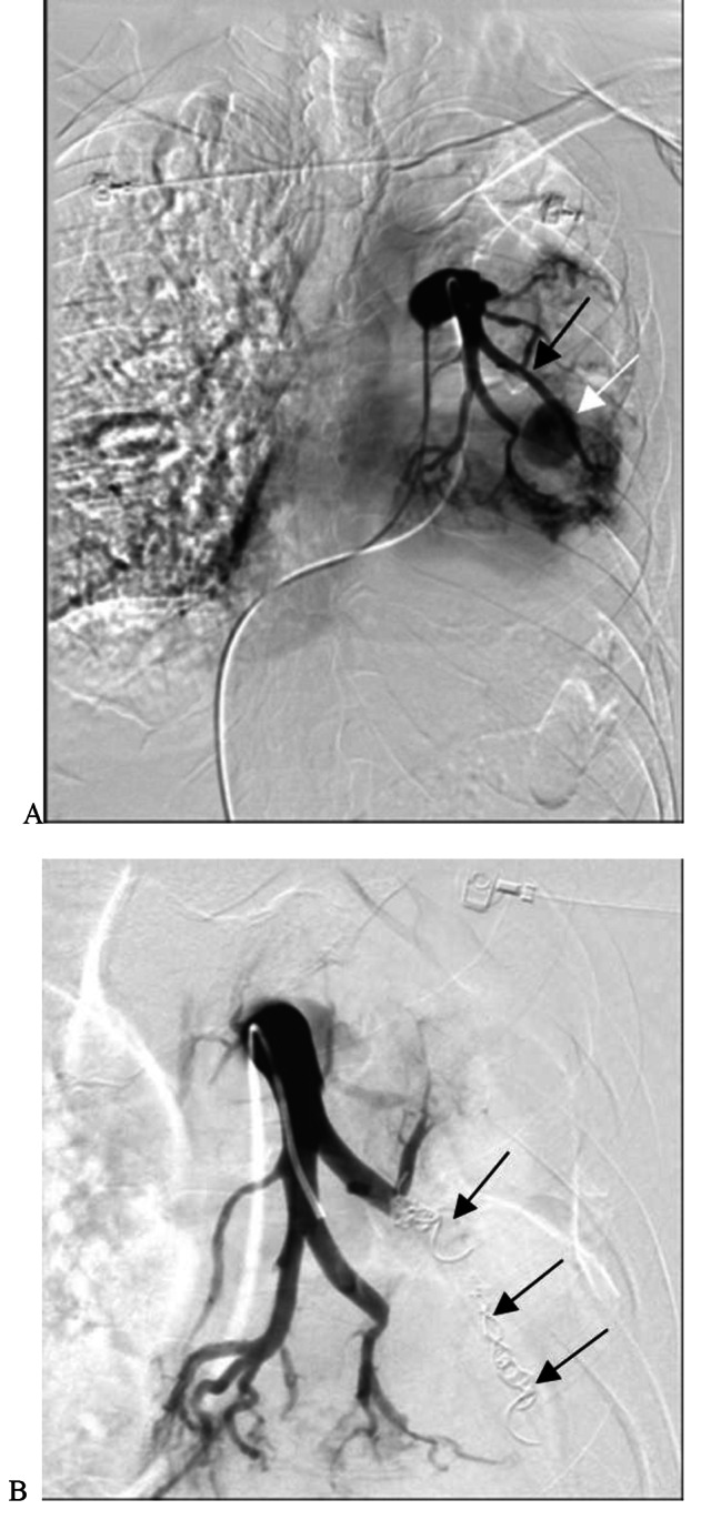
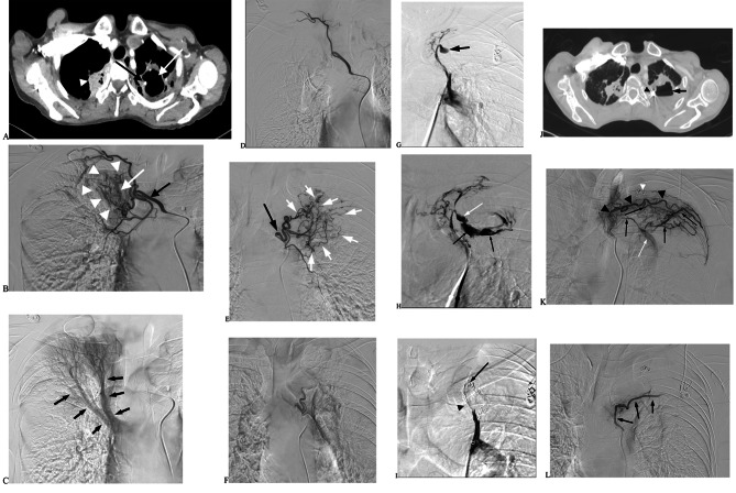
Results: Nineteen patients with a bronchial artery-pulmonary artery (BA-PA) and/or nonbronchial systemic artery-pulmonary artery (NBSA-PA) fistula underwent bronchial artery embolization (BAE) and/or nonbronchial systemic artery embolization (NBSAE) + pulmonary artery embolization (PAE). The pulmonary artery (PA) pressures before and after embolization were 52.11 ± 2.12 (35-69 cmH2O) and 33.58 ± 1.63 (22-44 cmH2O), respectively (P = 0.001). Four patients did not have a BA-PA and/or NBSA-PA fistula. Embolization was performed in two patients with a distal PAP of the pulmonalis lobar arteria. Bare stent-assisted microcoils embolization was performed in the other two patients with a PAP of the main pulmonary lobar arteries. The PA pressures of the four patients before and after treatment were 24.50 ± 1.32 (22-28 cmH2O) and 24.75 ± 1.70 (22-29 cmH2O), respectively (P = 0.850). The technique had a 100% success rate with no serious complications and a postoperative recurrence rate of 30%.

Conclusion: Endovascular treatment is safe and effective for massive haemoptysis caused by PAP. BAE and/or NBSAE can effectively reduce pulmonary hypertension in patients with a BA-PA and/or NBSA-PA fistula.

Keywords: Endovascular treatment; Haemoptysis; Pulmonary artery pseudoaneurysms.


Conflict of interest statement

The authors declare no competing interests.

Figures

Fig. 1
Fig. 1
Sudden massive hemoptysis (about 600ml) of unknown cause. Contrast-enhanced CT showed a pseudoaneurysm of the left inferior pulmonary artery. A. Left pulmonary artery angiography shows the left inferior pulmonary artery (black arrow) was PAP (white arrow). B. The distal pulmonary artery (white arrow) of the left lower lobe of the lung was superselected with a microcatheter. Complete embolization was performed from the distal end to the proximal end with interlock coils (black arrow)
Fig. 2
Fig. 2
A patient with old tuberculosis was hospitalized with sudden massive hemoptysis for 1 day, and the volume of one-time hemoptysis was about 500ml. A. Chest enhanced CT shows cavity formation in the lower lobe of the right lung and PAP formation in pulmonalis lobar arteria (white arrow). B. No PAP was found after the stent (black arrow) assisted PAPE with a micros coils (white arrow) for the right lower pulmonalis lobar arteria. C. Postoperative chest CT scan shows the position of the stent (white arrow) and coils (black arrow)
Fig. 3
Fig. 3
The patient with old pulmonary tuberculosis was hospitalized for 2 days with intermittent hemoptysis. The maximum hemoptysis volume in 24 h was approximately 700ml. A. Chest enhanced CT showing old lesions in the upper lobe of the right lung (white arrowhead). The left upper lobe cavity (white arrow) was formed, and PAP was observed in the cavity (black arrow). B. Right intercostal bronchial artery (black arrow) angiography shows obvious thickening, increased number, disorder, and structural abnormality of the bronchial artery (white arrowhead) and premature pulmonary artery branch (white arrow) in the upper lobe of the right lung. C. Right intercostal bronchial arteriography shows reverse flow in the late stage of the artery. The angiography also shows the pulmonary artery (black arrow) in the upper lobe of the right lung. D. After intercostal bronchial artery embolization with 350-560 μm PVA particles, the main intercostal artery remained intact. The bronchial artery and its branches disappeared completely, indicating complete embolization. E. Left bronchial artery (black arrow) angiography shows obvious thickening, increased number, disorder, and structural abnormality of the bronchial artery (white arrow). No obvious PA or PAP was observed. F. Left bronchial artery angiography again shows left BA branch vessels disappeared post BAE in 350-560 μm PVA particles. G. PAP showed on the left upper lobe of the lung by introducing a single curved catheter assisted by the long vascular sheath (black arrow) pulmonary angiography. H. PAP (white arrow) rupture, and contrast agent entering the cavity (black arrow) as observed in pulmonary angiography. I. Angiography after PAPE + PAE with micro coils, dense embolization with micro coils in parent artery (black arrowhead), and displacement of the micro coils at PAP rupture (black arrow). J. The patient was discharged from the hospital and admitted again with hemoptysis 13 months later. The amount of hemoptysis was about 100ml within 24 h. Enhanced chest CT examination showed that the cavity in the upper lobe of the left lung (black arrow) was smaller than before, and the shadow of the micro coils was visible within it (black arrowhead). K. Left intercostal artery (black arrowhead) angiography shows a large number of collateral vessels (black arrows) deviating into the lung, forming a large number of tortuous, thickened, malformed vessels, pulmonary artery fistula (white arrow), and micro coils shadow (white arrowhead). L. The presence of the main intercostal artery (black arrow) and the disappearance of collateral malformed vessels and pulmonary artery fistula were angiographed with 350-560 μm PVA embolization

References

    1. Spinu C, Castaner E, Gallrado X, et al. Multidetector computed tomography in life-threatening hemoptysis. Radiologia. 2013;55(6):483–98. doi: 10.1016/j.rx.2013.05.006. - DOI - PubMed
    1. Nguyen ET, Silva CI, Seely JM, et al. Pulmonary artery aneurysms and pseudoaneurysms in adults: findings at CT and radiography. AJR Am J Roentgenol. 2007;188:126–34. doi: 10.2214/AJR.05.1652. - DOI - PubMed
    1. Poplausky MR, Rozenblit G, Rundback JH, et al. Swan-ganz catheter-induced pulmonary artery pseudoaneurysm formation: three case reports and a review of the literature. Chest. 2001;120:2105–11. doi: 10.1378/chest.120.6.2105. - DOI - PubMed
    1. Jean-Baptiste E. Clinical assessment and management of massive hemoptysis. Crit Care Med. 2000;28:1642–7. doi: 10.1097/00003246-200005000-00066. - DOI - PubMed
    1. Hei jkoop B, Gillespie H, Kiroff G. Emergency management of massive haemoptysis. BMJ Case Rep. 2019;12(1):e225620. doi: 10.1136/bcr-2018-225620. - DOI - PMC - PubMed

Publication types

Substances