. 2023 Aug 14;15(15):7362-7380.
doi: 10.18632/aging.204959. Epub 2023 Aug 14.

Natural aging and ovariectomy induces parallel phosphoproteomic alterations in skeletal muscle of female mice

Affiliations

Natural aging and ovariectomy induces parallel phosphoproteomic alterations in skeletal muscle of female mice

Mina P Peyton et al. Aging (Albany NY). .

Abstract

The loss of skeletal muscle strength mid-life in females is associated with the decline of estrogen. Here, we questioned how estrogen deficiency might impact the overall skeletal muscle phosphoproteome after contraction, as force production induces phosphorylation of several muscle proteins. Phosphoproteomic analyses of the tibialis anterior muscle after contraction in two mouse models of estrogen deficiency, ovariectomy (Ovariectomized (Ovx) vs. Sham) and natural aging-induced ovarian senescence (Older Adult (OA) vs. Young Adult (YA)), identified a total of 2,593 and 3,507 phosphopeptides in Ovx/Sham and OA/YA datasets, respectively. Further analysis of estrogen deficiency-associated proteins and phosphosites identified 66 proteins and 21 phosphosites from both datasets. Of these, 4 estrogen deficiency-associated proteins and 4 estrogen deficiency-associated phosphosites were significant and differentially phosphorylated or regulated, respectively. Comparative analyses between Ovx/Sham and OA/YA using Ingenuity Pathway Analysis (IPA) found parallel patterns of inhibition and activation across IPA-defined canonical signaling pathways and physiological functional analysis, which were similarly observed in downstream GO, KEGG, and Reactome pathway overrepresentation analysis pertaining to muscle structural integrity and contraction, including AMPK and calcium signaling. IPA Upstream regulator analysis identified MAPK1 and PRKACA as candidate kinases and calcineurin as a candidate phosphatase sensitive to estrogen. Our findings highlight key molecular signatures and pathways in contracted muscle suggesting that the similarities identified across both datasets could elucidate molecular mechanisms that may contribute to skeletal muscle strength loss due to estrogen deficiency.

Keywords: CAST; MAPK; PKA; calcineurin; estrogen deficiency.


Conflict of interest statement

CONFLICTS OF INTEREST: The authors declare no conflicts of interest related to this study.

Figures

Figure 1
Figure 1
Schematic of experimental design created with https://www.biorender.com and mouse characteristics. (A) 6 mo C57BL/6J female mice were assigned to a Sham or Ovx group and underwent their respective surgeries. YA and OA mice were 4 mo and 24 mo, respectively. The left leg of anesthetized mice was subjected to in vivo contractions and then tibialis anterior muscles were immediately dissected. Frozen TA muscles underwent peptide extraction with trypsin digestion and TiO2 phosphopeptide enrichment. nLC-MS/MS was performed on the Orbitrap Fusion Tribrid mass spectrometer for label-free phosphoproteomic analysis. (B) Body mass of all four groups of female mice measured before the terminal contraction experiment. Data was analyzed by a one-way ANOVA with Tukey’s multiple comparison test (p < 0.001); n = 3–4/group. (C) Uterine mass of Ovx and Sham mice dissected and weighed after the terminal contraction experiment. Data was analyzed by a pooled t-test (Ovx vs. Sham, p = 0.016); n = 4/group. Values represents mean ± SD. *p < 0.05, ***p < 0.001, ****p < 0.0001.
Figure 2
Figure 2
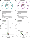
Characteristics of the Ovx/Sham and OA/YA phosphoproteomes. Proteome Discoverer (v2.4) was used for database search and identification of phosphopeptides and proteins. Venn diagram of identified phosphopeptides unique to each group and common to both groups in (A) Ovx/Sham and (B) OA/YA datasets. Prevalence of phosphorylation on amino acid residues serine (S), threonine (T), and tyrosine (Y) in (C) Ovx/Sham and (D) OA/YA datasets. Volcano plots of differentially regulated phosphopeptides (p < 0.05 and 1.4-fold change) in (E) Ovx/Sham and (F) OA/YA mice.
Figure 3
Figure 3
Comparative GO term enrichment analysis between Ovx/Sham and OA/YA. Phosphopeptides were mapped back to their precursor protein and submitted for Gene Ontology (GO) overrepresentation analysis using the clusterProfiler package in R. The top 10 overrepresented GO terms in the dataset for (A) molecular functions, (B) cellular components, and (C) biological processes are listed. P-value was adjusted using Benjamini – Hochberg post-hoc analysis for multiple comparison. Significant GO terms were accepted at p.adjusted < 0.05.
Figure 4
Figure 4
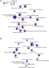
Comparative KEGG and Reactome pathway enrichment analysis between Ovx/Sham and OA/YA. Significant and differentially regulated phosphopeptides (adjusted p-value < 0.05 and |FC| ≥ 1.4) were mapped back to their precursor protein and the list was submitted to R for analysis of KEGG and Reactome pathways. The top 10 overrepresented (A) KEGG and (B) Reactome pathways were clustered using K-means clustering. The size of the circle represents the number of proteins associated to the pathway, and the connecting lines represents the strength of the similarity (i.e., shorter and thicker lines correspond to stronger similarity and inversely.
Figure 5
Figure 5
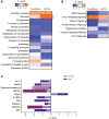
IPA’s predictive downstream effect and upstream regulator analytics between Ovx/Sham and OA/YA. Phosphopeptides identified in both the Ovx/Sham and OA/YA datasets were submitted to IPA for comparative analysis. (A) canonical pathways, (B) functions, and (C) kinases and phosphatases using IPA’s predictive activation Z-score to determine downstream and upstream effects of estrogen deficiency. Pathways, functions, and kinases identified in both datasets are represented. Significant Z-scores were accepted at |Z| ≥ 2. Red box denotes significant Z-scores.

References

    1. Haynes EMK, Neubauer NA, Cornett KMD, O'Connor BP, Jones GR, Jakobi JM. Age and sex-related decline of muscle strength across the adult lifespan: a scoping review of aggregated data. Appl Physiol Nutr Metab. 2020; 45:1185–96. 10.1139/apnm-2020-0081 - DOI - PubMed
    1. Phillips SK, Rook KM, Siddle NC, Bruce SA, Woledge RC. Muscle weakness in women occurs at an earlier age than in men, but strength is preserved by hormone replacement therapy. Clin Sci (Lond). 1993; 84:95–8. 10.1042/cs0840095 - DOI - PubMed
    1. Agostini D, Zeppa Donati S, Lucertini F, Annibalini G, Gervasi M, Ferri Marini C, Piccoli G, Stocchi V, Barbieri E, Sestili P. Muscle and Bone Health in Postmenopausal Women: Role of Protein and Vitamin D Supplementation Combined with Exercise Training. Nutrients. 2018; 10:1103. 10.3390/nu10081103 - DOI - PMC - PubMed
    1. Maltais ML, Desroches J, Dionne IJ. Changes in muscle mass and strength after menopause. J Musculoskelet Neuronal Interact. 2009; 9:186–97. - PubMed
    1. Asikainen TM, Kukkonen-Harjula K, Miilunpalo S. Exercise for health for early postmenopausal women: a systematic review of randomised controlled trials. Sports Med. 2004; 34:753–78. 10.2165/00007256-200434110-00004 - DOI - PubMed

Publication types