. 2023 Aug 1;13(8):5119-5129.
doi: 10.21037/qims-22-1319. Epub 2023 May 25.

Ridge operator-assisted delineation of capsulorhexis border for cataract surgery

Affiliations

Ridge operator-assisted delineation of capsulorhexis border for cataract surgery

Yishan Li et al. Quant Imaging Med Surg. .

Abstract

Background: With the continuous development of machine vision and imaging technology and its application in computer-aided diagnosis, it is clinically important to use computer technology to assist physicians in accurate cataract surgery. The capsulorhexis directly affects the outcome of cataract surgery, therefore, we design a method to automatically determine the virtual boundary of capsulorhexis for cataract surgery planning and tracking in-vivo to help surgeons achieve a more ideal capsulotomy geometry.

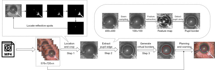
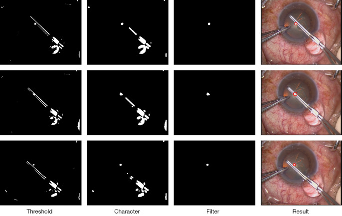
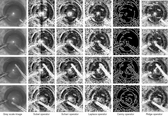
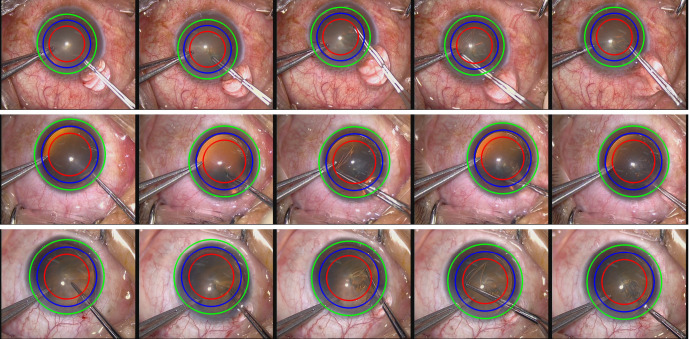
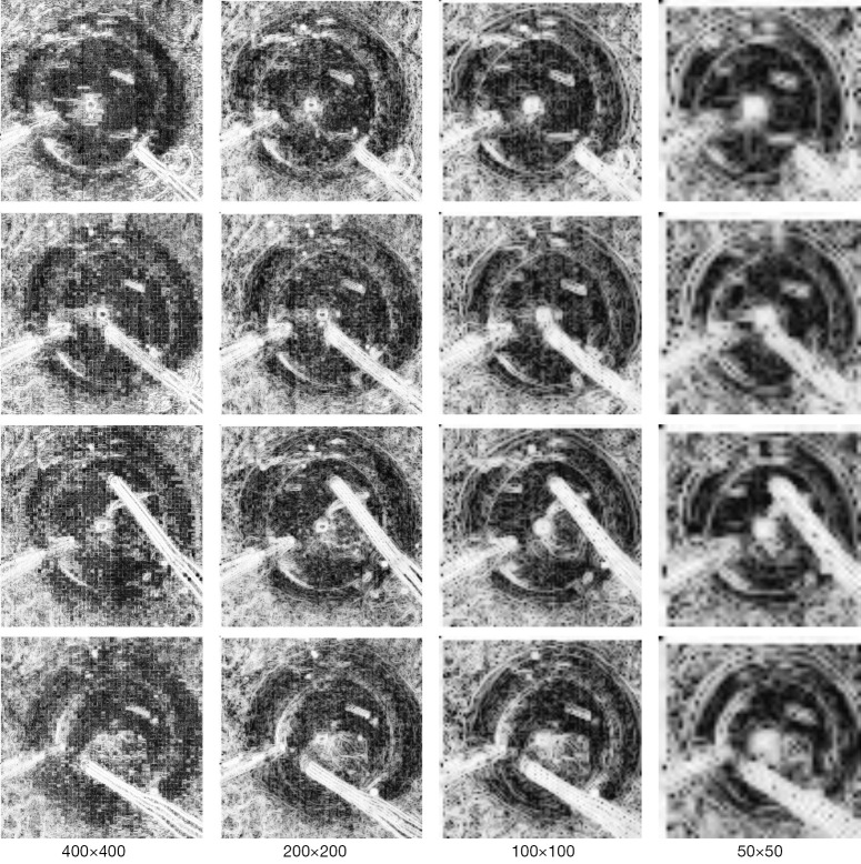
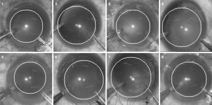
Methods: In this study, an effective method was proposed to detect and display the location of capsulorhexis in cataract videos in-vivo. The initial step was locating the entire eye area by analyzing the connected components of the mirror reflective points in the image in the cataract surgery video. Then, an operator was designed for ridge edge variation and used to extract pupil edge features. Lastly, circular Hough transform was used to detect the pupillary margin and calculate the boundary between the scleral limbus and the virtual capsulorhexis border in accordance with the pupillary margin and finally displayed it in-vivo during cataract surgery.

Results: The method was tested on eight videos of cataract surgery and the results showed that 98.52% accuracy was achieved in the localization of the specular reflection point. We compared the proposed operator with the Sobel, Scharr, Laplace and Canny operators and the results showed that our operator achieved the smallest mean square error with the greatest structural similarity.

Conclusions: The analysis demonstrated that the proposed operator outperformed other operators in detection and achieved satisfactory results in the videos of actual cataract surgeries.

Keywords: Continuous curvilinear capsulorhexis; cataract surgery planning; cataract video; pupillary margin; ridge edge.


Conflict of interest statement

Conflicts of Interest: All authors have completed the ICMJE uniform disclosure form (available at https://qims.amegroups.com/article/view/10.21037/qims-22-1319/coif). The authors have no conflicts of interest to declare.

Figures

Figure 1
Figure 1
Gradient change in pupil edge. (A) The original cataract surgery image; (B) the grayscale change image of (A); (C1,D1,E1) the magnified images of the pupil edge information in the grayscale image; (C2,D2,E2) the three-dimensional gradient magic figures of C1, D1, and E1, respectively.
Figure 2
Figure 2
Virtual capsulorhexis border detection and tracking process in cataract surgery.
Figure 3
Figure 3
Localization result of capsulorhexis forceps occluding the specular reflection point. The threshold image shows the filtered and denoised original image. Character image shows the feature map obtained by erosion and dilation of the image in the first column. Filter image shows the results after connected component filtering. Result image shows the minimum circle drawn on the third image, which is displayed on the original image.
Figure 4
Figure 4
Pupillary margin features extracted by different operators. The leftmost column shows the grayscale image of the original image. Each of the remaining columns displays the edge extraction results obtained using different operators on the grayscale image on the left. The Sobel, Scharr, Laplace, Canny, and proposed ridge operators were utilized for edge extraction.
Figure 5
Figure 5
Illustration of some key frames. The blue circle represents the pupillary margin, the green circle represents the scleral limbus, and the red circle represents the virtual capsulorhexis border.
Figure 6
Figure 6
Extraction effect of feature maps of different sizes.
Figure 7
Figure 7
Pupil edge extraction results of different cases.

Similar articles

References

    1. Joo CK, Shin JA, Kim JH. Capsular opening contraction after continuous curvilinear capsulorhexis and intraocular lens implantation. J Cataract Refract Surg 1996;22:585-90. 10.1016/S0886-3350(96)80014-9 - DOI - PubMed
    1. Dong J, Wang X, Wang X, Li J. A practical continuous curvilinear capsulorhexis self-training system. Indian J Ophthalmol 2021;69:2678-86. 10.4103/ijo.IJO_210_21 - DOI - PMC - PubMed
    1. Sharma B, Abell RG, Arora T, Antony T, Vajpayee RB. Techniques of anterior capsulotomy in cataract surgery. Indian J Ophthalmol 2019;67:450-60. 10.4103/ijo.IJO_1728_18 - DOI - PMC - PubMed
    1. Bang SP, Jun JH. Comparison of postoperative axial stability of intraocular lens and capsulotomy parameters between precision pulse capsulotomy and continuous curvilinear capsulotomy: A prospective cohort study. Medicine (Baltimore) 2019;98:e18224. 10.1097/MD.0000000000018224 - DOI - PMC - PubMed
    1. Li S, Li X, He S, Zheng Q, Chen X, Wu X, Xu W. Early Postoperative Rotational stability and its related factors of a single-piece acrylic toric intraocular lens. Eye (Lond) 2020;34:474-9. 10.1038/s41433-019-0521-0 - DOI - PMC - PubMed