Review
. 2023 Aug 12:14:20417314231191881.
doi: 10.1177/20417314231191881. eCollection 2023 Jan-Dec.

Fibrous hydrogels by electrospinning: Novel platforms for biomedical applications

Affiliations
Review

Fibrous hydrogels by electrospinning: Novel platforms for biomedical applications

Ji Woo Lee et al. J Tissue Eng. .

Abstract

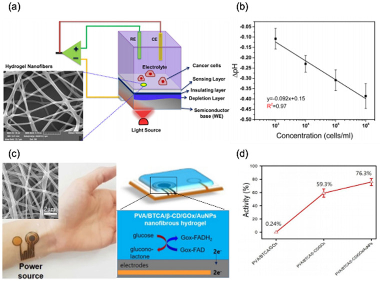
Hydrogels, hydrophilic and biocompatible polymeric networks, have been used for numerous biomedical applications because they have exhibited abilities to mimic features of extracellular matrix (ECM). In particular, the hydrogels engineered with electrospinning techniques have shown great performances in biomedical applications. Electrospinning techniques are to generate polymeric micro/nanofibers that can mimic geometries of natural ECM by drawing micro/nanofibers from polymer precursors with electrical forces, followed by structural stabilization of them. By exploiting the electrospinning techniques, the fibrous hydrogels have been fabricated and utilized as 2D/3D cell culture platforms, implantable scaffolds, and wound dressings. In addition, some hydrogels that respond to external stimuli have been used to develop biosensors. For comprehensive understanding, this review covers electrospinning processes, hydrogel precursors used for electrospinning, characteristics of fibrous hydrogels and specific biomedical applications of electrospun fibrous hydrogels and highlight their potential to promote use in biomedical applications.

Keywords: Hydrogels; biomedical applications; electrospinning; fibrous hydrogels.


Conflict of interest statement

The author(s) declared no potential conflicts of interest with respect to the research, authorship, and/or publication of this article.

Figures

Figure 1.
Figure 1.
Crosslinking methods for fibrous structure stabilization and biomedical applications (cell culture platforms, implantable scaffolds, wound dressings, and biosensors) of electrospun fibrous hydrogels.
Figure 2.
Figure 2.
A schematic illustration of general electrospinning setup and processes. (a) General electrospinning setup. It is mainly composed of a syringe pump ejecting hydrogel precursors in a controlled flow rate, a power supply to apply voltage to a metal needle and a collector to collect polymeric fibers. (b) Overall processes to generate electrospun fibers. A droplet of polymer precursor at a needle tip is ejected toward a grounded collector when electrostatic repulsion exceeds surface tension of the droplet. Reproduced from Han et al., Chen et al., and Marginean et al.
Figure 3.
Figure 3.
Various types of electrospinning techniques with crosslinking steps used for fabrication of electrospun hydrogel fibers. A power supply and a grounded collector are commonly required for electrospinning techniques: (a) co-axial electrospinning technique uses a co-axial needle to obtain fibers composed of one material at the core and another one at the sheath, (b) wet electrospinning technique uses a coagulation bath containing a grounded collector to fabricate fibrous hydrogel mats with relatively low fiber packing density, and (c) needleless electrospinning technique generates multiple jets to increase productivity of fibrous hydrogels. Reproduced from Chen et al.
Figure 4.
Figure 4.
Comparison between characteristic of bulk hydrogel and fibrous hydrogel. Bulk hydrogels were referred to hydrogels in millimeter to centimeter scales, and diameters of electrospun fibrous hydrogels were generally in nano/micrometer scales. Fabrication methods could determine overall shapes of bulk and fibrous hydrogels. Physical properties (Stiffness, porosity, alignment and deformability) of fibrous hydrogels might induce cellular behaviors different from cellular behaviors influenced by those properties of bulk hydrogels.
Figure 5.
Figure 5.
Comparisons of cellular behaviors on/within bulk and fibrous hydrogels. (a) Live/dead staining after 7 days of 3T3 cell culture in bulk hydrogels (top) and within fibrous hydrogels (bottom) (Scale bar: 200 µm). Reproduced from Xu et al. Myofibroblast differentication study in soft/stiff bulk hydrogels (top, scale bar:200 µm) and in hydrogels with 0.0% and 5.0% fiber density (bottom, FD: fiber density, scale bar: 100 µm). Stiffness of hydrogels was controlled by controlling concentration of crosslinkers (GCVPMS↓MRGGCG, VPMS). Reproduced from Matera et al. Distributed under the Creative Commons Attribution NonCommercial License 4.0 (CC BY-NC, https://creativecommons.org/licenses/by-nc/4.0/). (c) Infiltration thickness of hPASMCs on bulk (left) and fibrous (right) hydrogels after 1 day of seeding. Reproduced from Ding et al.
Figure 6.
Figure 6.
Cells on and within electrospun fibrous hydrogels were observed to investigate various cellular behaviors and responses. (a) Cell spreading on soft (top) and stiff (bottom) fibrous hydrogels after 7 days of cell culture. Reproduced from Davidson et al. (b) Cell infiltration through fibrous hydrogels after 3 days of cell culture. Reproduced form Wang et al. (c) Viability of cells on fibrous hydrogel membranes. Reproduced from Sun et al. (d) Cells on aligned fibrous hydrogels (top) and randomly oriented ones (bottom) after 1 day of cell culture. Reproduced from Du et al.
Figure 7.
Figure 7.
2D cell cultures on electrospun fibrous hydrogels. (a) Live/dead staining (left, middle) and F-actin staining (right) of human umbilical vein endothelial cells (HUVECs) cultured on fibrous gelatin and Gel-HPA hydrogels on Day 1 and 3. Fibrous gelatin hydrogel was stiffer than fibrous Gel-HPA hydrogels. Reproduced from Nie et al. Distributed under the Creative Commons Attribution 4.0 International License (CCBY 4.0, http://creativecommons.org/licenses/by/4.0/). (b) Activated stellate cells on soft/stiff MeHA hydrogel nanofibers. Representative masks of cell outlines on soft/stiff nanofibers (left, scale bar: 50 µm) and fluorescent images of soft/stiff nanofibers under the cells (right, scale bar: 50 µm). Reproduced from Davidson et al.
Figure 8.
Figure 8.
3D cell culture within fibrous hydrogels. (a) A schematic illustration of processes to encapsulate 3T3 mouse fibroblast cells within fibrous hydrogels. (b) 3D confocal images of the cells encapsulated in bulk hydrogel (left) and electrospun hydrogel nanofibers (right) after 3 and 7 days of incubation (scale bar: 200 µm). Reproduced from Xu et al. (c) Representative fluorescent images of NorHA nanofibers with varied nanofiber densities (75%, 50%, and 20%) (scale bar: 10 µm). (d) Representative inverted phase-contrast images of mesenchymal stem cells (MSCs) encapsulated within nanofibers on day 1 and 3 with varied nanofiber densities (75%, 50%, and 20%) (scale bar: 2 mm). (e) A schematic illustration (top) and representative images (bottom) of 3D-printed cell-laden nanofibers with 75% (gray) and 20% (pink) nanofiber densities (scale bar: 1 mm). Reproduced from Davidson et al. Distributed under the Creative Commons Attribution NonCommercial License 4.0 (CCBY-NC, https://creativecommons.org/licenses/by-nc/4.0/).
Figure 9.
Figure 9.
Implantable scaffolds developed with fibrous hydrogels for dense connective tissue regeneration. (a) A scheme of knee meniscus regeneration strategy using PEO and HA nanofibers releasing collagenase and PDGF-AB, respectively. (b) H&E staining and collagen type Ⅰ and Ⅱ immunostaining of the wounded meniscus tissues without scaffolds and with scaffolds containing PDGF-AB only and PDGF-AB with collagenase after 4 weeks of subcutaneous implantation (scale bar: 100 µm). Reproduced from Qu et al. Distributed under the Creative Commons Attribution 4.0 International License (CCBY, http://creativecommons.org/licenses/by/4.0). (c) Electrospun gelatin/PLGA fibers and (d) the fragmented fibers. (e) Scaffolds 3D printed with gelatin/PLGA hydrogel fibers. Analysis of (f) GAG content and (g) total collagen content for evaluation of cartilage regeneration after 8 weeks of implantation. Reproduced from Chen et al.
Figure 10.
Figure 10.
Implantable scaffolds developed with fibrous hydrogels to guide axonal regeneration. (a) Reinnervation of gastrocnemius at 12 weeks after implantation (left limb). Pictures of the isolated muscle and images of Masson’s trichrome staining of the sectioned muscles from the injured limbs. RFG: randomly oriented fibrin hydrogel nanofibers, AFG: aligned fibrin hydrogel nanofibers. (b) Images of regenerated nerve fibers isolated from middle of the implanted site at 12 weeks after surgery. Toluidine blue-stained transverse sections (top) and transmission electron microscopy (TEM) images (middle and bottom) of the regenerated sciatic nerve. Reproduced from Du et al. (c) Processes of spinal cord injury repair study using fibrous GelMA scaffold. (d) Evaluation of rat limb motor function with Basso, Beattie, and Bresnahan (BBB) score after implantation of the GelMA scaffold. Reproduced from Chen et al.
Figure 11.
Figure 11.
Wound dressings developed with fibrous hydrogels. (a) Pictures of wounds covered with gauze (Control) and GelMA-PDA-ASP nanofibers on day 0, 4, 8, and 12. (b) H&E images of sectioned wound site after 12 days of treatment. Reproduced from Zhang et al. (c) Pictures of wounds treated with PVA/CS (PC) nanofibers or PC nanofibers loaded with DFO (PCD) on diabetic rats on day 0, 6, 12, and 18. (d) mRNA expression of VEGF and SDF-1α after 6 days of PC and PCD treatment. Reproduced from Chen et al. (e) Pictures of the wounds from control, PNS microfibers with GS-Rg3 and cell adhesion peptide (PNS-G-RGDC) and PNS microfibers with cell adhesion peptide (PNS-RGDC) groups on day 0, 4, 8, and 16. (f) Immunohistochemical staining analysis of VEGF (top) and CD31 (bottom) after 16 days of treatment. Reproduced from Xu et al.
Figure 12.
Figure 12.
Illustration of working principles of pH-sensitive and glucose-sensitive biosensors fabricated with fibrous hydrogels.
Figure 13.
Figure 13.
Biosensors developed with electrospun fibrous hydrogels. (a) A structure of a light addressable potentiometric sensor integrated with PVA/PAA nanofibers (NF-LAPS). (b) Measurement of pH shift with varied concentration of cancer cells. Reproduced from Shaibani et al. (c) A wearable biosensor fabricated with PVA/BTCA/β-CD/GOx/AuNPs hydrogel nanofibers. (d) Analysis of enzymatic activity of PVA/BTCA/GOx, PVA/BTCA/β-CD/GOx, and PVA/BTCA/β-CD/GOx/AuNPs hydrogel nanofibers. Reproduced from Kim and Kim. Distributed under the Creative Commons Attribution License 4.0 (CCBY, http://creativecommons.org/licenses/by/4.0).

References

    1. Frantz C, Stewart KM, Weaver VM. The extracellular matrix at a glance. J Cell Sci 2010; 123: 4195–4200. - PMC - PubMed
    1. Chen Y, Lee K, Kawazoe N, et al.. ECM scaffolds mimicking extracellular matrices of endochondral ossification for the regulation of mesenchymal stem cell differentiation. Acta Biomater 2020; 114: 158–169. - PubMed
    1. Geckil H, Xu F, Zhang X, et al.. Engineering hydrogels as extracellular matrix mimics. Nanomed 2010; 5: 469–484. - PMC - PubMed
    1. Zhu C, Lei H, Fan D, et al.. Novel enzymatic crosslinked hydrogels that mimic extracellular matrix for skin wound healing. J Mater Sci 2018; 53: 5909–5928.
    1. Lee KY, Mooney DJ. Hydrogels for tissue engineering. Chem Rev 2001; 101: 1869–1879. - PubMed

LinkOut - more resources