. 2023 Oct 31;14(5):e0158723.
doi: 10.1128/mbio.01587-23. Epub 2023 Aug 15.

Pyronaridine tetraphosphate is an efficacious antiviral and anti-inflammatory active against multiple highly pathogenic coronaviruses

Affiliations

Pyronaridine tetraphosphate is an efficacious antiviral and anti-inflammatory active against multiple highly pathogenic coronaviruses

Jeremy Ardanuy et al. mBio. .

Abstract

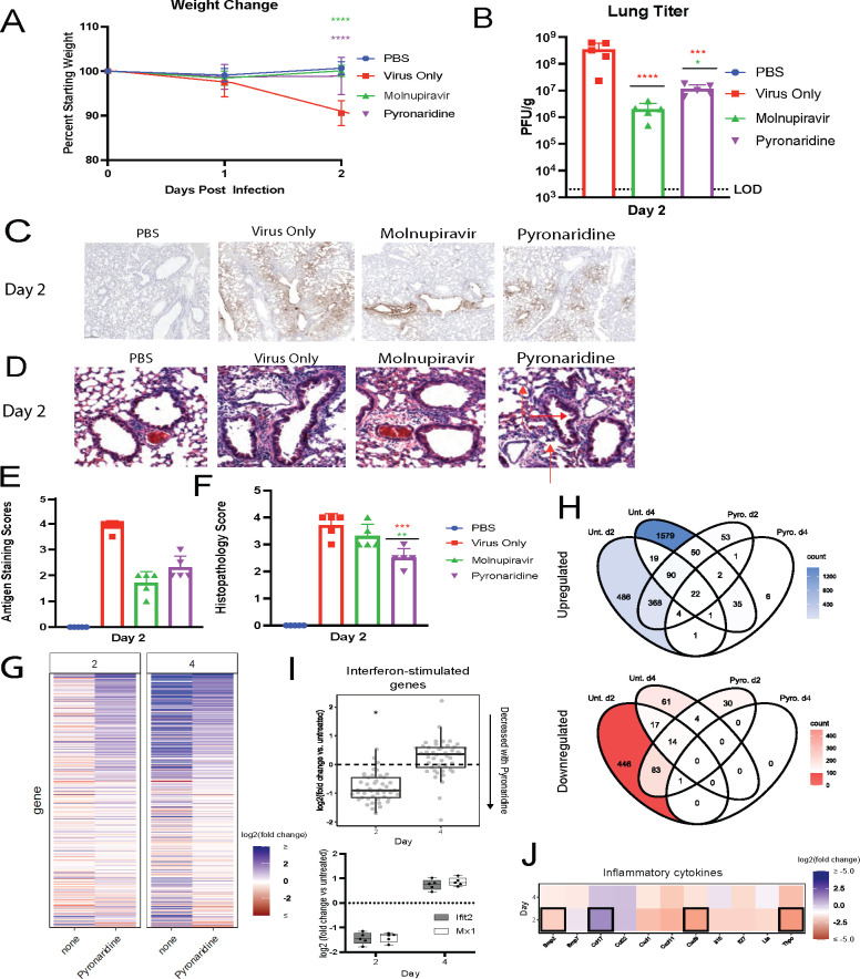
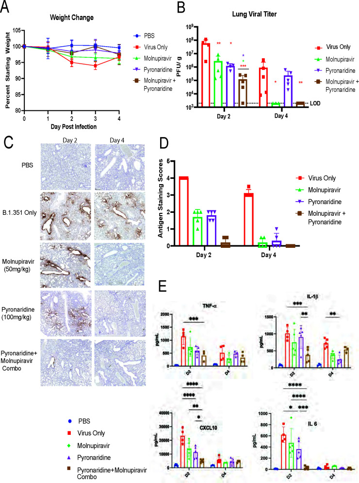
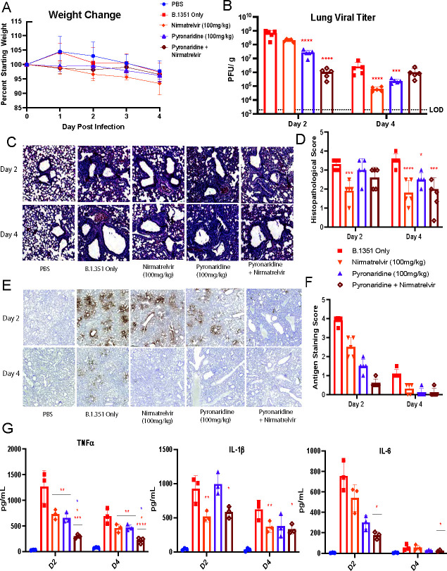
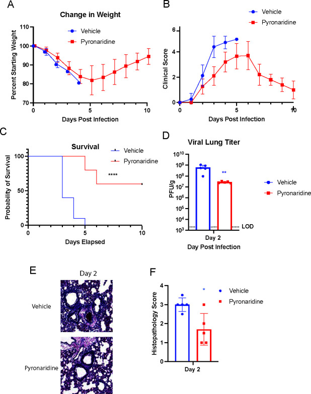
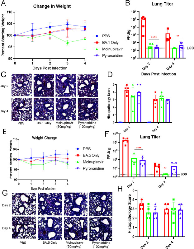
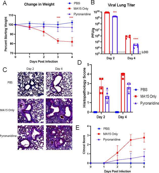
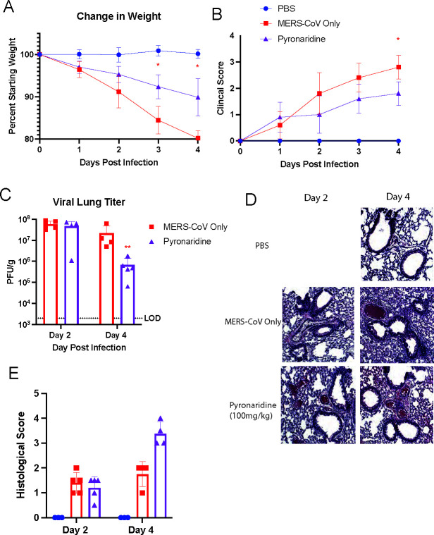
Pyronaridine tetraphosphate is on the WHO Essential Medicine List for its importance as a widely available and safe treatment for malaria. We find that pyronaridine is a highly effective antiviral therapeutic across mouse models using multiple variants of severe acute respiratory syndrome coronavirus-2 (SARS-CoV-2), and the highly pathogenic viruses SARS-CoV-1 and Middle East respiratory syndrome coronavirus responsible for previous coronavirus outbreaks. Additionally, we find that pyronaridine additively combines with current COVID-19 treatments such as nirmatrelvir (protease inhibitor in Paxlovid) and molnupiravir to further inhibit SARS-CoV-2 infections. There are many antiviral compounds that demonstrate efficacy in cellular models, but few that show this level of impact in multiple mouse models and represent a promising therapeutic for the current coronavirus pandemic as well as future outbreaks as well.

Keywords: antiviral agents; coronavirus; preclinical drug studies.


Conflict of interest statement

The authors declare no conflict of interest.

Figures

Fig 1
Fig 1
Prophylactic pyronaridine reduces SARS-CoV-2 infection and inflammation in mice. Eight- to 10-week-old wild-type Balb/c mice (n = 5 per group) were challenged with 1 × 10⁵ PFU of SARS-CoV-2 Beta variant (B.1.351). And treated with either nothing, molnupiravir, or pyronaridine orally beginning 12 h before infection. (A) Mouse weights were measured each day and lungs were harvested at 2 dpi for (B) viral titer quantification, (D) lung histology staining with red arrows indicating reduced lung inflammatory infiltrates and bronchovascular cuffing and (F) scoring analysis, and (C) nucleocapsid staining with hematoxylin and eosin and (E) scoring quantification for interstitial inflammation. Lungs were harvested at 2 dpi and 4 dpi for RNA extraction and RNA-sequencing analysis including (G) heatmap showing fold changes of genes relative to uninfected mice. Only genes differentially expressed at any time point (at least twofold expression change in either direction, adjusted P-value <0.01). (H) Venn diagrams showing sets of significantly upregulated (top, blue) or downregulated (bottom, red) genes, relative to mock, across the time points and treatments tested. Unt. = untreated. (I) Fold change of interferon-stimulated genes (ISGs) with pyronaridine treatment (treated vs untreated, all infected). Day 2 mean ISG levels are lower with pyronaridine treatment (P = 9.3e−15). (J) Heatmap showing the most different cytokines with pyronaridine treatment, relative to mock. Heatmap tiles outlined in black are significantly increased or decreased (adjusted P-value <0.01). n = 5 mice per group, mean ± SD is shown. *P < 0.05, **P < 0.01, and ***P < 0.001, using one-way analysis of variance with Dunnett’s multiple comparison test. The red asterisks are compared to virus-only group, whereas the green and purple asterisks are compared to molnupiravir and pyronaridine groups, respectively.
Fig 2
Fig 2
Combination of molnupiravir and pyronaridine treatment synergistically reduces SARS-CoV-2 infection and inflammation in vivo compared to single drug treatment. Eight- to 10-week-old wild-type BALB/c mice (n = 5 per group) were challenged with 1 × 10⁵ PFU of SARS-CoV-2 Beta variant (B.1.351) and treated with either nothing, molnupiravir, pyronaridine, or combination treatment of molnupiravir and pyronaridine orally beginning 12 h before infection. (A) Mouse weights were measured each day and lungs were harvested at 2 dpi for (B) mouse lungs that were analyzed for viral titer quantification by plaque assay on day 2 and day 4. (C) Lungs were imaged using nucleocapsid staining via IHC antibody on the lungs on day 2 and day 4 as well as by H&E staining for inflammatory pathology (Fig. S1). (D) Nucleocapsid staining was scored by percentage stained. (E) Lungs were harvested and analyzed for protein levels using a Bio-Plex Pro Mouse Chemokine Panel, a complete heat map of the chemokine/cytokine panel is available (Fig. S1). n = 5 mice per group, mean ± SD is shown. *P < 0.05, **P < 0.01, and ***P < 0.001, lung titers were analyzed for significance by log transformation and mixed-effect analysis with the Tukey test for multiple comparisons. Cytokine comparisons were analyzed by mixed-effect analysis followed by the Sidak test for multiple comparisons. The red asterisks are compared to virus-only group, whereas the green and purple asterisks are compared to molnupiravir and pyronaridine groups, respectively.
Fig 3
Fig 3
Combination of nirmatrelvir and pyronaridine treatment additively reduces SARS-CoV-2 infection and inflammation in vivo. Eight- to 10-week-old wild-type BALB/c mice were treated with nirmatrelvir (oral administration), and/or nirmatrelvir (oral administration) daily at the indicated concentrations starting 12 h before infection. Mice (n = 5 per group) were intranasally inoculated with 1 × 10⁵ PFU per mouse of the SARS-CoV-2 Beta variant (B.1.351). (A–G) Mice were weighed daily (A), lungs were analyzed for viral titer 2 and 4 days after infection by plaque assay (B), or fixed in 4% paraformaldehyde for H&E staining and quantified for interstitial and bronchovascular inflammation (C and D), or stained for SARS-CoV-2 nucleocapsid via IHC (E) and the staining was quantified by the percentage of tissue (F). Lung homogenate was also used in a Bio-Plex chemokine and cytokine protein quantification assay and select cytokines are shown (G), while the entire assay heatmap is shown separately(Fig. S3). n = 5 mice per group, mean ± SD is shown. *P < 0.05, **P < 0.01, and ***P < 0.001, lung titers were analyzed for significance by log transformation and mixed-effect analysis with the Tukey test for multiple comparisons. Cytokine comparisons were analyzed by mixed-effect analysis followed by the Sidak test for multiple comparisons. The red asterisks are compared to virus-only group.
Fig 4
Fig 4
Therapeutic pyronaridine protects against lethal infection by mouse-adapted SARS-CoV-2 in old mice. Six-month-old wild-type BALB/c mice were treated with pyronaridine(oral administration), or a phosphate-buffered saline vehicle control daily at the indicated concentrations starting 1 h post-infection. Mice (n = 5 per group) were intranasally inoculated with 1 × 10⁴ PFU per mouse of mouse-adapted SARS-CoV-2 (MA-10). (A–F) Mice were weighed daily (A), clinical scores for disease severity were recorded daily (B), lethality or survival was recorded daily (C), lungs were analyzed for viral titer 2 days after infection by plaque assay (D), or fixed in 4% paraformaldehyde for H&E staining and quantified for interstitial and bronchovascular inflammation (E and F). Mean ± SD is shown. *P < 0.05, **P < 0.01, and ***P < 0.001, with blue asterisks indicating differences with the vehicle control group. Lung titers were analyzed for significance by log transformation and paired t-test analysis. A comparison of survival curves was done with log-rank (Mantel-Cox) test.
Fig 5
Fig 5
Pyronaridine reduces infection against multiple Omicron subvariants of SARS-CoV-2. Wild-type 8- to 10-week-old BALB/c mice were treated with molnupiravir (oral administration) or pyronaridine(oral administration) daily at the indicated concentrations starting 12 h before infection. Mice (n = 5 per group over two independent experiments) were intranasally inoculated with 1 × 10⁵ PFU per mouse of SARS-CoV-2 Omicron (BA.1 for A–D, and BA.5 for E–H). Mice were weighed daily (A and E), and lungs were analyzed for viral titer 2 and 4 days after infection by plaque assay (B and F), or fixed in 4% paraformaldehyde for H&E staining (C and G) and quantified for lung inflammation (D and H). n = 5 mice per group. Mean ± SD is shown. *P < 0.05, **P < 0.01, and ***P < 0.001, using two-way analysis of variance with Tukey’s multiple comparison test. The red asterisks are compared to virus-only group.
Fig 6
Fig 6
Pyronaridine reduces infection against mouse-adapted SARS-CoV-1 in vivo. Wild type 8- to 10-week-old BALB/c mice were treated with pyronaridine (oral administration) daily at the indicated concentrations starting 12 h before infection. Mice (n = 5 per group) were intranasally inoculated with 1 × 10⁵ PFU per mouse of SARS-CoV-1 mouse-adapted strain (MA-15). (A–E) Mice were weighed daily (A), lungs were analyzed for viral titer 2 and 4 days after infection by plaque assay (B), or fixed in 4% paraformaldehyde for H&E staining (C) and quantified for lung inflammation scoring (D). Mice were monitored and given a clinical score for disease severity daily (E). n = 5 mice per group. Mean ± SD is shown. *P < 0.05, **P < 0.01, and ***P < 0.001, using two-way analysis of variance with Tukey’s multiple comparison tests on log-transformed virus titers. The red asterisks are compared to virus-only group.
Fig 7
Fig 7
Pyronaridine reduces infection against mouse-adapted MERS-CoV in vivo. hDPP4 knock-in mice aged 10–12 weeks were treated with pyronaridine (oral administration) daily at 100 mg/kg starting 12 h before infection. Mice (n = 5 per group) were intranasally inoculated with 5 × 10³ PFU per mouse of MERS-CoV mouse-adapted strain. (A–E) Mice were weighed daily (A), mice were monitored and given a clinical score for disease severity daily (B), lungs were analyzed for viral titer 2 and 4 days after infection by plaque assay (C), or fixed in 4% paraformaldehyde for H&E staining (D) and quantified for lung inflammation scoring (E). n = 5 mice per group, mean ± SD is shown. *P < 0.05, **P < 0.01, and ***P < 0.001, red asterisks indicate comparisons with virus-only group. Lung titers were analyzed for significance by log transformation and mixed-effect analysis with the Tukey test for multiple comparisons.

References

    1. Wu F, Zhao S, Yu B, Chen Y-M, Wang W, Song Z-G, Hu Y, Tao Z-W, Tian J-H, Pei Y-Y, Yuan M-L, Zhang Y-L, Dai F-H, Liu Y, Wang Q-M, Zheng J-J, Xu L, Holmes EC, Zhang Y-Z. 2020. A new coronavirus associated with human respiratory disease in China. Nature 579:265–269. doi:10.1038/s41586-020-2008-3 - DOI - PMC - PubMed
    1. Al-Tawfiq JA, Memish ZA. 2020. Middle East respiratory syndrome coronavirus and severe acute respiratory syndrome coronavirus. Semin Respir Crit Care Med 41:568–578. doi:10.1055/s-0040-1709160 - DOI - PMC - PubMed
    1. Stefan N, Birkenfeld AL, Schulze MB. 2021. Global pandemics interconnected - obesity, impaired metabolic health and COVID-19. Nat Rev Endocrinol 17:135–149. doi:10.1038/s41574-020-00462-1 - DOI - PubMed
    1. Zhu Z, Lian X, Su X, Wu W, Marraro GA, Zeng Y. 2020. From SARS and MERS to COVID-19: a brief summary and comparison of severe acute respiratory infections caused by three highly pathogenic human coronaviruses. Respir Res 21:224. doi:10.1186/s12931-020-01479-w - DOI - PMC - PubMed
    1. Hansen J, Baum A, Pascal KE, Russo V, Giordano S, Wloga E, Fulton BO, Yan Y, Koon K, Patel K, Chung KM, Hermann A, Ullman E, Cruz J, Rafique A, Huang T, Fairhurst J, Libertiny C, Malbec M, Lee W-Y, Welsh R, Farr G, Pennington S, Deshpande D, Cheng J, Watty A, Bouffard P, Babb R, Levenkova N, Chen C, Zhang B, Romero Hernandez A, Saotome K, Zhou Y, Franklin M, Sivapalasingam S, Lye DC, Weston S, Logue J, Haupt R, Frieman M, Chen G, Olson W, Murphy AJ, Stahl N, Yancopoulos GD, Kyratsous CA. 2020. Studies in humanized mice and convalescent humans yield a SARS-CoV-2 antibody cocktail. Science 369:1010–1014. doi:10.1126/science.abd0827 - DOI - PMC - PubMed