Review
. 2023 Aug;308(2):e230531.
doi: 10.1148/radiol.230531.

MRI Advancements in Musculoskeletal Clinical and Research Practice

Affiliations
Review

MRI Advancements in Musculoskeletal Clinical and Research Practice

Darryl B Sneag et al. Radiology. 2023 Aug.

Abstract

Over the past decades, MRI has become increasingly important for diagnosing and longitudinally monitoring musculoskeletal disorders, with ongoing hardware and software improvements aiming to optimize image quality and speed. However, surging demand for musculoskeletal MRI and increased interest to provide more personalized care will necessitate a stronger emphasis on efficiency and specificity. Ongoing hardware developments include more powerful gradients, improvements in wide-bore magnet designs to maintain field homogeneity, and high-channel phased-array coils. There is also interest in low-field-strength magnets with inherently lower magnetic footprints and operational costs to accommodate global demand in middle- and low-income countries. Previous approaches to decrease acquisition times by means of conventional acceleration techniques (eg, parallel imaging or compressed sensing) are now largely overshadowed by deep learning reconstruction algorithms. It is expected that greater emphasis will be placed on improving synthetic MRI and MR fingerprinting approaches to shorten overall acquisition times while also addressing the demand of personalized care by simultaneously capturing microstructural information to provide greater detail of disease severity. Authors also anticipate increased research emphasis on metal artifact reduction techniques, bone imaging, and MR neurography to meet clinical needs.


Conflict of interest statement

Disclosures of conflicts of interest: D.B.S. Institutional research agreements with GE Healthcare and Siemens Medical Solutions; research support from AMAG Pharmaceuticals and the HSS Innovation Institute; consulting fees from GE Healthcare; provisional patents in system and apparatus for overlapping phased-array coils for curved surfaces, system and method for MR neurography, and system and apparatus for simplified diffusion imaging. F.A. No relevant relationships. H.G.P. Grant from the National Institutes of Health. J.F. Grants from GE Healthcare, Siemens, QED, and SyntheticMR; patents planned, issued, or pending with Siemens Healthcare, Johns Hopkins University, and New York University; participation on a data safety monitoring board or advisory board for Siemens, SyntheticMR, GE Healthcare, QED, ImageBiopsy Lab, Boston Scientific, Mirata Pharmaceuticals, and Guerbet; member of Radiology editorial board. M.F.K. Institutional research agreements with GE Healthcare and Siemens Healthineers. C.B.C. Grants from the National Institutes of Health and VA Merit; secretary of the International Skeletal Society. V.P. Member of Radiology editorial board. E.T.T. Institutional research support from GE Healthcare, Siemens Healthcare, Medtronic, and AMAG Pharmaceuticals.

Figures

None
Graphical abstract
Acute-on-chronic low-grade pectoralis major strain in a 42-year-old male
patient with right chest pain after bench-pressing 1 week before imaging. (A)
Oblique coronal short-tau inversion-recovery (STIR) and (B) T1-weighted MRI
scans demonstrate a small amount of fatty deposition (arrowheads) along the
pectoralis major sternal head myotendinous junction (arrow) compatible with an
old tear. (C) Immediately posterior oblique coronal short-tau inversion-recovery
image demonstrates evidence of more recent, low-grade myotendinous junction
(arrowheads) and intramuscular (arrow) tear.
Figure 1:
Acute-on-chronic low-grade pectoralis major strain in a 42-year-old male patient with right chest pain after bench-pressing 1 week before imaging. (A) Oblique coronal short-tau inversion-recovery (STIR) and (B) T1-weighted MRI scans demonstrate a small amount of fatty deposition (arrowheads) along the pectoralis major sternal head myotendinous junction (arrow) compatible with an old tear. (C) Immediately posterior oblique coronal short-tau inversion-recovery image demonstrates evidence of more recent, low-grade myotendinous junction (arrowheads) and intramuscular (arrow) tear.
The top row shows commercially available coils. (A) A rigid 18-channel
transmit/receive coil is primarily used for knee imaging, whereas (B) a
24-channel flexible “multipurpose” receive-only coil can be
used for the knee, (C) brachial plexus or shoulder, and (D) elbow regions,
among others. The bottom row shows prototype receive-only coils. (E) A
screen-printed 12-channel array has been developed for pediatric
applications; note flexibility by the anterior component being folded in the
longitudinal direction (arrows). (Adapted, with permission, from reference
19.) (F) A 23-channel array designed for cervical imaging comprises
high-impedance coil elements embedded in a flexible mask and a rigid
posterior head-rest housing. (Courtesy of Tesla Dynamic Coils.) (G, H) A
dual-channel array using a liquid metal conductor and self-tuning
stretchable capacitor (arrows) demonstrates form-fitting conformability for
different degrees of knee flexion. (Adapted, with permission, from reference
101.)
Figure 2:
The top row shows commercially available coils. (A) A rigid 18-channel transmit/receive coil is primarily used for knee imaging, whereas (B) a 24-channel flexible “multipurpose” receive-only coil can be used for the knee, (C) brachial plexus or shoulder, and (D) elbow regions, among others. The bottom row shows prototype receive-only coils. (E) A screen-printed 12-channel array has been developed for pediatric applications; note flexibility by the anterior component being folded in the longitudinal direction (arrows). (Adapted, with permission, from reference .) (F) A 23-channel array designed for cervical imaging comprises high-impedance coil elements embedded in a flexible mask and a rigid posterior head-rest housing. (Courtesy of Tesla Dynamic Coils.) (G, H) A dual-channel array using a liquid metal conductor and self-tuning stretchable capacitor (arrows) demonstrates form-fitting conformability for different degrees of knee flexion. (Adapted, with permission, from reference .)
Acute osteomyelitis of the distal tibia in a 5-year-old boy
experiencing lower leg pain. (A) Cinematic three-dimensional (3D) MRI
surface rendering shows a markedly swollen (arrow) ankle and foot. (B)
Sagittal contrast-enhanced 3D T1-weighted fat-suppressed volumetric
interpolated breath-hold examination (T1FS VIBE) and (C) corresponding
cinematic rendering (CR) image demonstrate an intraosseous Brodie-type
abscess (black arrow, B) decompressing into a subperiosteal abscess (white
arrows). (D) The corresponding cinematic rendering vascular map shows marked
hyperemia of the distal tibia (arrow).
Figure 3:
Acute osteomyelitis of the distal tibia in a 5-year-old boy experiencing lower leg pain. (A) Cinematic three-dimensional (3D) MRI surface rendering shows a markedly swollen (arrow) ankle and foot. (B) Sagittal contrast-enhanced 3D T1-weighted fat-suppressed volumetric interpolated breath-hold examination (T1FS VIBE) and (C) corresponding cinematic rendering (CR) image demonstrate an intraosseous Brodie-type abscess (black arrow, B) decompressing into a subperiosteal abscess (white arrows). (D) The corresponding cinematic rendering vascular map shows marked hyperemia of the distal tibia (arrow).
Osteochondral defect in a 42-year-old male patient with left knee pain
after osteosynthesis of the proximal tibia. Compared with the conventional,
high–receiver bandwidth (HBW) (A) proton density (PD) and (C)
short-tau inversion-recovery (STIR) images, (B) compressed sensing (CS)
slice encoding for metal artifact correction (SEMAC) proton density and (D)
short-tau inversion-recovery turbo spin-echo (TSE) images correct the image
distortion, unmasking a focally subsided osteochondral defect (arrows). The
time stamps indicate the acquisition times of each pulse
sequence.
Figure 4:
Osteochondral defect in a 42-year-old male patient with left knee pain after osteosynthesis of the proximal tibia. Compared with the conventional, high–receiver bandwidth (HBW) (A) proton density (PD) and (C) short-tau inversion-recovery (STIR) images, (B) compressed sensing (CS) slice encoding for metal artifact correction (SEMAC) proton density and (D) short-tau inversion-recovery turbo spin-echo (TSE) images correct the image distortion, unmasking a focally subsided osteochondral defect (arrows). The time stamps indicate the acquisition times of each pulse sequence.
Osteoarthritis in a 49-year-old male patient with chronic left knee
pain. (A) Routine two-dimensional sagittal proton density (Sag PD) MRI scan
demonstrates chondral high signal intensity (blue arrows), and (B) T2 maps
obtained from a sixfold-accelerated GRAPPATINI acquisition (10 echoes,
twofold parallel imaging, threefold acceleration in echo time) demonstrate
mild surface chondral wear over posterior margins of the lateral femoral
condyle with T2 prolongation (black arrows). GRAPPATINI is a combination of
a k-based method for undersampling called generalized autocalibrating
partial parallel acquisition, or GRAPPA, and the reconstruction technique
model-based accelerated relaxometry by iterative nonlinear inversion, or
MARTINI (33).
Figure 5:
Osteoarthritis in a 49-year-old male patient with chronic left knee pain. (A) Routine two-dimensional sagittal proton density (Sag PD) MRI scan demonstrates chondral high signal intensity (blue arrows), and (B) T2 maps obtained from a sixfold-accelerated GRAPPATINI acquisition (10 echoes, twofold parallel imaging, threefold acceleration in echo time) demonstrate mild surface chondral wear over posterior margins of the lateral femoral condyle with T2 prolongation (black arrows). GRAPPATINI is a combination of a k-based method for undersampling called generalized autocalibrating partial parallel acquisition, or GRAPPA, and the reconstruction technique model-based accelerated relaxometry by iterative nonlinear inversion, or MARTINI (33).
A 3.0-T two-dimensional turbo spin-echo MRI examination was performed
in a 58-year-old female patient presenting with ankle pain and swelling by
using a 4-minute combined, sixfold acceleration protocol (with simultaneous
multislice, parallel imaging, and superresolution deep learning
reconstructions). (A) Axial (Ax) proton density (PD), (B) T2-weighted
fat-suppressed (T2FS), (C) sagittal (Sag) T1-weighted, (D) sagittal
T2-weighted fat-suppressed, and (E) coronal (Cor) proton density
fat-suppressed (PDFS) images show an osteochondral lesion within the medial
apex of the talar dome (white arrows) characterized by subchondral cyst
formation, flattening of the subchondral plate, and proton density and T2
signal hyperintensity of the overlying articular cartilage. The axial (A)
proton density and (B) T2-weighted fat-suppressed images also demonstrate
midsubstance Achilles tendinopathy (black arrows) characterized by expansion
of the anterior-posterior tendon diameter and increased proton density and
T2 intrasubstance signal.
Figure 6:
A 3.0-T two-dimensional turbo spin-echo MRI examination was performed in a 58-year-old female patient presenting with ankle pain and swelling by using a 4-minute combined, sixfold acceleration protocol (with simultaneous multislice, parallel imaging, and superresolution deep learning reconstructions). (A) Axial (Ax) proton density (PD), (B) T2-weighted fat-suppressed (T2FS), (C) sagittal (Sag) T1-weighted, (D) sagittal T2-weighted fat-suppressed, and (E) coronal (Cor) proton density fat-suppressed (PDFS) images show an osteochondral lesion within the medial apex of the talar dome (white arrows) characterized by subchondral cyst formation, flattening of the subchondral plate, and proton density and T2 signal hyperintensity of the overlying articular cartilage. The axial (A) proton density and (B) T2-weighted fat-suppressed images also demonstrate midsubstance Achilles tendinopathy (black arrows) characterized by expansion of the anterior-posterior tendon diameter and increased proton density and T2 intrasubstance signal.
Peripheral nerve-sheath tumor in a 76-year-old male patient with
chronic right leg weakness. Oblique coronal three-dimensional multiecho in
steady-state acquisition (MENSA)/dual-echo steady state MR neurography
images of the lumbosacral plexus reconstructed (A) without and (B) with a
commercially available deep learning (DL) algorithm (AIR Recon DL, GE
Healthcare) demonstrate a peripheral nerve-sheath tumor (thick arrows)
arising from the lower right lumbosacral plexus. Note the increased
sharpness and conspicuity of other nerve branches (eg, obturator nerve [thin
arrows]) and less noise of the surrounding soft tissues in the
Dl-reconstructed image.
Figure 7:
Peripheral nerve-sheath tumor in a 76-year-old male patient with chronic right leg weakness. Oblique coronal three-dimensional multiecho in steady-state acquisition (MENSA)/dual-echo steady state MR neurography images of the lumbosacral plexus reconstructed (A) without and (B) with a commercially available deep learning (DL) algorithm (AIR Recon DL, GE Healthcare) demonstrate a peripheral nerve-sheath tumor (thick arrows) arising from the lower right lumbosacral plexus. Note the increased sharpness and conspicuity of other nerve branches (eg, obturator nerve [thin arrows]) and less noise of the surrounding soft tissues in the DL-reconstructed image.
Multiacquisition variable-resonance image combination, or MAVRIC,
three-dimensional techniques for morphologic and quantitative imaging in a
67-year-old female patient with pain after left total hip arthroplasty. (A)
Coronal MAVRIC proton-density (PD) image demonstrates the total hip arthroplasty
in situ. (B) Conventional high-bandwidth coronal proton density image anterior
to the total hip arthroplasty shows signal dropout, pileup, and through-plane
distortion (arrows) within the iliopsoas bursa, which prevents full
visualization of the synovial reaction (arrowheads); these artifacts are
mitigated with (C) a MAVRIC acquisition, which also demonstrates intermediate
signal intensity–dependent debris (presumed metallosis) (black arrow).
(D) MAVRIC T2 mapping and (E) diffusion-weighted imaging (DWI) sequences provide
quantitative T2 and apparent diffusion coefficient (ADC) values, which help to
characterize this abnormal synovial reaction.
Figure 8:
Multiacquisition variable-resonance image combination, or MAVRIC, three-dimensional techniques for morphologic and quantitative imaging in a 67-year-old female patient with pain after left total hip arthroplasty. (A) Coronal MAVRIC proton-density (PD) image demonstrates the total hip arthroplasty in situ. (B) Conventional high-bandwidth coronal proton density image anterior to the total hip arthroplasty shows signal dropout, pileup, and through-plane distortion (arrows) within the iliopsoas bursa, which prevents full visualization of the synovial reaction (arrowheads); these artifacts are mitigated with (C) a MAVRIC acquisition, which also demonstrates intermediate signal intensity–dependent debris (presumed metallosis) (black arrow). (D) MAVRIC T2 mapping and (E) diffusion-weighted imaging (DWI) sequences provide quantitative T2 and apparent diffusion coefficient (ADC) values, which help to characterize this abnormal synovial reaction.
Quantitative T2* maps, derived from a multiecho, three-dimensional
ultrashort-echo-time radial-cones acquisition, in an asymptomatic 19-year-old
male collegiate basketball player. (A) T2* prolongation of cartilage over
posterior margins of the lateral tibial plateau and femoral condyle (blue
arrows) are noted, suggesting early chondral degeneration. (B) The lateral
meniscus anterior horn also demonstrates early degeneration, reflected by
T2* prolongation (white arrows) relative to the posterior
horn.
Figure 9:
Quantitative T2* maps, derived from a multiecho, three-dimensional ultrashort-echo-time radial-cones acquisition, in an asymptomatic 19-year-old male collegiate basketball player. (A) T2* prolongation of cartilage over posterior margins of the lateral tibial plateau and femoral condyle (blue arrows) are noted, suggesting early chondral degeneration. (B) The lateral meniscus anterior horn also demonstrates early degeneration, reflected by T2* prolongation (white arrows) relative to the posterior horn.
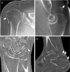
Zero-echo-time MRI scans in four patients after acute trauma. (A) Oblique
coronal image of the glenohumeral joint in a 50-year-old male patient
demonstrates a comminuted greater tuberosity fracture (arrow). (B) Sagittal
image of the left elbow in a 33-year-old male patient demonstrates a superiorly
displaced triceps avulsion fracture (arrow). (C) Coronal wrist image in a
61-year-old male patient shows small capsular triquetral avulsion fracture
(arrow). (D) Sagittal ankle image in a 39-year-old male patient demonstrates
Achilles tendon avulsion fracture off the calcaneus, with retraction of osseous
fragments (arrows). Images courtesy of Ryan Breighner, PhD, Hospital for Special
Surgery.
Figure 10:
Zero-echo-time MRI scans in four patients after acute trauma. (A) Oblique coronal image of the glenohumeral joint in a 50-year-old male patient demonstrates a comminuted greater tuberosity fracture (arrow). (B) Sagittal image of the left elbow in a 33-year-old male patient demonstrates a superiorly displaced triceps avulsion fracture (arrow). (C) Coronal wrist image in a 61-year-old male patient shows small capsular triquetral avulsion fracture (arrow). (D) Sagittal ankle image in a 39-year-old male patient demonstrates Achilles tendon avulsion fracture off the calcaneus, with retraction of osseous fragments (arrows). Images courtesy of Ryan Breighner, PhD, Hospital for Special Surgery.
Three-dimensional (3D) ultrashort-echo-time (UTE) MRI scans of the
osteochondral junction in a (A–D) normal cadaveric knee from a
62-year-old male donor and an (E–H) osteoarthritic cadaveric knee from a
87-year-old male donor. (A, E) Two-dimensional T2-weighted (T2) fast spin-echo
(FSE), (B, F) 3D T1-weighted fat-saturated (T1-FS) UTE, (C, G) 3D
inversion-recovery fat-saturated (IR-FS) UTE, and (D, H) 3D dual
inversion-recovery (DIR) UTE images. In the (A–D) normal knee, the
osteochondral junction region was dark on the T2-weighted FSE image due to its
fast signal decay. The normal osteochondral junction regions were highlighted as
bright signal lines in T1-weighted fat-saturated UTE, inversion-recovery
fat-saturated UTE, and dual inversion-recovery UTE images. In the (E–H)
osteoarthritic knee, the osteochondral junction signals were reduced or
completely lost in the abnormal cartilage regions (arrows) of central osteophyte
formation. Of note, there was preservation of overlying cartilage in these
regions. Images courtesy of Yajun Ma, PhD, University of California San
Diego.
Figure 11:
Three-dimensional (3D) ultrashort-echo-time (UTE) MRI scans of the osteochondral junction in a (A–D) normal cadaveric knee from a 62-year-old male donor and an (E–H) osteoarthritic cadaveric knee from a 87-year-old male donor. (A, E) Two-dimensional T2-weighted (T2) fast spin-echo (FSE), (B, F) 3D T1-weighted fat-saturated (T1-FS) UTE, (C, G) 3D inversion-recovery fat-saturated (IR-FS) UTE, and (D, H) 3D dual inversion-recovery (DIR) UTE images. In the (A–D) normal knee, the osteochondral junction region was dark on the T2-weighted FSE image due to its fast signal decay. The normal osteochondral junction regions were highlighted as bright signal lines in T1-weighted fat-saturated UTE, inversion-recovery fat-saturated UTE, and dual inversion-recovery UTE images. In the (E–H) osteoarthritic knee, the osteochondral junction signals were reduced or completely lost in the abnormal cartilage regions (arrows) of central osteophyte formation. Of note, there was preservation of overlying cartilage in these regions. Images courtesy of Yajun Ma, PhD, University of California San Diego.
Parsonage-Turner syndrome (acute brachial neuropathy) in a 38-year-old
female patient with severe left shoulder pain that began 2 weeks before imaging,
followed by weakness. (A) Coronal two-dimensional T2-weighted Dixon water MRI
scan demonstrates denervation edema of the supraspinatus (SS) and infraspinatus
(IS) muscles. Due to improved vascular suppression, (C) gadolinium-enhanced
three-dimensional (3D) oblique coronal short-tau inversion-recovery (STIR)
maximum intensity projection (20-mm thick) shows severe intrinsic constrictions
(arrows) more clearly compared with the (B) noncontrast projection using an
otherwise identical 3D short-tau inversion-recovery protocol.
Figure 12:
Parsonage-Turner syndrome (acute brachial neuropathy) in a 38-year-old female patient with severe left shoulder pain that began 2 weeks before imaging, followed by weakness. (A) Coronal two-dimensional T2-weighted Dixon water MRI scan demonstrates denervation edema of the supraspinatus (SS) and infraspinatus (IS) muscles. Due to improved vascular suppression, (C) gadolinium-enhanced three-dimensional (3D) oblique coronal short-tau inversion-recovery (STIR) maximum intensity projection (20-mm thick) shows severe intrinsic constrictions (arrows) more clearly compared with the (B) noncontrast projection using an otherwise identical 3D short-tau inversion-recovery protocol.

References

    1. Smith-Bindman R , Kwan ML , Marlow EC , et al. . Trends in use of medical imaging in US health care systems and in Ontario, Canada, 2000-2016 . JAMA 2019. ; 322 ( 9 ): 843 – 856 . - PMC - PubMed
    1. Ekstrand J , Healy JC , Waldén M , Lee JC , English B , Hägglund M . Hamstring muscle injuries in professional football: the correlation of MRI findings with return to play . Br J Sports Med 2012. ; 46 ( 2 ): 112 – 117 . - PubMed
    1. Nattiv A , Kennedy G , Barrack MT , et al. . Correlation of MRI grading of bone stress injuries with clinical risk factors and return to play: a 5-year prospective study in collegiate track and field athletes . Am J Sports Med 2013. ; 41 ( 8 ): 1930 – 1941 . - PMC - PubMed
    1. Kaeding CC , Yu JR , Wright R , Amendola A , Spindler KP . Management and return to play of stress fractures . Clin J Sport Med 2005. ; 15 ( 6 ): 442 – 447 . - PubMed
    1. Pedoia V , Lee J , Norman B , Link TM , Majumdar S . Diagnosing osteoarthritis from T2 maps using deep learning: an analysis of the entire Osteoarthritis Initiative baseline cohort . Osteoarthritis Cartilage 2019. ; 27 ( 7 ): 1002 – 1010 . - PMC - PubMed

Publication types