. 2023 Aug;308(2):e230079.
doi: 10.1148/radiol.230079.

Extramural Venous Invasion and Tumor Deposit at Diffusion-weighted MRI in Patients after Neoadjuvant Treatment for Rectal Cancer

Affiliations

Extramural Venous Invasion and Tumor Deposit at Diffusion-weighted MRI in Patients after Neoadjuvant Treatment for Rectal Cancer

Tae-Hyung Kim et al. Radiology. 2023 Aug.

Abstract

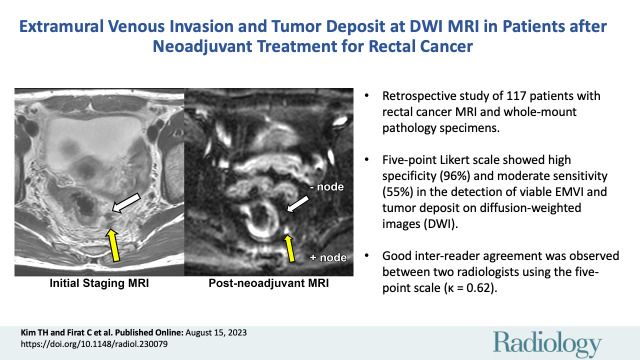
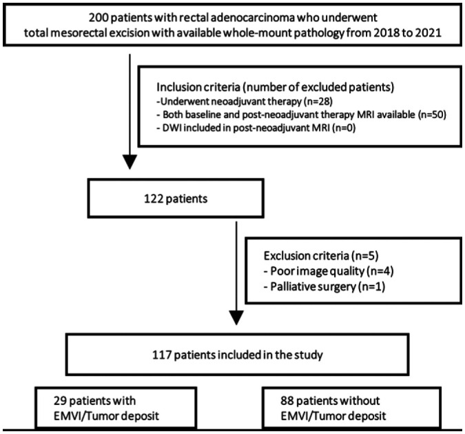
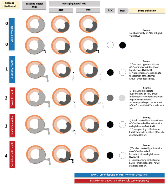
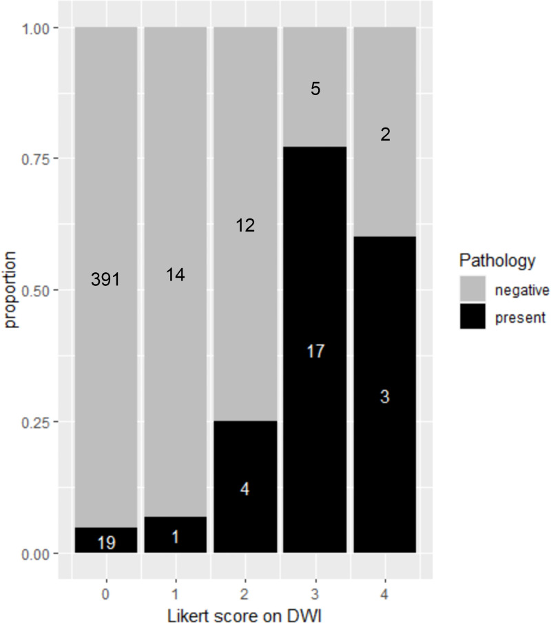
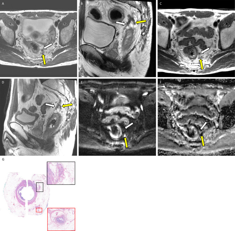
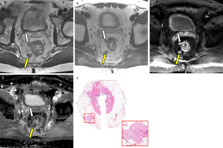
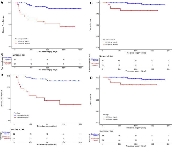
Background Diffusion-weighted (DW) imaging is useful in detecting tumor in the primary tumor bed in locally advanced rectal cancer (LARC) after neoadjuvant therapy, but its value in detecting extramural venous invasion (EMVI) and tumor deposit is not well validated. Purpose To evaluate diagnostic accuracy and association with patient prognosis of viable EMVI and tumor deposit on DW images in patients with LARC after neoadjuvant therapy using whole-mount pathology specimens. Materials and Methods This retrospective study included patients who underwent neoadjuvant therapy and surgery from 2018 to 2021. Innovative five-point Likert scale was used by two radiologists to independently evaluate the likelihood of viable EMVI and tumor deposit on restaging DW MRI scans in four axial quadrants (12 to 3 o'clock, 3 to 6 o'clock, 6 to 9 o'clock, and 9 to 12 o'clock). Diagnostic accuracy was assessed at both the per-quadrant and per-patient level, with whole-mount pathology as the reference standard. Weighted κ values for interreader agreement and Cox regression models for disease-free survival and overall survival analyses were used. Results A total of 117 patients (mean age, 56 years ± 12 [SD]; 70 male, 47 female) were included. Pathologically proven viable EMVI and tumor deposit was detected in 29 of 117 patients (25%) and in 44 of 468 quadrants (9.4%). Per-quadrant analyses showed an area under the receiver operating characteristics curve of 0.75 (95% CI: 0.68, 0.83), with sensitivity and specificity of 55% and 96%, respectively. Good interreader agreement was observed between the radiologists (κ = 0.62). Per-patient analysis showed sensitivity and specificity of 62% and 93%, respectively. The presence of EMVI and tumor deposit on restaging DW MRI scans was associated with worse disease-free survival (hazard ratio [HR], 5.6; 95% CI: 2.4, 13.3) and overall survival (HR, 8.9; 95% CI: 1.6, 48.5). Conclusion DW imaging using the five-point Likert scale showed high specificity and moderate sensitivity in the detection of viable extramural venous invasion and tumor deposits in LARC after neoadjuvant therapy, and its presence on restaging DW MRI scans is associated with worse prognosis. Published under a CC BY 4.0 license. Supplemental material is available for this article. See also the editorial by Méndez and Ayuso in this issue.


Conflict of interest statement

Disclosures of conflicts of interest: T.H.K. No relevant relationships. C.F. No relevant relationships. H.M.T. No relevant relationships. N.G. No relevant relationships. J.Z. No relevant relationships. M.C. No relevant relationships. D.D.B.B. Consultant for Boston Imaging Core Lab and GE Healthcare. V.P. Grand rounds at Rutgers University, diagnostic imaging symposium, continuing medical education, gave lectures in Orlando; compensated for consultation on legal cases; compensated through academic allowance budget to attend Society of Abdominal Radiology meeting in March 2022 and 2023, Society for Advanced Body Imaging meeting in 2022, and American Roentgen Ray Society meeting in May 2022. J.G.A. Consultant and speaker for and stockholder in Intuitive Surgical, consultant to Medtronic; speaker for and receives honoraria from Ethicon J&J. J.S. Consulting fees from Paige AI. M.J.G. Consultant for GlaxoSmithKline. N.H. Consulting fees from Guerbet; payment or honoraria for lectures, presentations, speakers bureaus, manuscript writing or educational events from Bayer; support for attending Society of Abdominal Radiology 2023 meeting from Guerbet.

Figures

None
Graphical abstract
Flowchart of patient inclusion and exclusion criteria. DWI =
diffusion-weighted imaging, EMVI = extramural venous invasion.
Figure 1:
Flowchart of patient inclusion and exclusion criteria. DWI = diffusion-weighted imaging, EMVI = extramural venous invasion.
Diagram of the five-point Likert scale scoring system used to assess
extramural venous invasion (EMVI) and tumor deposit on diffusion-weighted
images with apparent diffusion coefficient (ADC) mapping. DWI =
diffusion-weighted imaging.
Figure 2:
Diagram of the five-point Likert scale scoring system used to assess extramural venous invasion (EMVI) and tumor deposit on diffusion-weighted images with apparent diffusion coefficient (ADC) mapping. DWI = diffusion-weighted imaging.
Stacked bar plot shows the per-quadrant distribution of extramural
venous invasion and tumor deposit as assessed with the diffusion-weighted
imaging (DWI) five-point Likert scale and whole-mount pathology. Gray area
of the stacked bar represents quadrants with nonviable cells at pathology,
and the black area of the stacked bars represents quadrants with viable
cells at pathology. A total of 410 quadrants were assigned a Likert score of
0 (unlikely viable), 15 quadrants were assigned a Likert score of 1 (less
likely viable), 16 quadrants were assigned a Likert score of 2 (possibly
viable), 22 quadrants were assigned a Likert score of 3 (suspicious viable),
and five quadrants were assigned a Likert score of 4 (consistent with
viable).
Figure 3:
Stacked bar plot shows the per-quadrant distribution of extramural venous invasion and tumor deposit as assessed with the diffusion-weighted imaging (DWI) five-point Likert scale and whole-mount pathology. Gray area of the stacked bar represents quadrants with nonviable cells at pathology, and the black area of the stacked bars represents quadrants with viable cells at pathology. A total of 410 quadrants were assigned a Likert score of 0 (unlikely viable), 15 quadrants were assigned a Likert score of 1 (less likely viable), 16 quadrants were assigned a Likert score of 2 (possibly viable), 22 quadrants were assigned a Likert score of 3 (suspicious viable), and five quadrants were assigned a Likert score of 4 (consistent with viable).
Example application of the diffusion-weighted (DW) image five-point
Likert scale for assessing extramural venous invasion (EMVI) or tumor
deposit at MRI with corresponding whole-mount pathology in a 46-year-old man
with locally advanced rectal adenocarcinoma. Baseline (A) axial and (B)
sagittal T2-weighted MRI scans show EMVI and tumor deposit in the 12 to 3
o’clock quadrant (white arrow) and in the 3 to 6 o’clock
quadrant (yellow arrow). Postneoadjuvant (C) axial and (D) sagittal
T2-weighted MRI scans show regressed EMVI and tumor deposit in both the 12
to 3 o’clock quadrant (white arrow) and the 3 to 6 o’clock
quadrant (yellow arrow). Postneoadjuvant therapy (E) axial DW image and (F)
apparent diffusion coefficient map yielded a score of 0 (unlikely viable) in
the 12 to 3 o’clock quadrant (white arrow) and a score of 3
(suspicious viable) in the 3 to 6 o’clock quadrant (yellow arrow).
(G) Photomicrograph (hematoxylin-eosin stain; original magnification,
×2) shows acellular mucin corresponding to the 12 to 3 o’clock
quadrant, consistent with posttreatment change without viable malignant
cells (black box; magnification, ×50). On the contrary, there is
viable EMVI corresponding to the 3 to 6 o’clock quadrant (red box;
magnification, ×50).
Figure 4:
Example application of the diffusion-weighted (DW) image five-point Likert scale for assessing extramural venous invasion (EMVI) or tumor deposit at MRI with corresponding whole-mount pathology in a 46-year-old man with locally advanced rectal adenocarcinoma. Baseline (A) axial and (B) sagittal T2-weighted MRI scans show EMVI and tumor deposit in the 12 to 3 o’clock quadrant (white arrow) and in the 3 to 6 o’clock quadrant (yellow arrow). Postneoadjuvant (C) axial and (D) sagittal T2-weighted MRI scans show regressed EMVI and tumor deposit in both the 12 to 3 o’clock quadrant (white arrow) and the 3 to 6 o’clock quadrant (yellow arrow). Postneoadjuvant therapy (E) axial DW image and (F) apparent diffusion coefficient map yielded a score of 0 (unlikely viable) in the 12 to 3 o’clock quadrant (white arrow) and a score of 3 (suspicious viable) in the 3 to 6 o’clock quadrant (yellow arrow). (G) Photomicrograph (hematoxylin-eosin stain; original magnification, ×2) shows acellular mucin corresponding to the 12 to 3 o’clock quadrant, consistent with posttreatment change without viable malignant cells (black box; magnification, ×50). On the contrary, there is viable EMVI corresponding to the 3 to 6 o’clock quadrant (red box; magnification, ×50).
Example application of the diffusion-weighted (DW) image five-point
Likert scale for assessing extramural venous invasion (EMVI) or tumor
deposit on MRI scans with corresponding whole-mount pathology in a
36-year-old man with locally advanced rectal adenocarcinoma. (A) Baseline
axial T2-weighted MRI scan shows EMVI and tumor deposit in the 6 to 9
o’clock quadrant (yellow arrow) and 9 to 12 o’clock quadrant
(white arrow). (B) Postneoadjuvant therapy axial T2-weighted MRI scan shows
partially regressed EMVI and tumor deposit in the 6 to 9 o’clock
quadrant (yellow arrow) and 9–12 o’clock quadrant (white
arrow). Postneoadjuvant therapy (C) axial DW image and (D) apparent
diffusion coefficient map with a score of 4 (consistent with viable) in the
6 to 9 o’clock quadrant (yellow arrow) and in the 9 to 12
o’clock quadrant (white arrow). (E) Photomicrograph
(hematoxylin-eosin stain, original magnification, ×2) shows viable
tumor deposits corresponding to the 6 to 9 o’clock quadrant and 9 to
12 o’clock quadrant (red box; original magnification, ×50).
Note that on A the lesion in the 9–12 o’clock quadrant
presented similar to EMVI, contiguous from primary tumor bed; however, these
turned out to be tumor deposits separate from primary tumor bed on
E.
Figure 5:
Example application of the diffusion-weighted (DW) image five-point Likert scale for assessing extramural venous invasion (EMVI) or tumor deposit on MRI scans with corresponding whole-mount pathology in a 36-year-old man with locally advanced rectal adenocarcinoma. (A) Baseline axial T2-weighted MRI scan shows EMVI and tumor deposit in the 6 to 9 o’clock quadrant (yellow arrow) and 9 to 12 o’clock quadrant (white arrow). (B) Postneoadjuvant therapy axial T2-weighted MRI scan shows partially regressed EMVI and tumor deposit in the 6 to 9 o’clock quadrant (yellow arrow) and 9–12 o’clock quadrant (white arrow). Postneoadjuvant therapy (C) axial DW image and (D) apparent diffusion coefficient map with a score of 4 (consistent with viable) in the 6 to 9 o’clock quadrant (yellow arrow) and in the 9 to 12 o’clock quadrant (white arrow). (E) Photomicrograph (hematoxylin-eosin stain, original magnification, ×2) shows viable tumor deposits corresponding to the 6 to 9 o’clock quadrant and 9 to 12 o’clock quadrant (red box; original magnification, ×50). Note that on A the lesion in the 9–12 o’clock quadrant presented similar to EMVI, contiguous from primary tumor bed; however, these turned out to be tumor deposits separate from primary tumor bed on E.
Survival analyses using the Kaplan-Meier curve. (A) Disease-free
survival based on extramural venous invasion (EMVI) and tumor deposit at
postneoadjuvant MRI, and (B) disease-free survival based on EMVI and tumor
deposit at pathology. Survival analyses using the Kaplan-Meier curve. (C)
Overall survival based on EMVI and tumor deposit at postneoadjuvant MRI, and
(D) overall survival based on EMVI and tumor deposit at
pathology.
Figure 6:
Survival analyses using the Kaplan-Meier curve. (A) Disease-free survival based on extramural venous invasion (EMVI) and tumor deposit at postneoadjuvant MRI, and (B) disease-free survival based on EMVI and tumor deposit at pathology. Survival analyses using the Kaplan-Meier curve. (C) Overall survival based on EMVI and tumor deposit at postneoadjuvant MRI, and (D) overall survival based on EMVI and tumor deposit at pathology.

Comment in

References

    1. Lord AC , D’Souza N , Shaw A , et al. . MRI-Diagnosed Tumor Deposits and EMVI Status Have Superior Prognostic Accuracy to Current Clinical TNM Staging in Rectal Cancer . Ann Surg 2022. ; 276 ( 2 ): 334 – 344 . - PubMed
    1. Schaap DP , Voogt ELK , Burger JWA , et al. . Prognostic Implications of MRI-Detected EMVI and Tumor Deposits and Their Response to Neoadjuvant Therapy in cT3 and cT4 Rectal Cancer . Int J Radiat Oncol Biol Phys 2021. ; 111 ( 3 ): 816 – 825 . - PubMed
    1. Kim TH , Woo S , Han S , Suh CH , Vargas HA . The Diagnostic Performance of MRI for Detection of Extramural Venous Invasion in Colorectal Cancer: A Systematic Review and Meta-Analysis of the Literature . AJR Am J Roentgenol 2019. ; 213 ( 3 ): 575 – 585 . - PMC - PubMed
    1. Atre ID , Eurboonyanun K , Noda Y , et al. . Utility of texture analysis on T2-weighted MR for differentiating tumor deposits from mesorectal nodes in rectal cancer patients, in a retrospective cohort . Abdom Radiol (NY) 2021. ; 46 ( 2 ): 459 – 468 . - PubMed
    1. Altinmakas E , Dogan H , Taskin OC , et al. . Extramural venous invasion (EMVI) revisited: a detailed analysis of various characteristics of EMVI and their role as a predictive imaging biomarker in the neoadjuvant treatment response in rectal cancer . Abdom Radiol (NY) 2022. ; 47 ( 6 ): 1975 – 1987 . - PubMed

Publication types

LinkOut - more resources