. 2023 Nov 1;29(21):4464-4478.
doi: 10.1158/1078-0432.CCR-23-1439.

SPOP Mutations Target STING1 Signaling in Prostate Cancer and Create Therapeutic Vulnerabilities to PARP Inhibitor-Induced Growth Suppression

Affiliations

SPOP Mutations Target STING1 Signaling in Prostate Cancer and Create Therapeutic Vulnerabilities to PARP Inhibitor-Induced Growth Suppression

Chuandong Geng et al. Clin Cancer Res. .

Abstract

Purpose: Speckle-type POZ protein (SPOP) is important in DNA damage response (DDR) and maintenance of genomic stability. Somatic heterozygous missense mutations in the SPOP substrate-binding cleft are found in up to 15% of prostate cancers. While mutations in SPOP predict for benefit from androgen receptor signaling inhibition (ARSi) therapy, outcomes for patients with SPOP-mutant (SPOPmut) prostate cancer are heterogeneous and targeted treatments for SPOPmut castrate-resistant prostate cancer (CRPC) are lacking.

Experimental design: Using in silico genomic and transcriptomic tumor data, proteomics analysis, and genetically modified cell line models, we demonstrate mechanistic links between SPOP mutations, STING signaling alterations, and PARP inhibitor vulnerabilities.

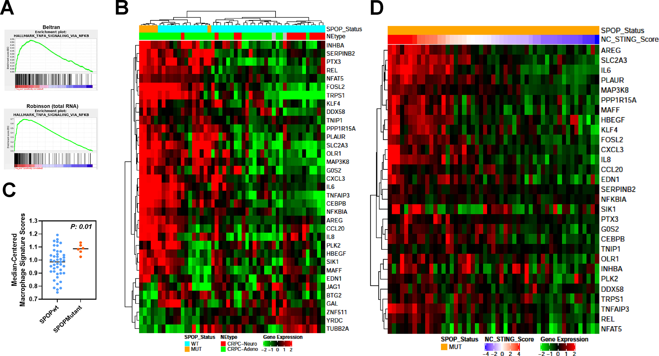
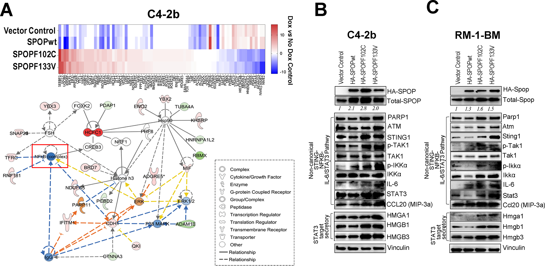
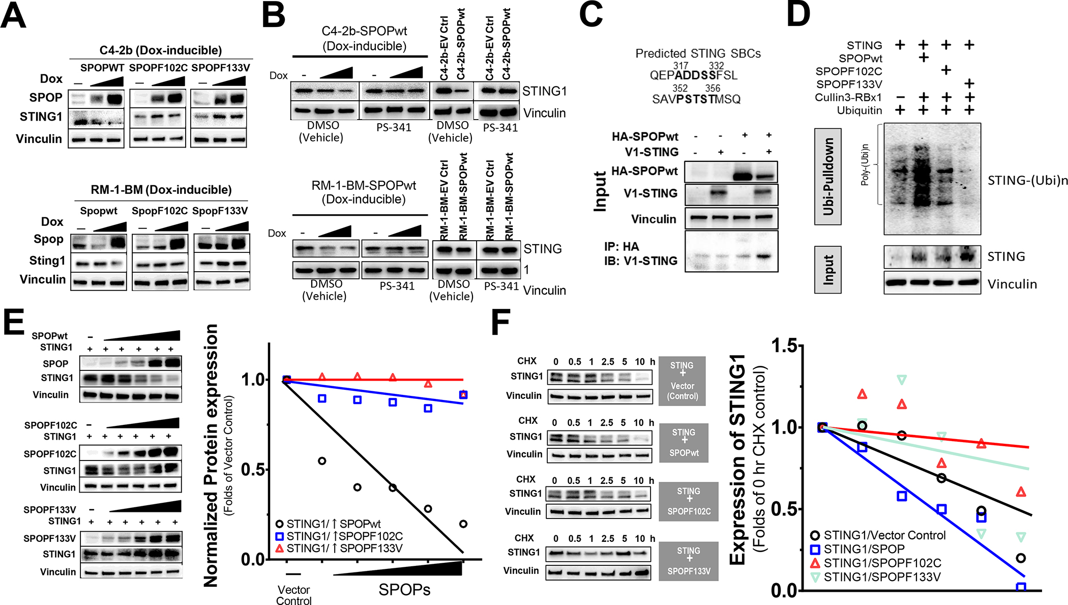
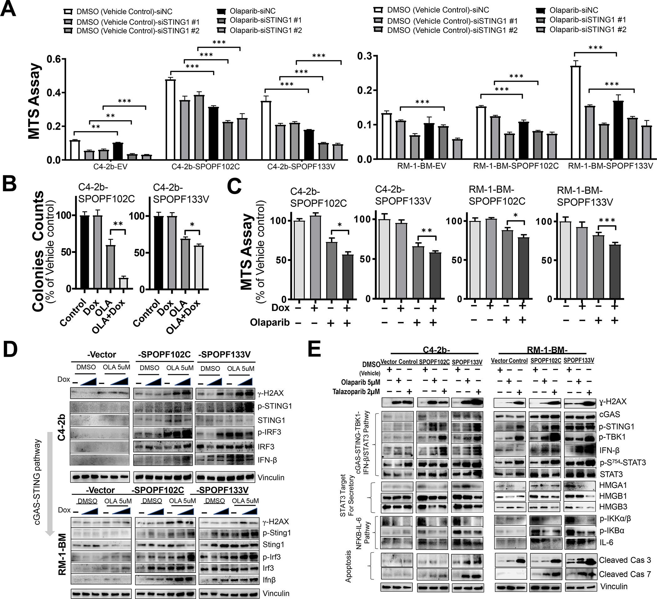
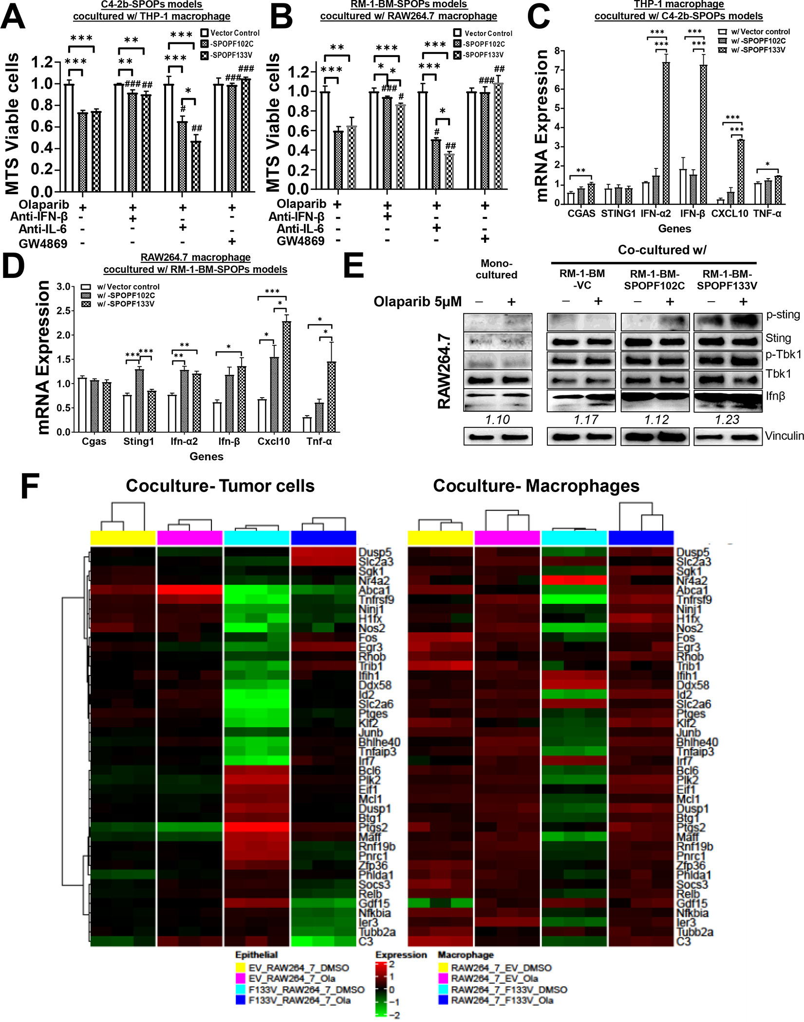
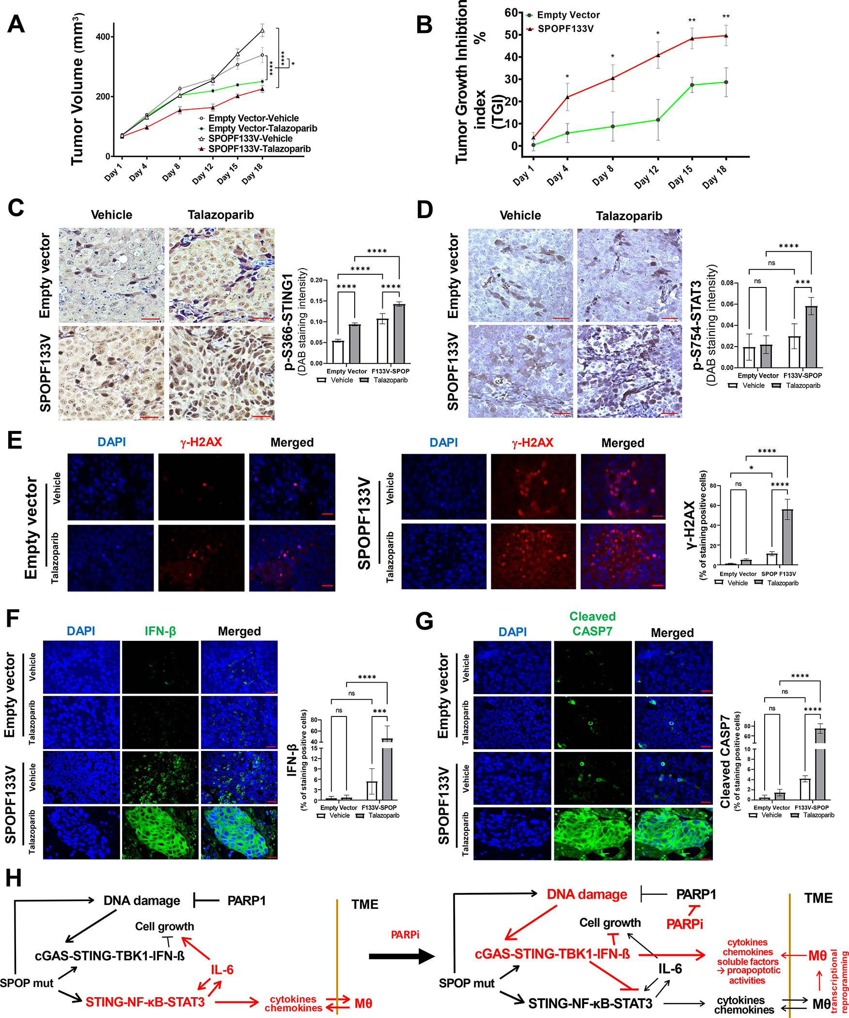
Results: We demonstrate that SPOP mutations are associated with upregulation of a 29-gene noncanonical (NC) STING (NC-STING) signature in a subset of SPOPmut, treatment-refractory CRPC patients. We show in preclinical CRPC models that SPOP targets and destabilizes STING1 protein, and prostate cancer-associated SPOP mutations result in upregulated NC-STING-NF-κB signaling and macrophage- and tumor microenvironment (TME)-facilitated reprogramming, leading to tumor cell growth. Importantly, we provide in vitro and in vivo mechanism-based evidence that PARP inhibitor (PARPi) treatment results in a shift from immunosuppressive NC-STING-NF-κB signaling to antitumor, canonical cGAS-STING-IFNβ signaling in SPOPmut CRPC and results in enhanced tumor growth inhibition.

Conclusions: We provide evidence that SPOP is critical in regulating immunosuppressive versus antitumor activity downstream of DNA damage-induced STING1 activation in prostate cancer. PARPi treatment of SPOPmut CRPC alters this NC-STING signaling toward canonical, antitumor cGAS-STING-IFNβ signaling, highlighting a novel biomarker-informed treatment strategy for prostate cancer.


Conflict of interest statement

The authors declare no potential conflicts of interest.

Figures

Fig. 1.
Fig. 1.. Upregulation of TNF-α-STING-NF-κB and non-canonical STING-NF-κB gene expression in SPOP mutant prostate cancer patients.
A. GSEA analysis of castration-resistant prostate cancer data sets (Beltran, CRPC-Adeno and CRPC-Neuro; Robinson-total RNA; Robinson- polyA RNA) with a curated gene set (Hallmark) shows enrichment of TNF-α-NFκB signaling genes. B. Heatmap of significantly differentially regulated genes from unsupervised analysis of the same data set using a 259 gene set comprised of canonical cGAS-STING-TBK1 and NF-κB signaling genes identified a cluster of differentially expressed genes in SPOPmut prostate cancer patients compared to SPOP wild-type patients. C. “Immunome” analysis of Beltran CRPC cohort using a compendium of publicly available data from purified immune subsets. D. Heatmap of significantly upregulated genes in SPOPmut prostate cancer from Beltran dataset in B (NC-STING signature genes) using TCGA SPOPmut data set ordered by the z-score of NC-STING signature (NC-STING Score).
Fig. 2.
Fig. 2.. Upregulation of non-canonical STING-NF-κB/STAT3, canonical cGAS-STING and activation STAT3-HMG secretory pathways in SPOP mutant (SPOPF102C/SPOPF133V)-expressing prostate cancer cells.
A. Targeted proteomics analysis of Dox-inducible SPOPwt and SPOPmut C4–2b and RM-1-BM prostate cancer models yielded 81 protein features that were differentially expressed between SPOPmut (F102C and F133V) C4–2b prostate cancer cells compared to SPOPwt or empty vector controls (upper panel). Ingenuity Pathway Analyses of these 81 proteins revealed enriched representation of NF-κB-centric protein network features (lower panel). B, C. Immunoblot analysis of proteins involved in non-canonical STING-NF-κB signaling, cGAS-STING signaling and STAT3-HMG secretory regulation pathways in stably transduced SPOPwt- and SPOPmut-(F102C, F133V) expressing or empty vector control C4–2b and RM-1-BM prostate cancer cells.
Fig. 3.
Fig. 3.. STING1 is a putative SPOP-interacting substrate and is functionally regulated by expression of substrate-binding deficient SPOP mutation (SPOPF102C, SPOPF133V).
A. Analysis of STING1 protein following induced expression of SPOPwt, SPOPF102C or SPOPF133V in doxycycline-inducible C4–2b and RM-1-BM cells (induced with the dose of doxycycline at 0, 10 or 200 for C4–2b-SPOPs and 0, 20 or 200 for RM-1-BM-SPOPs, respectively). B. Analysis of STING1 protein in proteosome inhibitor (PS-341) treated, Dox-induced and stably transfected SPOPwt models. C. Co-IP analysis of protein-protein interactions of SPOP and STING1. D. In vivo ubiquitination assay to examine the polyubiquitination of STING1 protein by SPOP E3 ligase complex (SPOP-Culin3- RBX1) or SPOPmuts (SPOPF102C, SPOPF133V) cotranfected in 293T cells. E. Analysis of SPOPwt and STING1 following cotransfection in 293T cells. F. Cycloheximide (CHX) chase protein half-life assay of STING1 following SPOPwt, SPOPF102C or SPOPF133V co-expression.
Fig. 4.
Fig. 4.. SPOP mutants potentiate growth inhibition and induction of cGAS-STING by PARP inhibitor treatment, associated with increased inhibitory STAT3 phosphorylation and suppression of secretory signaling targets and non-canonical STING-NF-κB signaling in prostate cancer models.
A. MTS analysis of C4–2b and RM-1-BM (SPOPF102C or SPOPF133V) treated with siSTING or siNC in the presence and absence of olaparib (OLA), *, P<0.05, and **, P<0.01. B. Colony formation and C. MTS analysis of cell proliferation in doxycycline (Dox)-induced SPOP mutant-expressing C4–2b and RM-1-BM (SPOPF102C or SPOPF133V) in the presence and absence of OLA. T-test was used for statistical analysis, *, P<0.05, and **, P<0.01. D. Immunoblot analysis of OLA-induced DNA damage and canonical STING1 signaling activities (phosphorylation of STING1 and IRF3, and IFN-β) in Dox-induced C4–2b and RM-1-BM SPOP mutants compared to empty vector controls in a dose-dependent manner (Dox 10 and 200 ng/mL for C4–2b-SPOP models, or 20 and 200 ng/mL for RM-1-BM-SPOP models). E. Analysis of the effects of PARP inhibitor (OLA and talazoparib, TALA) on DNA damage (γH2AX), cGAS-STING-TBK1 signaling, IFN-β protein expression, p-S754-STAT3 expression, HMGA1/HMGBs secretory signaling protein expression, NF-κB signaling, IL-6 expression and expression of proapoptotic signaling proteins (cleaved caspases 3 and 7) in human and mouse SPOPmut models compared to empty vector control cells.
Fig. 5.
Fig. 5.. Expression of SPOP mutations in prostate cancer cells enhance PARP inhibitor-mediated growth inhibition through paracrine activities in prostate cancer-macrophage coculture models.
A, B. Expression of SPOP mutant (F102C or F133V) sensitized C4–2b and RM-1-BM prostate cancer models to OLA-mediated growth inhibition. Data were normalized to monocultures of the designated cells following DMSO (control) and OLA treatment. C, D. RT-qPCR analysis of cGAS-STING signaling and IFN-β production specifically in macrophages (THP-1 or RAW264.7) that were cocultured with OLA-treated C4–2b and RM-1-BM cells expressing SPOP mutants (SPOPF102C or SPOPF133V) or vector controls. E. IB analysis of p-Sting-p-Tbk1-IFN-β signaling in RAW264.7 cells in monoculture and coculture [with RM-1-BM prostate cancer models expressing SPOPmuts (SPOPF102C and SPOPF133V) or empty Vector Control (VC)]. A-D. * One-way ANOVA was used to compare the differences in one group, #T test for compare the differences between OLA and other OLA combination treatments, respectively. *or # P<0.05, **or ## P<0.01, ***or ### P<0.005. Bar legend for panels A-D is shown in panel C. F. Heatmaps of common differentially expressed genes in cocultured RM-1-BM-SPOPF133V prostate cancer cells and macrophages to show significant (FDR < 0.05) interaction between treatment [OLA or vehicle control (DMSO)] and SPOP phenotype.
Fig. 6.
Fig. 6.. SPOPmut prostate cancer demonstrates enhanced sensitivity to PARP inhibitor therapy.
A. Tumor growth during vehicle or talazoparib (TALA) treatment in C4–2b vector controls and C4–2b-SPOP-F133V mutant expressing xenograft models. Two-way ANOVA was used to analyze the statistical significance of control vehicle vs TALA-treated C4–2b-empty vector tumor growth or C4–2b-SPOPF133V tumor growth, respectively, ****, P<0.0001. In addition, three-way ANOVA test was used to analyze TALA treatment affected growth of C4–2b-SPOPF133V tumors (−/+TALA) vs C4–2b-empty vector tumors (−/+TALA), *, P=0.0105. B. Tumor growth inhibition (TGI) index analysis of talazoparib-induced growth suppression in C4–2b-SPOP-F133V mutant expressing xenograft models compared to vector controls. For each individual day point, t-test is used to determine the significance of C4–2b-SPOPF133V vs C4–2b-empty vector tumor TGI %, **, P<0.01 and *, P<0.05. C, D. Immunostaining analysis shows significantly increased p-S366-STING1 and inhibitory p-S754-STAT3 in tumors from A and B, respectively (Bar inset=200 μm). E, F, G. Immunofluorescence (IF) analysis to examine ɣ-H2AX (E), Interferon-β (F), and cleaved caspase 7 (G) in tumors from A and B, respectively (Bar inset=25 μm). Quantitative data of immune staining or IF signals shown in C-G are MEAN ± SD (n=5), ***p<0.001, ****p<0.0005, two-way ANOVA. H. Mechanistic interactions that underlie the “switch” from non-canonical STING signaling to canonical STING signaling shown before and after PARP inhibitor treatment in SPOPmut prostate cancer.

References

    1. Robinson D, Van Allen EM, Wu YM, Schultz N, Lonigro RJ, Mosquera JM, et al. Integrative clinical genomics of advanced prostate cancer. Cell 2015;161(5):1215–28 doi 10.1016/j.cell.2015.05.001. - DOI - PMC - PubMed
    1. Barbieri CE, Baca SC, Lawrence MS, Demichelis F, Blattner M, Theurillat JP, et al. Exome sequencing identifies recurrent SPOP, FOXA1 and MED12 mutations in prostate cancer. Nat Genet 2012;44(6):685–9 doi 10.1038/ng.2279. - DOI - PMC - PubMed
    1. Geng C, He B, Xu L, Barbieri CE, Eedunuri VK, Chew SA, et al. Prostate cancer-associated mutations in speckle-type POZ protein (SPOP) regulate steroid receptor coactivator 3 protein turnover. Proc Natl Acad Sci U S A 2013;110(17):6997–7002 doi 10.1073/pnas.1304502110. - DOI - PMC - PubMed
    1. Zhuang M, Calabrese MF, Liu J, Waddell MB, Nourse A, Hammel M, et al. Structures of SPOP-substrate complexes: insights into molecular architectures of BTB-Cul3 ubiquitin ligases. Mol Cell 2009;36(1):39–50 doi 10.1016/j.molcel.2009.09.022. - DOI - PMC - PubMed
    1. Boysen G, Barbieri CE, Prandi D, Blattner M, Chae SS, Dahija A, et al. SPOP mutation leads to genomic instability in prostate cancer. Elife 2015;4 doi 10.7554/eLife.09207. - DOI - PMC - PubMed

Publication types

MeSH terms