. 2023 Nov;66(11):2075-2086.
doi: 10.1007/s00125-023-05991-8. Epub 2023 Aug 15.

IFNɣ but not IFNα increases recognition of insulin defective ribosomal product-derived antigen to amplify islet autoimmunity

Affiliations

IFNɣ but not IFNα increases recognition of insulin defective ribosomal product-derived antigen to amplify islet autoimmunity

Sofia Thomaidou et al. Diabetologia. 2023 Nov.

Abstract

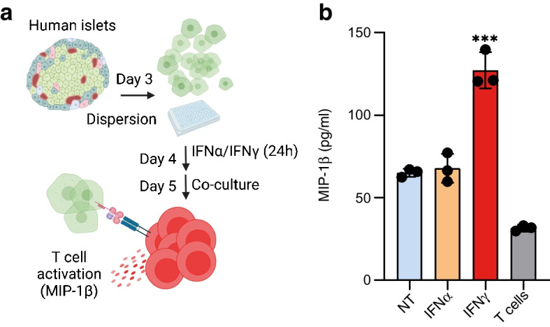
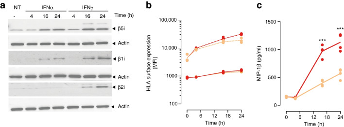
Aims/hypothesis: The inflammatory milieu characteristic of insulitis affects translation fidelity and generates defective ribosomal products (DRiPs) that participate in autoimmune beta cell destruction in type 1 diabetes. Here, we studied the role of early innate cytokines (IFNα) and late immune adaptive events (IFNɣ) in insulin DRiP-derived peptide presentation to diabetogenic CD8+ T cells.

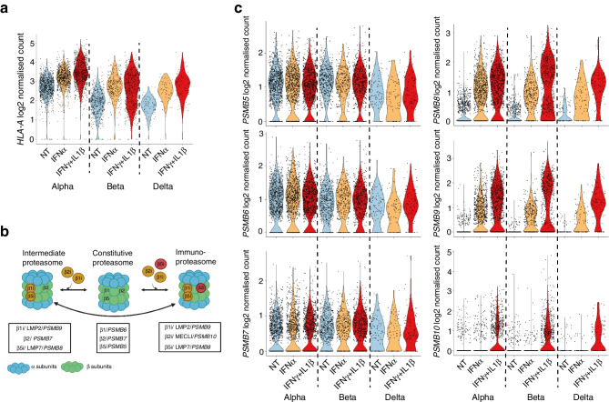
Methods: Single-cell transcriptomics of human pancreatic islets was used to study the composition of the (immuno)proteasome. Specific inhibition of the immunoproteasome catalytic subunits was achieved using siRNA, and antigenic peptide presentation at the cell surface of the human beta cell line EndoC-βH1 was monitored using peptide-specific CD8 T cells.

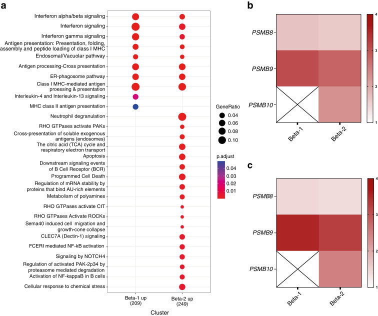
Results: We found that IFNγ induces the expression of the PSMB10 transcript encoding the β2i catalytic subunit of the immunoproteasome in endocrine beta cells, revealing a critical role in insulin DRiP-derived peptide presentation to T cells. Moreover, we showed that PSMB10 is upregulated in a beta cell subset that is preferentially destroyed in the pancreases of individuals with type 1 diabetes.

Conclusions/interpretation: Our data highlight the role of the degradation machinery in beta cell immunogenicity and emphasise the need for evaluation of targeted immunoproteasome inhibitors to limit beta cell destruction in type 1 diabetes.

Data availability: The single-cell RNA-seq dataset is available from the Gene Expression Omnibus (GEO) using the accession number GSE218316 ( https://www.ncbi.nlm.nih.gov/geo/query/acc.cgi?acc=GSE218316 ).

Keywords: Autoantigens; Degradation; Inflammation; Proteasome; Type 1 diabetes.


Figures

Fig. 1
Fig. 1
IFNα and IFNγ differentially regulate INS-DRiP presentation in human islets. (a) Schematic of the islet cells/INS-DRiP-specific CTLs co-culture experiment in the presence of 2000 U/ml IFNα or 1000 U/ml IFNγ. Created with BioRender.com. (b) MIP-1β secretion by INS-DRiP-specific CTLs after co-culture with HLA-A2+ primary human islet cells treated with IFNα or IFNγ for 24 h (n=3). NT, non-treated
Fig. 2
Fig. 2
Cytokines differentially regulate immunoproteasome catalytic subunit expression in primary human endocrine cells. (a) Violin plots showing the expression levels of HLA-A after IFNα or IFNγ/IL1β treatment of endocrine cells. (b) Schematic representation of the composition of the proteasome and immunoproteasome. (c) Violin plots showing the expression levels of the mRNAs encoding the constitutive (PSMB5, PSMB6 and PSMB7) and induced (PSMB8, PSMB9 and PSMB10) catalytic subunits of the (immuno)proteasome. Expression levels correspond to log2 normalised counts/cell, as obtained in single-cell RNA-seq of human islets treated with IFNα (2000 U/ml) or IFNγ (1000 U/ml)+IL1β (2 ng/ml) for 24 h. NT, non-treated
Fig. 3
Fig. 3
Proteasome composition in EndoC-βH1 cells after exposure to cytokines and sensitivity to INS-DRiP-specific CTLs. (a) β5i (encoded by PSMB8), β1i (PSMB9) and β2i (PSMB10) protein expression after incubation with 2000 U/ml IFNα or 1000 U/ml IFNγ determined by Western blot analysis. Actin was used as an internal control. Treatment duration was 4, 16 or 24 h, as indicated. (b) Surface expression of HLA-A/B/C (dashed lines) and HLA-A2 (solid lines) on EndoC-βΗ1-HLA-A2 cells treated with IFNα (orange lines) or IFNγ (red lines) for 4, 16 and 24 h. Values are presented as mean fluorescence intensity (MFI). (c) INS-DRiP-specific CTL activation determined by MIP-1β secretion after co-culture with EndoC-βΗ1-HLA-A2 cells treated with IFNα (orange line) or IFNγ (red line) for 4, 16 and 24 h. n=3 independent experiments. ***p≤ 0.001. NT, non-treated
Fig. 4
Fig. 4
Immunoproteasome silencing in the EndoC-βΗ1 cell line. (a) PSMB8, (b) PSMB9 and (c) PSMB10 mRNA expression in EndoC-βΗ1-HLA-A2 cells transfected with non-targeted siRNA (siCTRL) and siRNAs specific for PSMB8, PSMB9 and PSMB10. Cells were treated with 2000 U/ml IFNα (orange bars), 1000 U/ml IFNγ (red bars) or control medium (blue bars) 72 h post transfection for 24 h. Gene expression levels were corrected for levels of the housekeeping gene GAPDH and are presented as the induction ratio (relative to levels for siCTRL(1) in IFNα samples and siCTRL(2) for IFNγ samples) (n=3). *p≤ 0.05, **p≤ 0.01, ***p≤ 0.001
Fig. 5
Fig. 5
PSMB10 silencing exclusively reduces INS-DRiP-specific CTL activation. (a) HLA-A2 (genetically introduced) surface expression of EndoC-βΗ1-HLA-A2 cells transfected with non-targeted siRNA (siCTRL) and siRNAs specific for PSMB8, PSMB9 and PSMB10. Cells were left untreated (blue bars) or treated with 2000 U/ml IFNα (orange bars) or 1000 U/ml IFNγ (red bars) 72 h post transfection for 24 h. Values are presented as mean fluorescence (FITC) intensity (MFI) (n=3). (b, c) MIP-1β secretion by (b) INS-DRiP1–9-specific CTLs and (c) PPI15–24 after co-culture with EndoC-βΗ1-HLA-A2 cells. Prior to co-culture, target cells were transfected with non-targeted siRNA and siRNAs specific for PSMB8, PSMB9 and PSMB10. Cells were left untreated (blue circles) or treated with IFNα (orange circles) or IFNγ (red circles) 72 h post transfection for 24 h (n=3). Statistical analyses are comparing IFNγ to IFNα treatments. ***p≤0.001. NT, non-treated
Fig. 6
Fig. 6
Immunoproteasome composition of beta cells from individuals with type 1 diabetes. (a) Gene ontology plot of transcripts upregulated in two distinct beta cell subsets (p<0.05 and logFC>1) in individuals with type 1 diabetes compared with control individuals (donors without diabetes). The plot was generated with clusterProfiler using the enrichPathway classification. (b) Heatmap showing the logFC (p<0.05) of PSMB8, PSMB9 and PSMB10 in the beta-1 and beta-2 populations from individuals with type 1 diabetes compared with control donors. (c) Heatmap showing the logFC (p<0.05) of PSMB8, PSMB9 and PSMB10 in the beta-1 and beta-2 populations from individuals with type 1 diabetes patients compared with AAb+ individuals. This figure has been generated using data described in Fasolino et al, 2022 [29]

References

    1. Pugliese A. Autoreactive T cells in type 1 diabetes. J Clin Invest. 2017;127(8):2881–2891. doi: 10.1172/JCI94549. - DOI - PMC - PubMed
    1. Abreu JR, Martina S, Verrijn Stuart AA, et al. CD8 T cell autoreactivity to preproinsulin epitopes with very low human leucocyte antigen class I binding affinity. Clin Exp Immunol. 2012;170(1):57–65. doi: 10.1111/j.1365-2249.2012.04635.x. - DOI - PMC - PubMed
    1. Bender C, Rodriguez-Calvo T, Amirian N, Coppieters KT, von Herrath MG. The healthy exocrine pancreas contains preproinsulin-specific CD8 T cells that attack islets in type 1 diabetes. Sci Adv. 2020;6(42):eabc5586. doi: 10.1126/sciadv.abc5586. - DOI - PMC - PubMed
    1. Culina S, Lalanne AI, Afonso G, et al. Islet-reactive CD8(+) T cell frequencies in the pancreas, but not in blood, distinguish type 1 diabetic patients from healthy donors. Sci Immunol. 2018;3(20):eaao4013. doi: 10.1126/sciimmunol.aao4013. - DOI - PMC - PubMed
    1. Thomaidou S, Slieker RC, van der Slik AR, et al. Long RNA sequencing and ribosome profiling of inflamed beta-cells reveal an extensive translatome landscape. Diabetes. 2021;70(10):2299–2312. doi: 10.2337/db20-1122. - DOI - PubMed

Publication types

MeSH terms