. 2023 Sep 22;8(18):e167210.
doi: 10.1172/jci.insight.167210.

Maternal acellular pertussis vaccination in mice impairs cellular immunity to Bordetella pertussis infection in offspring

Maternal acellular pertussis vaccination in mice impairs cellular immunity to Bordetella pertussis infection in offspring

Violaine Dubois et al. JCI Insight. .

Abstract

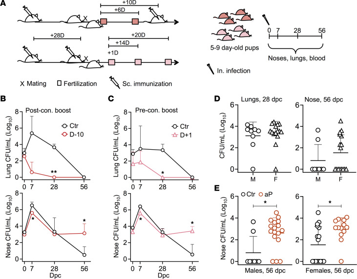
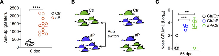
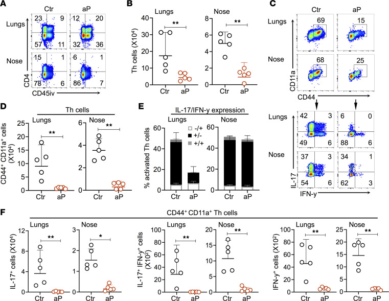
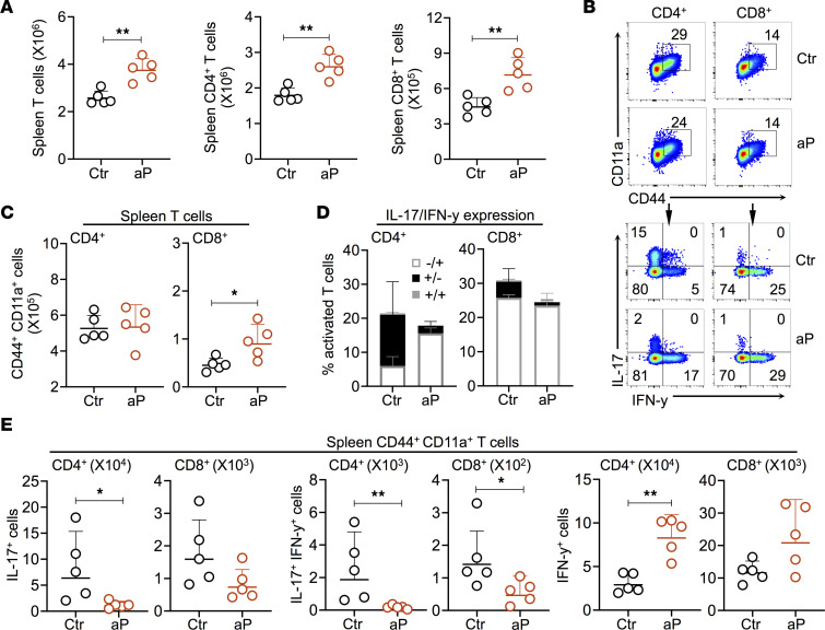
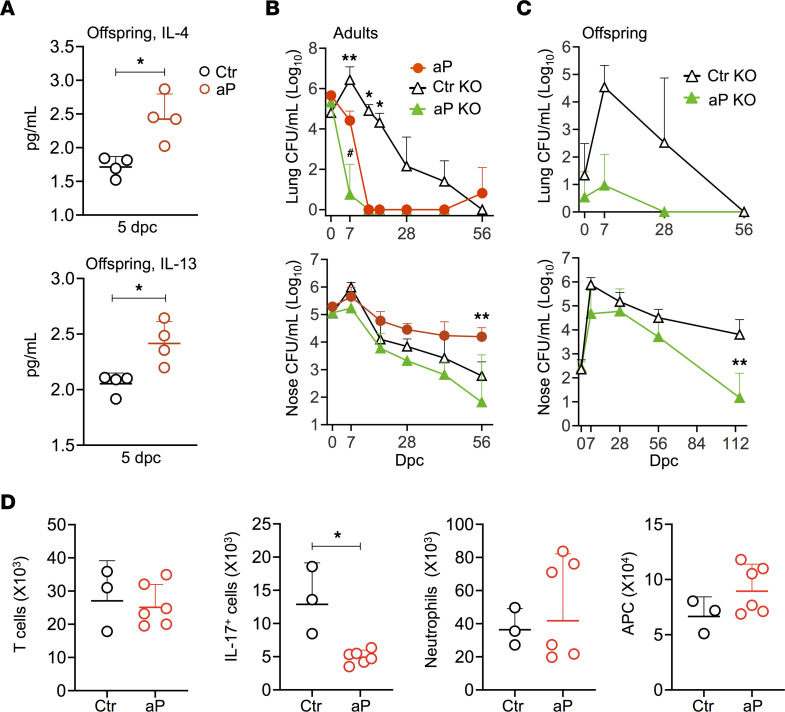
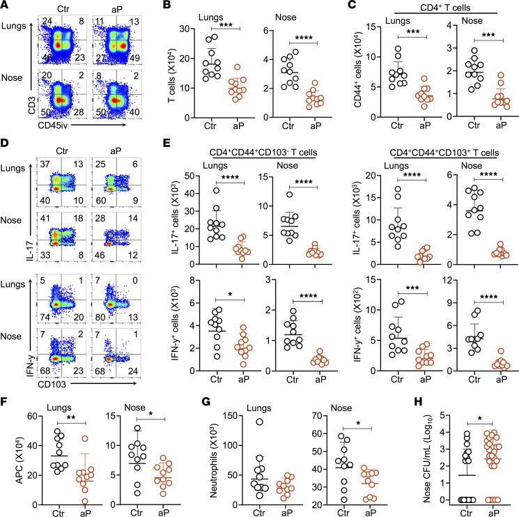
Given the resurgence of pertussis, several countries have introduced maternal tetanus, diphtheria, and acellular pertussis (aP) vaccination during pregnancy to protect young infants against severe pertussis. Although protective against the disease, the effect of maternal aP vaccination on bacterial colonization of the offspring is unknown. Here, we used a mouse model to demonstrate that maternal aP immunization, either before or during pregnancy, protects pups from lung colonization by Bordetella pertussis. However, maternal aP vaccination resulted in significantly prolonged nasal carriage of B. pertussis by inhibiting the natural recruitment of IL-17-producing resident memory T cells and ensuing neutrophil influx in the nasal tissue, especially of those with proinflammatory and cytotoxic properties. Prolonged nasal carriage after aP vaccination is due to IL-4 signaling, as prolonged nasal carriage is abolished in IL-4Rα-/- mice. The effect of maternal aP vaccination can be transferred transplacentally to the offspring or via breastfeeding and is long-lasting, as it persists into adulthood. Maternal aP vaccination may, thus, augment the B. pertussis reservoir.

Keywords: Bacterial vaccines; Cellular immune response; Infectious disease; Mouse models; Vaccines.


Conflict of interest statement

Conflict of interest: The authors have declared that no conflict of interest exists.

Figures

Figure 1
Figure 1. Antenatal aP immunization prevents lung colonization in offspring while enhancing nasal carriage.
(A) Female BALB/c mice were immunized twice s.c. with 1/10 human dose of Infanrix (aP). Control mice received PBS. Mice were mated 28 days after priming and boosted on the day of the mating (preconception [pre-con.] boost) or 10 days later (post-con. boost). For post-con. boosting, fertilization occurred at the day of mating — i.e., 10 days before boosting (D–10) — or 6 days after mating — i.e., 4 days before boosting (D–4). For the preconception boosting, fertilization occurred 1 (D+1), 14 (D+14), or 20 (D+20) after boosting. Five to 9 days after birth, the pups were nasally infected with 5 × 10³ CFU B1917. At the indicated time points after challenge, lungs and noses were harvested for CFU counting and blood was collected to measure maternally transferred anti–B. pertussis IgG. (B) CFU counts in lungs (upper panel) and noses (lower panel) from pups born to mothers boosted 10 days after fertilization (D–10) compared with control mice (Ctr) at indicated time points after challenge. (C) CFU counts in lungs (upper panel) and noses (lower panel) from pups born to mothers boosted 1 day before fertilization (D+1) compared with control mice (Ctr) at indicated time points after challenge. (D) CFU counts in lungs (left panel) and noses (right panel) of male (M) and female (F) pups born to nonvaccinated mothers 28 days (left panel) or 56 days (right panel) after challenge. (E) CFU counts in the noses of male (left panel) and female (right panel) pups born to nonvaccinated (Ctr) or aP-vaccinated (aP) mothers 56 days after challenge. Results shown are geometric means ± SD. n = 3–6 for the Ctr groups and n = 3–5 for the aP groups in (B and C). Mann-Whitney tests were performed to compare Ctr and aP offspring. *P < 0.05; **P < 0.01.
Figure 2
Figure 2. Placental and breast milk–derived inhibition of nasal B. pertussis clearance in the offspring.
(A) Anti–B. pertussis IgG titers in the serum of control (Ctr) and aP offspring 3 hours after infection (n = 12–15). (B and C) Effect of nursing on nasal colonization of the offspring. Offspring from control (Ctr, in green) and aP-vaccinated mothers (aP, in blue) were switched within 12 hours after birth. Control pups nursed by aP-immunized mice (Ctr/aP) and pups from aP-vaccinated mothers nursed by control dams (aP/Ctr) were infected with 5 × 10³ CFU B1917 and sacrificed 56 dpc to assess the bacterial burden in the noses. Data on Ctr pups (Ctr/Ctr) are derived from the results shown in Figure 1 (n = 4–19). Mann-Whitney tests were performed to compare Ctr/Ctr with Ctr/aP or aP/Ctr offspring. **P < 0.01; ***P < 0.005; ****P < 0.001.
Figure 3
Figure 3. Effect of maternal aP immunization on recruitment and activation of T cells in lungs and noses.
Neonatal mice born to control (Ctr) or aP-vaccinated mothers (aP) were infected with 5 × 10³ CFU B1917 6–7 days after birth and sacrificed 28 dpc. Ten minutes before euthanasia, mice were i.v. injected with anti–CD45-PE (CD45iv) antibody. (A) Representative plots showing the recruitment of CD4+CD45iv T cells in the lungs (upper plots) and noses (lower plots). (B) Absolute numbers of CD4+CD45iv T cells in the lungs (left panel) and nose (right panel) of offspring. (C) Representative plots showing the expression of CD11a and CD44 on CD4+ T cells (upper panel) and expression of IL-17 and IFN-γ upon stimulation with PMA and ionomycin (lower panel). (D) Absolute numbers of pulmonary (left) and nasal (right) CD4+ T cells expressing CD44 and CD11a. (E) Percentages of CD4+ T cells producing IL-17 and/or IFN-γ in lungs (left) and noses (right). (F) Absolute numbers of CD44+CD11a+CD4+ T cells expressing IL-17 (left panel), IFN-γ (right panel), or both (middle panel). Results shown are geometric means ± SD. n = 4–5. Mann-Whitney tests were performed to compare Ctr and aP offspring. *P < 0.05; **P < 0.01.
Figure 4
Figure 4. Effect of maternal aP immunization on recruitment of neutrophils and phagocytic cells in lungs and noses of the offspring.
Neonatal mice born to control (Ctr) or aP-vaccinated mothers (aP) were infected with 5 × 10³ CFU B1917 6– 7 days after birth and sacrificed 28 dpc. Ten minutes before euthanasia, mice were i.v. injected with anti–CD45-PE antibody (CD45iv). (A) Absolute numbers of neutrophils in lungs and noses. (B) Representative plots showing high–FSC-A (FSChi) Ly-6G+ neutrophils in lungs and noses (left panel) and corresponding percentages (right panel). (C) Fold increases of phagocytic cells with the indicated phenotype in Ctr over aP offspring in lungs (upper panel) and noses (lower panel). (D) Percentages of phagocytic cells with the indicated phenotypes in lungs (left) and noses (right). Results shown are geometric means ± SD. n = 4–5. Mann-Whitney tests were performed to compare Ctr and aP offspring. **P < 0.01.
Figure 5
Figure 5. Effect of maternal aP immunization on the natural induction of Th17 cells in secondary lymphoid tissues.
(A) Absolute numbers of T cells (left panel), CD4+ (middle panel), and CD8+ T cells in the spleen of aP (red dots) and control (black dots) offspring 28 dpc. (B) Representative plots showing the expression of CD11a and CD44 on splenic T cells (upper panel) and expression of IL-17 and IFN-γ upon stimulation with PMA and ionomycin (lower panel) by splenic T cells from pups born to control (Ctr) or aP-vaccinated (aP) mothers. (C) Absolute numbers of splenic CD4+ (left panel) and CD8+ T cells expressing CD44 and CD11a from pups born to control (Ctr) or aP-vaccinated (aP) mothers. (D) Percentages of activated splenic CD4+ (left panel) and CD8+ (right panel) T cells from pups born to control (Ctr) or aP-vaccinated (aP) mothers producing IL-17 and/or IFN-γ. (E) Absolute numbers of splenic CD44+CD11a+CD4+ and CD44+CD11a+CD8+ T cells expressing IL-17 (left panel), IFN-γ (right panel), or both (middle panel). Results shown are geometric means ± SD. n = 5. Mann-Whitney tests were performed to compare Ctr and aP offspring. *P < 0.05; **P < 0.01.
Figure 6
Figure 6. Role of the IL-4 pathway in prolonged nasal B. pertussis carriage in aP-immunized BALB/c mice.
(A) Concentrations of IL-4 and IL-13 in the serum of neonates born to aP-immunized (aP) or control mice (Ctr) 5 days after challenge. (B and C) Six- to 8 week-old BALB/c and IL-4Ra–/– (KO) mice, were immunized twice s.c. with 1/10 human dose of aP or PBS and then challenged with 1 × 10⁶ CFU B1917 (B). Neonates were nasally infected with 5 × 10³ CFU B1917 (C). Bacterial burden was determined by CFU counting on homogenized lungs (upper panels) and noses (lower panels) at indicated time points after challenge. (D) Absolute numbers of T cells (left panel), IL-17+ T cells (middle left panel), neutrophils (middle right panel), and APCs (right panel) in the noses of aP (red dots) and control (black dots) IL-4Rα–/– mice 28 dpc. Results shown are geometric means ± SD. n = 5. Mann-Whitney tests were performed to compare Ctr and aP offspring and control KO with aP mice and aP KO mice. *P < 0.05; **P < 0.01.
Figure 7
Figure 7. Long-term effect of maternal aP immunization on offspring T cell responses.
Ten-week-old mice born to control (Ctr) or aP-immunized mice (aP) were infected with 1 × 10⁶ CFU B1917 and sacrificed 28 dpc. Ten minutes before euthanasia, mice were i.v. injected with anti–CD45-PE (CD45iv) antibodies. (A) Representative plots showing the recruitment of CD3+ T cells in the lungs and noses of aP and Ctr offspring. (B) Absolute numbers of T cells in the lungs (left panel) and noses (right panel) of aP and Ctr offspring. (C) Absolute numbers of CD44+CD4+ T cells in the lungs (left panel) and noses (right panel) of aP and Ctr offspring. (D) Representative plots showing the expression of IL-17 (upper panel) and IFN-γ (lower panel) by CD103+ T cells upon stimulation with PMA and ionomycin. (E) Absolute numbers of lung and noses CD4+CD44+CD103 (left panels) and CD4+CD44+CD103+ T cells (right panels) expressing IL-17 (upper panels) or IFN-γ (lower panels). (F) Absolute numbers of APC in the lungs (left panel) and noses (right panel) of Ctr and aP offspring. (G) Absolute numbers of neutrophils in the lungs (left panel) and noses (right panel) of Ctr and aP offspring. (H) CFU counts on nose homogenates 56 dpc of Ctr and aP offspring. Results shown are geometric means ± SD. n = 4–10. Mann-Whitney tests were performed to compare Ctr and aP. *P < 0.05; **P < 0.01; ***P < 0.005; ****P < 0.001.
Figure 8
Figure 8. Effect of maternal aP immunization on colonization by K. pneumoniae and S. pneumoniae in the offspring.
Neonatal mice born to control (Ctr) or aP-vaccinated mothers (aP) were i.n. infected with K. pneumoniae (A and B) or S. pneumoniae (C) 7 days after birth. One and 3 dpc with 50 CFU, K. pneumoniae CFU numbers were measured in nose, lungs, and spleen (A) (n = 3–4), and survival was monitored over time after K. pneumoniae infection (B) (n = 6). S. pneumoniae CFU numbers were measured in the lungs and noses (C) at indicated time points after challenge with 2 × 10³ CFU S. pneumoniae (n = 6–8). *P < 0.05.

Similar articles

Cited by

References

    1. Cherry JD. Epidemic pertussis in 2012--the resurgence of a vaccine-preventable disease. N Engl J Med. 2012;367(9):785–787. doi: 10.1056/NEJMp1209051. - DOI - PubMed
    1. Peck M, et al. Global routine vaccination coverage, 2018. MMWR Morb Mortal Wkly Rep. 2019;68(42):937–942. doi: 10.15585/mmwr.mm6842a1. - DOI - PMC - PubMed
    1. Rocha G, et al. Pertussis in the newborn: certainties and uncertainties in 2014. Paediatr Respir Rev. 2015;16(2):112–118. - PubMed
    1. Von Koenig CHW, Guiso N. Global burden of pertussis: signs of hope but need for accurate data. Lancet Infect Dis. 2017;17:889–890. doi: 10.1016/S1473-3099(17)30357-2. - DOI - PubMed
    1. Blain AE, et al. An assessment of the cocooning strategy for preventing infant pertussis-United States, 2011. Clin Infect Dis. 2016;63(suppl 4):S221–S226. doi: 10.1093/cid/ciw528. - DOI - PMC - PubMed

Publication types