. 2023 Sep 15;4(3):102511.
doi: 10.1016/j.xpro.2023.102511. Epub 2023 Aug 14.

Quantification of membrane-bound cytokine receptors by calibrated flow cytometry

Affiliations

Quantification of membrane-bound cytokine receptors by calibrated flow cytometry

Niloufarsadat Miri et al. STAR Protoc. .

Abstract

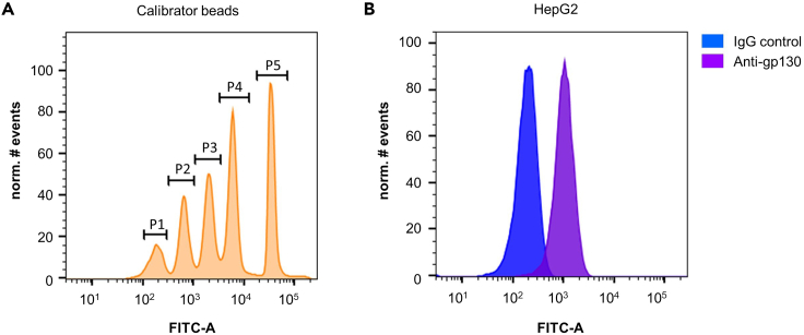
We present a protocol for quantifying the expression of the receptor gp130 using a calibrated flow cytometric approach. We describe pitfalls for receptor quantification such as titration of primary antibodies and standardizing cell culture. Receptors are stained with primary antibodies and fluorophore-coupled secondary antibodies. Beads covered with defined numbers of immunoglobulin G stained with fluorophore-coupled secondary antibodies serve as calibrators. In this way, the fluorescence intensity of cells is converted to the number of receptors on the cell surface. For complete details on the use and execution of this protocol, please refer to Reeh et al. (2019).1.

Keywords: Flow Cytometry/Mass Cytometry; Signal Transduction; Single Cell.


Conflict of interest statement

Declaration of interests The authors declare no competing interests.

Figures

None
Graphical abstract
Figure 1
Figure 1
Gating of single cells (A) SSC-A and FSC-A of HepG2 cells were analyzed by flow cytometry. The depicted gate marks living cells outside of bigger aggregates. (B) FSC-H and FSC-W of living HepG2 cells from Figure 1A. The depicted gate marks single cells. Figure 1 A) and B) are modified from Reeh et al. with permission from Springer Nature. (C) HepG2 cells were stained with anti-gp130 primary antibody followed by staining with FITC-conjugate. Unstained cells and cells only incubated with FITC-conjugate serve as control.
Figure 2
Figure 2
Determination of optimal gain of FITC detector channel (A) SSC-A and FSC-A of Set-up beads were analyzed by flow cytometry. The depicted gate marks single beads. (B) HepG2 cells were treated with IgG isotype control and FITC-conjugate (blue). Set-up beads were stained with FITC-conjugate (green). Analysis was performed by flow cytometry. Figure 2 is modified from Reeh et al. with permission from Springer Nature.
Figure 3
Figure 3
Quantification of gp130 surface expression by calibrated flow cytometry (A) Calibrator beads stained with FITC-conjugate were analyzed by flow cytometry. The five bead populations with different amounts of IgG were marked with gates p1-p5. (B) HepG2 cells were incubated with IgG isotype control and FITC-conjugate or anti-gp130 antibody and FITC-conjugate, respectively. Analysis was performed by flow cytometry. Figure 3 is modified from Reeh et al. with permission from Springer Nature.
Figure 4
Figure 4
Calibration curve Linear dependency of Log10 (corrected mean FITC intensity) and Log10 (IgG/bead) (Table 1). The equation displayed is the result of a linear regression. Figure 4 is modified from Reeh et al. with permission from Springer Nature.

References

    1. Reeh H., Rudolph N., Billing U., Christen H., Streif S., Bullinger E., Schliemann-Bullinger M., Findeisen R., Schaper F., Huber H.J., Dittrich A. Response to IL-6 trans- and IL-6 classic signalling is determined by the ratio of the IL-6 receptor alpha to gp130 expression: fusing experimental insights and dynamic modelling. Cell Commun. Signal. 2019;17:46. doi: 10.1186/s12964-019-0356-0. - DOI - PMC - PubMed
    1. Dittrich A., Hessenkemper W., Schaper F. Systems biology of IL-6, IL-12 family cytokines. Cytokine Growth Factor Rev. 2015;26:595–602. doi: 10.1016/j.cytogfr.2015.07.002. - DOI - PubMed
    1. Baumann H., Isseroff H., Latimer J.J., Jahreis G.P. Phorbol ester modulates interleukin 6- and interleukin 1-regulated expression of acute phase plasma proteins in hepatoma cells. J. Biol. Chem. 1988;263:17390–17396. doi: 10.1016/S0021-9258(19)77848-X. - DOI - PMC - PubMed
    1. Pietzko D., Zohlnhöfer D., Graeve L., Fleischer D., Stoyan T., Schooltink H., Rose-John S., Heinrich P.C. The hepatic interleukin-6 receptor. Studies on its structure and regulation by phorbol 12-myristate 13-acetate-dexamethasone. J. Biol. Chem. 1993;268:4250–4258. doi: 10.1016/S0021-9258(18)53603-6. - DOI - PubMed
    1. Dittrich A., Quaiser T., Khouri C., Görtz D., Mönnigmann M., Schaper F. Model-driven experimental analysis of the function of SHP-2 in IL-6-induced Jak/STAT signaling. Mol. Biosyst. 2012;8:2119–2134. doi: 10.1039/c2mb05488d. - DOI - PubMed

Publication types

LinkOut - more resources