. 2023 Oct 23;58(20):2163-2180.e9.
doi: 10.1016/j.devcel.2023.07.013. Epub 2023 Aug 14.

Single-cell census of human tooth development enables generation of human enamel

Affiliations

Single-cell census of human tooth development enables generation of human enamel

Ammar Alghadeer et al. Dev Cell. .

Abstract

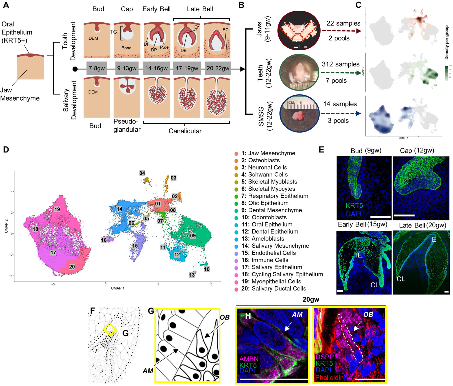
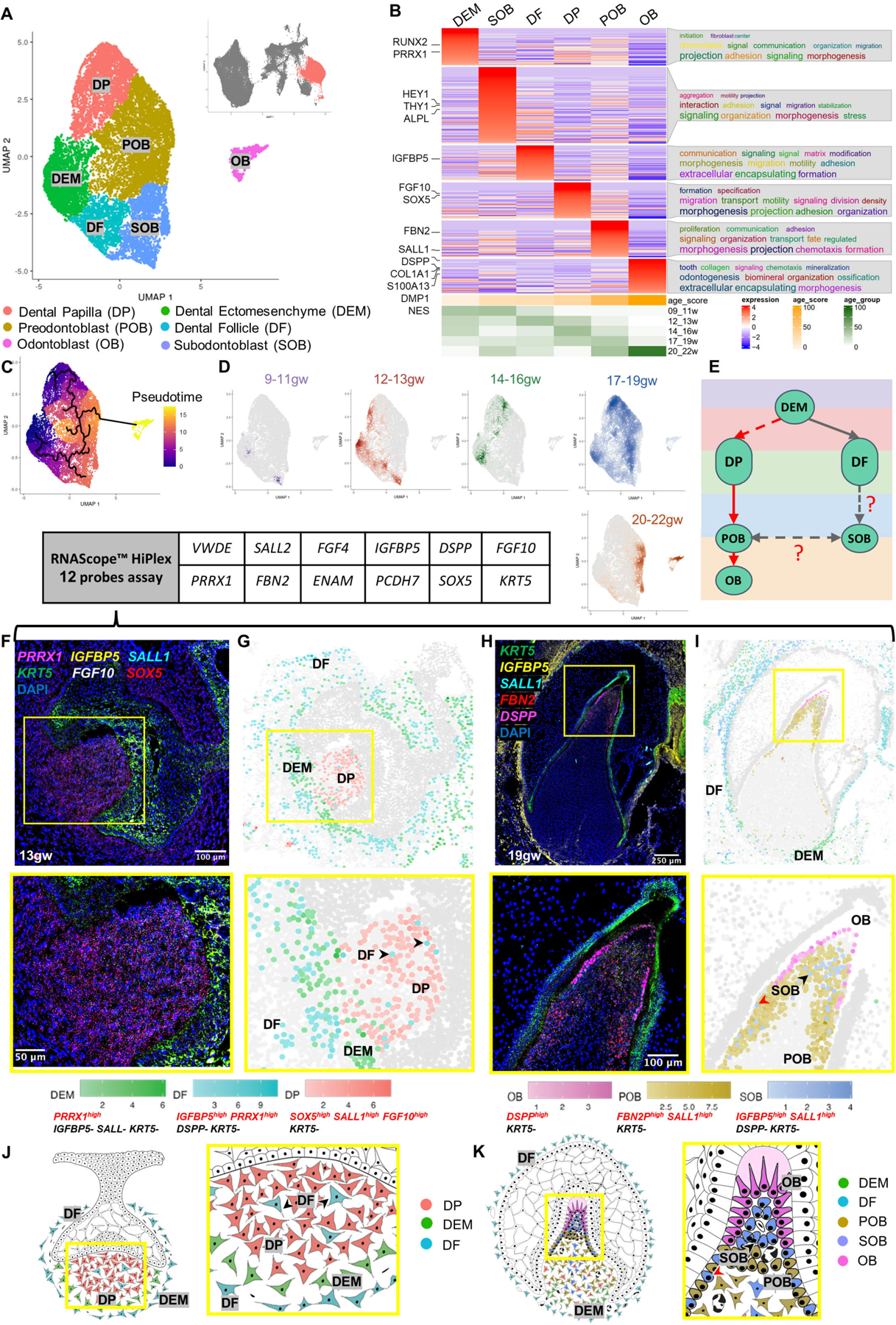
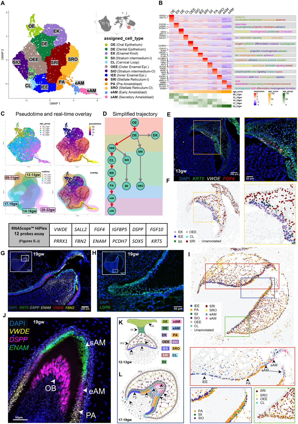
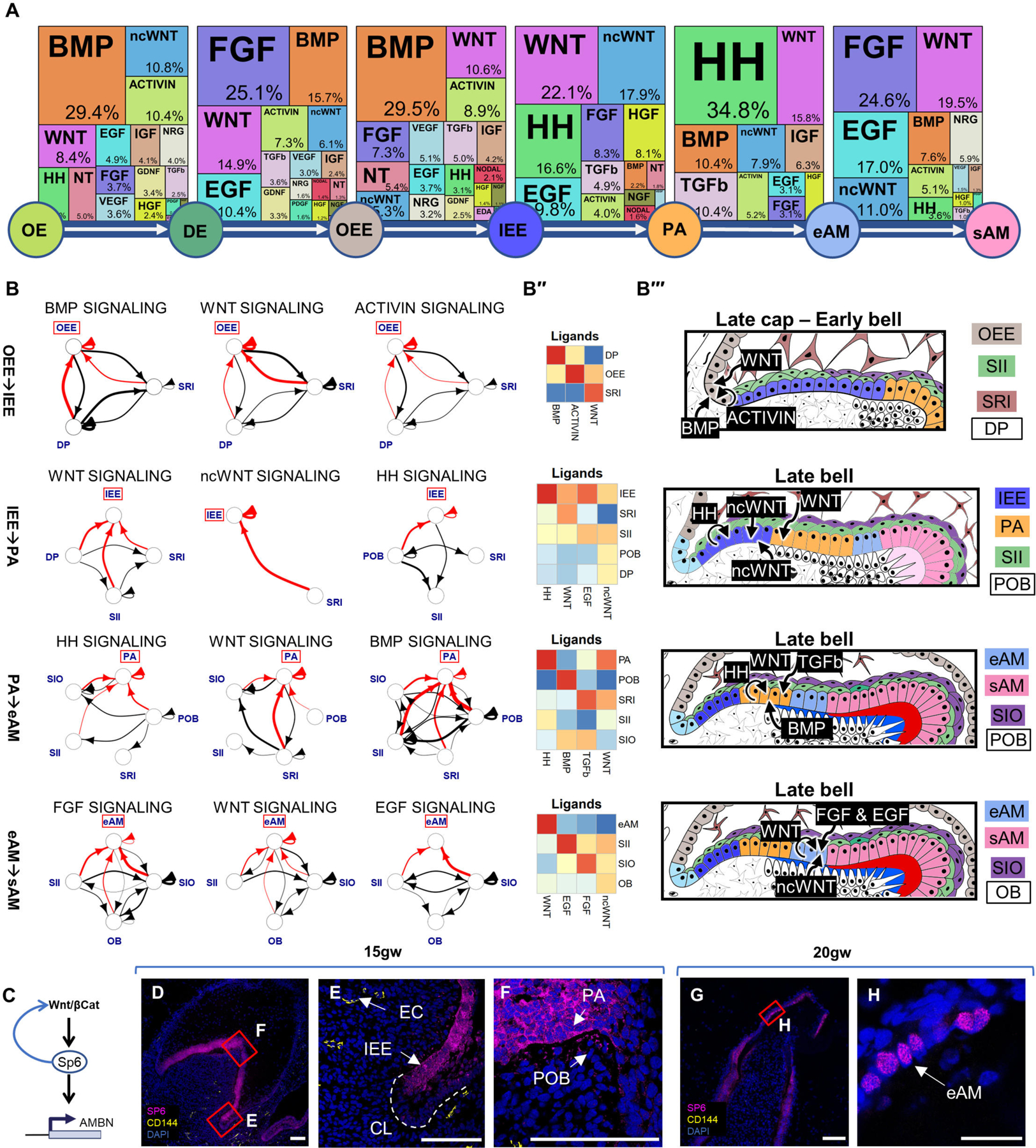
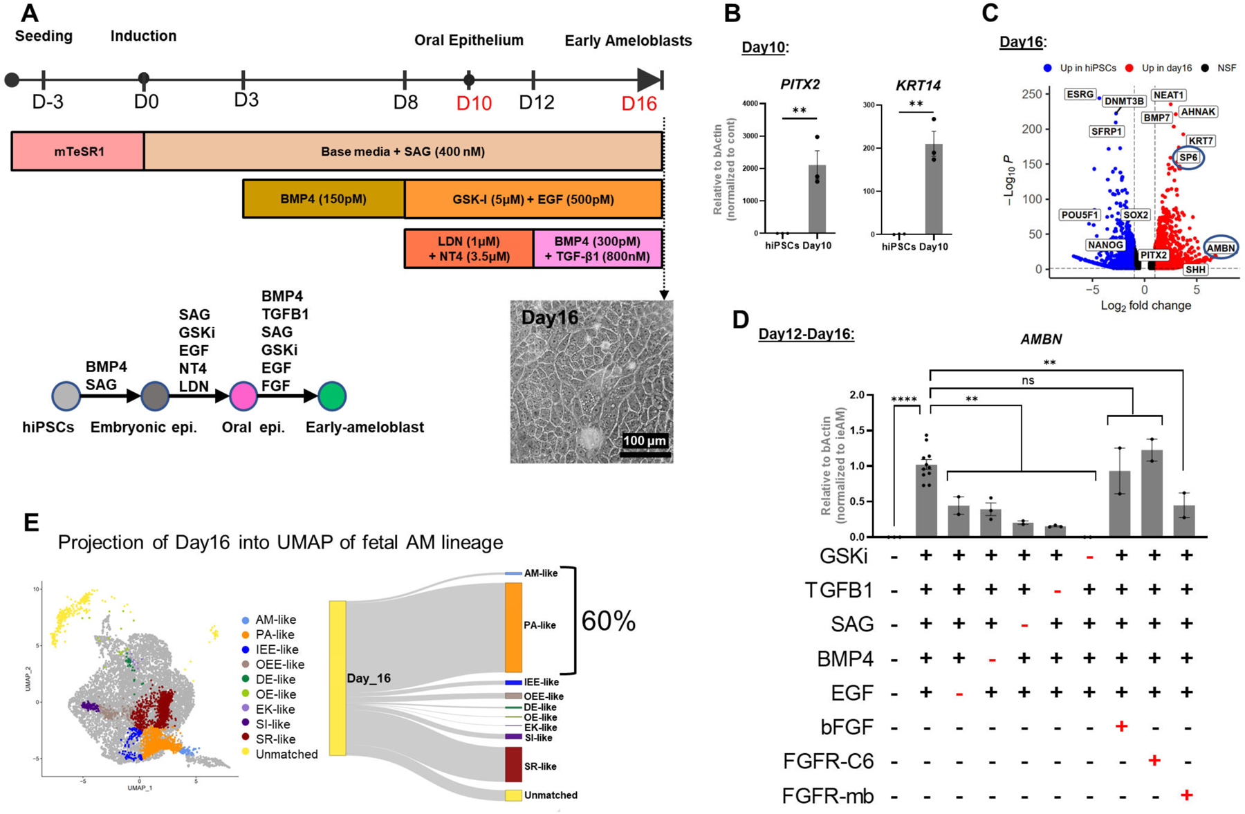
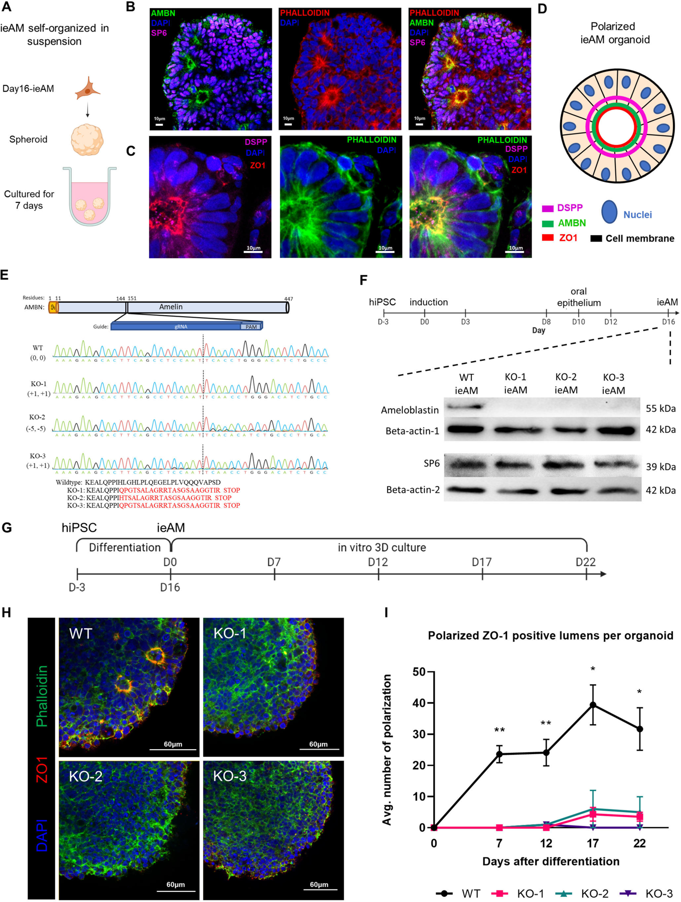
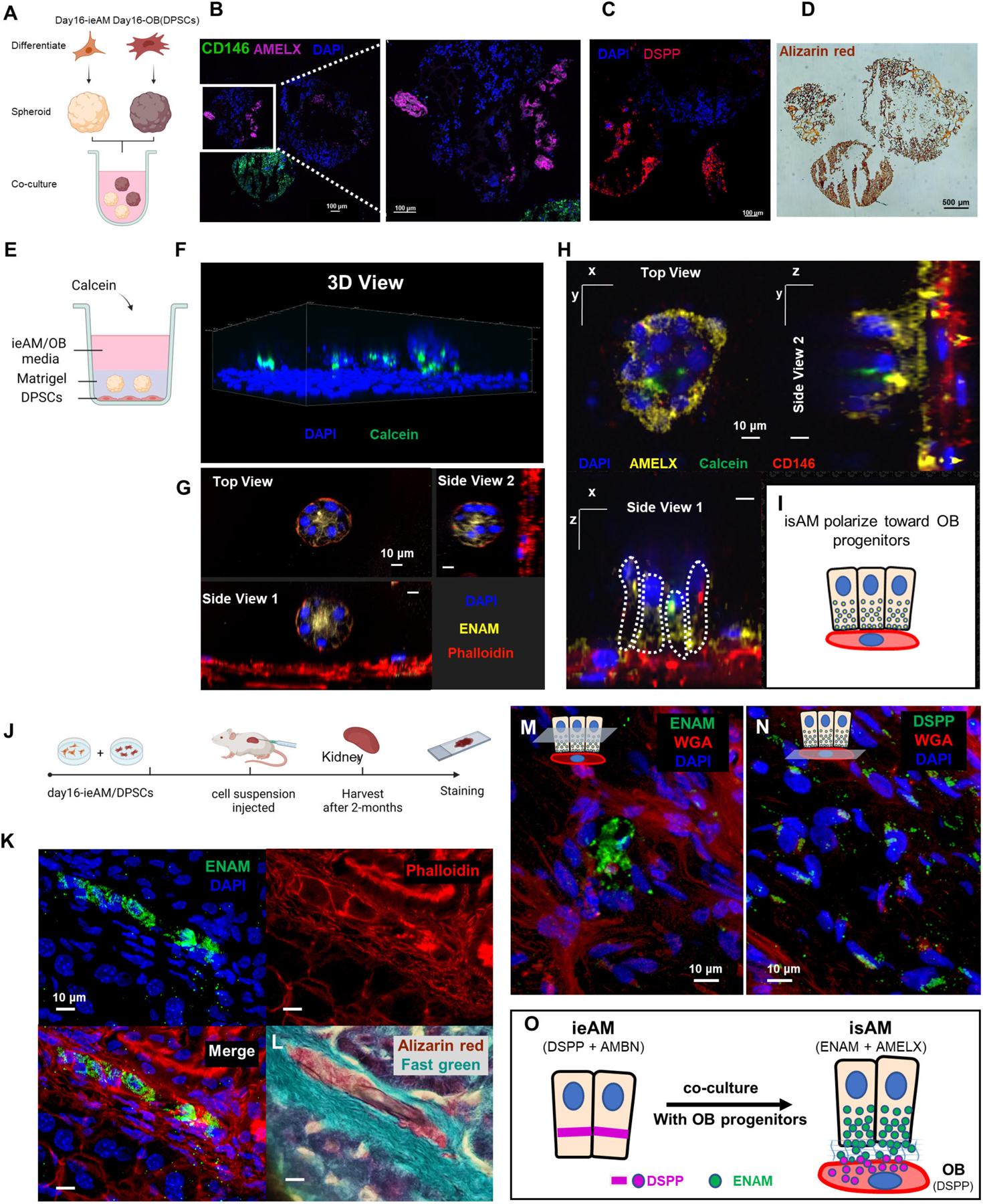
Tooth enamel secreted by ameloblasts (AMs) is the hardest material in the human body, acting as a shield to protect the teeth. However, the enamel is gradually damaged or partially lost in over 90% of adults and cannot be regenerated due to a lack of ameloblasts in erupted teeth. Here, we use single-cell combinatorial indexing RNA sequencing (sci-RNA-seq) to establish a spatiotemporal single-cell census for the developing human tooth and identify regulatory mechanisms controlling the differentiation process of human ameloblasts. We identify key signaling pathways involved between the support cells and ameloblasts during fetal development and recapitulate those findings in human ameloblast in vitro differentiation from induced pluripotent stem cells (iPSCs). We furthermore develop a disease model of amelogenesis imperfecta in a three-dimensional (3D) organoid system and show AM maturation to mineralized structure in vivo. These studies pave the way for future regenerative dentistry.

Keywords: ameloblast; amelogenesis imperfecta; directed differentiation; hiPSC; odontoblast; organoids; pathway analysis; regenerative dentistry; sci-RNA-seq; tooth development.


Conflict of interest statement

Declaration of interests A.A., S.H.-D., Y.T.Z., D.D.E., Y.W., A.P.P., H.Z., J.M., D.B., and H.R.-B. are co-inventors on a patent application entitled “Human IPSC Derived Ameloblasts and Uses Thereof” (PCT/US2022/053517 filed 12/20/2022 and published 7/6/2023).

Figures

Figure 1.
Figure 1.. Single-cell census of developing human fetal jaws, teeth, and salivary glands using sci-RNA-seq.
(A) Stepwise development of oral epithelium (red) and dental ectomesenchyme (grey) leading to tooth and salivary gland formation. TG: toothgerm, DF: dental follicle, DP: dental papilla, P-de: pre-dentin, De: dentin, En: enamel matrix. (B) Dissection of toothgerms and salivary glands from fetal jaw tissue. (C) Density plots showing tissue types in UMAP coordinate. (D) UMAP graph with 20 annotated clusters. (E) Immunofluorescence staining of toothgerms with Krt5 (green) and nuclear stain DAPI (blue). Abbreviations: incisal edge (IE), cervical loop (CL). (F, G) Simplified illustration. Immunofluorescence of toothgerms with DSPP (odontoblast marker) and AMBN (ameloblast marker) at 20gw (H, I). Scale bars: 50μm.
Figure 2.
Figure 2.. Dental Mesenchyme Developmental Trajectory.
(A) UMAP graph showing subclustered dental mesenchyme derived cells from molar and incisor toothgerms. Six transcriptionally defined clusters are identified: DP, POB, OB, SOB, OB, DEM, and DF. (B) Custom heatmap reveals marker genes, associated GO-terms, and age scores per cluster. (C) Pseudotime trajectory analysis suggests two branches (DP and DF) in dental mesenchyme. (D) Real-time density plots indicate cell migration from early progenitors to differentiated cell types. (E) Simplified differentiation trajectory tree illustrating common DEM progenitor giving rise to DP and DF. (F) RNAScope in situ hybridization image and inset showing marker probes at 13gw. (G) RNAScope map for marker combinations corresponding to dental mesenchyme clusters at 13gw. (H) RNAScope in situ hybridization image and inset showing marker probes at 19gw. (I) RNAScope map for marker combinations corresponding to dental mesenchyme clusters at 19gw. (J) Diagram illustrating the developing dental mesenchyme cell types in the human toothgerm.
Figure 3.
Figure 3.. Ameloblast Developmental Trajectory.
(A) UMAP graph showing subclustered dental epithelium derived cells from molar and incisor toothgerms. Thirteen transcriptionally defined clusters are identified, including OE, DE, EK, OEE, IEE, CL, SII, SIO, SR, SRI, PA, eAM, and sAM. (B) Custom heatmap reveals marker genes, associated GO-terms, and age scores per cluster. (C) Pseudotime trajectory analysis and real-time overlay suggest the DE gives rise to three branch lineages. (D) Simplified differentiation trajectory tree illustrating separate lineages originating from the DE. (E) RNAScope in situ hybridization image and inset for VWDE and FGF4 probes at 13gw of incisor. (F) RNAScope map of individual dental epithelium-derived clusters at 13gw of incisor. (G) RNAScope in situ hybridization image and inset for DSPP, ENAM, VWDE, and FBN2 probes at 19gw of central incisor. (H) RNAScope in situ hybridization image for LGR6 marking the CL at 19gw. (I) RNAScope map of individual dental epithelium derived clusters at 19gw. (J) RNAScope in situ hybridization of 19gw lateral incisor showing the transition of PA to eAM to sAM. (K) Diagram of developing dental epithelium derived cell types at 12–13gw. (L) Diagram of developing dental epithelium derived cell types at 17–19gw.
Figure 4.
Figure 4.. Signaling Pathway Analysis of the Ameloblast Trajectory.
(A) Identification of the most active signaling pathways involved in ameloblast differentiation: BMP, WNT, HH, and FGF. (B) Sources of critical signaling ligands for the top pathways at each developmental stage, originating from both dental epithelium and mesenchyme-derived tissues. (B’’) Heatmaps showing pathway ligand gene expression averaged per cluster. (B’’’) Diagrams illustrating the suggested ligand sources for each pathway at different stages of tooth development. (C) Proposed involvement of the WNT pathway in activating SP6 expression, leading to AMBN expression. Immunofluorescence staining of SP6 in 15gw toothgerm mainly in cytosol of IEE (D-F) and 20gw toothgerm (G-H), mainly localized to the nuclei of AM. Scale bars: 50μm.
Figure 5.
Figure 5.. Human Induced Pluripotent Stem Cells (HiPSC) Derived Pre-Ameloblast Differentiation Protocol Guided by sci-RNA-seq.
(A) Schematic of the 16-day differentiation protocol targeting signaling pathways using growth factors and small molecules. (B) QRT-PCR analysis showing upregulated expression of oral epithelium markers PITX2 and KRT14 at Day 10 of differentiation. (C) Bulk RNA-seq analysis demonstrating upregulation of ameloblast markers SP6 and AMBN at Day 16 of differentiation compared to undifferentiated hiPSC control. (D) Evaluation of pathway efficiency during differentiation by removing each agonist and or adding FGFR-mb to inhibit the FGFR1/2c pathway and assessing AMBN expression in QRT-PCR. Each performed in duplicates or more. (E) Projection of Day 16 differentiated cells onto in vivo dental epithelium-derived cell types, showing that 60% of the cells resemble the gene expression pattern of PA and eAM. Error bars represent SEM. Statistical significance was determined using one-way ANOVA; ***p < 0.001; ****p < 0.0001.
Figure 6.
Figure 6.. Characterization of ieAM and formation of ieAM organoids
(A) Schematic of iAM organoids formation while cultured in suspension in ultra-low attachment plate. The formed iAM organoids express SP6 in the nuclei and AMBN (B), DSPP and ZO-1 (C) toward the apical side of the polarized ameloblasts. (D) A diagram simplifying the ameloblast organoid polarized structure toward a central lumen marked by ZO1, DSPP and AMBN. The markers observed indicate that the ameloblasts are in early stage of development (ieAM; high expression of AMBN and low expression of DSPP). (E) Protein structure of ameloblastin with guide RNA location indicated, as well as DNA sequencing chromatograms to compare the wild-type AMBN with the three AMBN mutant DNA sequences. The protein sequences below show that the three mutant cell lines have an early stop codon in their AMBN gene. KO-2 and KO-3 have a small population (<5%) of +1 and −11 respectively. (F) Western blot analysis showing AMBN protein knocked out. SP6 protein is a transcription factor of the AMBN gene, which acts as a marker for early-ameloblasts. The timeline of ieAM differentiation is shown above, indicating that the Western blot analysis was done on day 16. (G) Timeline of the ieAM differentiation plus in vitro 3D organoids culturing. (H) Representative confocal microscopy images showing cross-sections of in vitro 3D-cultured organoids derived from wild-type and mutant early-ameloblast lines stained with DAPI (blue), Phalloidin (F-actin, green) and ZO-1 (tight junction protein 1, red). Scale: 60μm. (I): Quantification of polarized ZO-1 positive lumens among in vitro 3D-cultured wild-type and mutant organoids at various time points after the 16-day ameloblast differentiation. *p ≤ 0.05 and ** p ≤ 0.01
Figure 7:
Figure 7:. Ameloblast and Odontoblast Co-culture Allows Further Maturation of ieAM into isAM.
(A) Schematic representation of the co-culture experiment between iAM organoids and DPSC organoids in suspension culture. The organoids were formed separately and then combined for 14 days in iAM base media. Immunofluorescence analysis revealed the expression of AMELX in iAM organoids (B) and the expression of CD146 (mesenchymal marker) and DSPP in DPSC/OB organoids (C). Alizarin red staining (D) indicated positive calcification in both organoid types, with DPSC/OB organoids showing more calcifications. (E) Schematic representation of the co-culture experiment between DPSCs as a monolayer and iAM embedded in Matrigel above it. Calcein, a fluorescent dye that binds to calcium, was added to the media containing a mixture of iAM base media and odontogenic media. (F) Three-dimensional reconstructed image from confocal images of the co-cultured organoids, showing association with Calcein and expression of ENAM at the center after 7 days (G). After 14 days, the organoids close to CD146-expressing DPSC/OB started to revert polarity towards DPSCs/OB while expressing AMELX (H), as depicted in the simplified diagram (I). (J) Schematic representation of the in vivo mouse experiment, involving the injection of day16-ieAM combined with DPSCs beneath the capsule of the right kidney in adult SCID mice. Kidneys were dissected and cryosectioned for further analysis. (K) Immunofluorescence staining showing ENAM-positive isAM in engrafted areas beneath the capsule. (L) Alizarin red staining indicating calcifications associated with isAM. (M) Immunofluorescence staining showing ENAM-positive isAM and high DSPP-positive DPSCs/OB (N). (O) Summary model proposing the interaction between ieAM and DPSCs/OB leading to the maturation of isAM.

References

    1. Park SJ, Bae HS, Cho YS, Lim SR, Kang SA, and Park JC (2013). Apoptosis of the reduced enamel epithelium and its implications for bone resorption during tooth eruption. Journal of molecular histology 44, 65–73. 10.1007/s10735-012-9465-4. - DOI - PubMed
    1. Fugolin APP, and Pfeifer CS (2017). New Resins for Dental Composites. Journal of dental research 96, 1085–1091. 10.1177/0022034517720658. - DOI - PMC - PubMed
    1. Smith CEL, Poulter JA, Antanaviciute A, Kirkham J, Brookes SJ, Inglehearn CF, and Mighell AJ (2017). Amelogenesis Imperfecta; Genes, Proteins, and Pathways. Frontiers in physiology 8, 435. 10.3389/fphys.2017.00435. - DOI - PMC - PubMed
    1. Jernvall J, and Thesleff I (2012). Tooth shape formation and tooth renewal: evolving with the same signals. Development (Cambridge, England) 139, 3487–3497. 10.1242/dev.085084. - DOI - PubMed
    1. Yajima-Himuro S, Oshima M, Yamamoto G, Ogawa M, Furuya M, Tanaka J, Nishii K, Mishima K, Tachikawa T, Tsuji T, and Yamamoto M (2014). The junctional epithelium originates from the odontogenic epithelium of an erupted tooth. Scientific reports 4, 4867. 10.1038/srep04867. - DOI - PMC - PubMed

Publication types

LinkOut - more resources