. 2023 Aug 15;10(6):e200147.
doi: 10.1212/NXI.0000000000200147. Print 2023 Nov.

IgG1-3 MuSK Antibodies Inhibit AChR Cluster Formation, Restored by SHP2 Inhibitor, Despite Normal MuSK, DOK7, or AChR Subunit Phosphorylation

Affiliations

IgG1-3 MuSK Antibodies Inhibit AChR Cluster Formation, Restored by SHP2 Inhibitor, Despite Normal MuSK, DOK7, or AChR Subunit Phosphorylation

Michelangelo Cao et al. Neurol Neuroimmunol Neuroinflamm. .

Erratum in

  • Missing Full Disclosures.
    [No authors listed] [No authors listed] Neurol Neuroimmunol Neuroinflamm. 2025 Jan;12(1):e200342. doi: 10.1212/NXI.0000000000200342. Epub 2024 Oct 30. Neurol Neuroimmunol Neuroinflamm. 2025. PMID: 39475708 Free PMC article. No abstract available.

Abstract

Background and objectives: Up to 50% of patients with myasthenia gravis (MG) without acetylcholine receptor antibodies (AChR-Abs) have antibodies to muscle-specific kinase (MuSK). Most MuSK antibodies (MuSK-Abs) are IgG4 and inhibit agrin-induced MuSK phosphorylation, leading to impaired clustering of AChRs at the developing or mature neuromuscular junction. However, IgG1-3 MuSK-Abs also exist in MuSK-MG patients, and their potential mechanisms have not been explored fully.

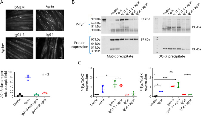
Methods: C2C12 myotubes were exposed to MuSK-MG plasma IgG1-3 or IgG4, with or without purified agrin. MuSK, Downstream of Kinase 7 (DOK7), and βAChR were immunoprecipitated and their phosphorylation levels identified by immunoblotting. Agrin and agrin-independent AChR clusters were measured by immunofluorescence and AChR numbers by binding of 125I-α-bungarotoxin. Transcriptomic analysis was performed on treated myotubes.

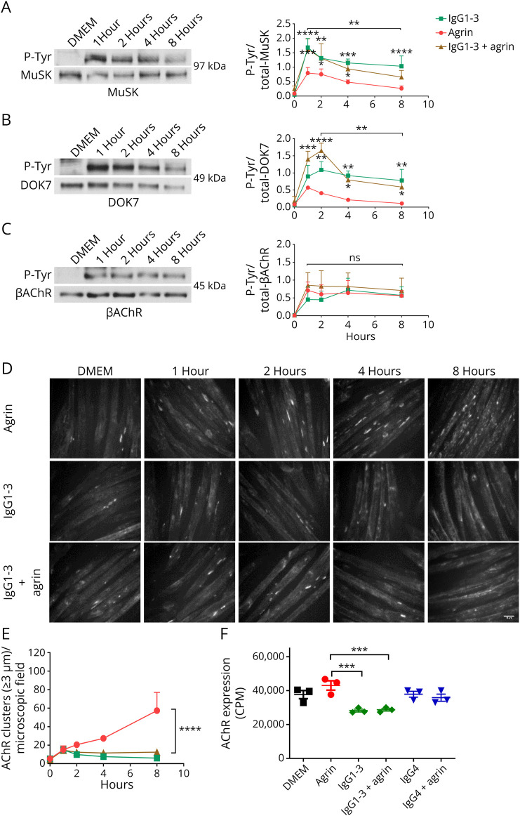
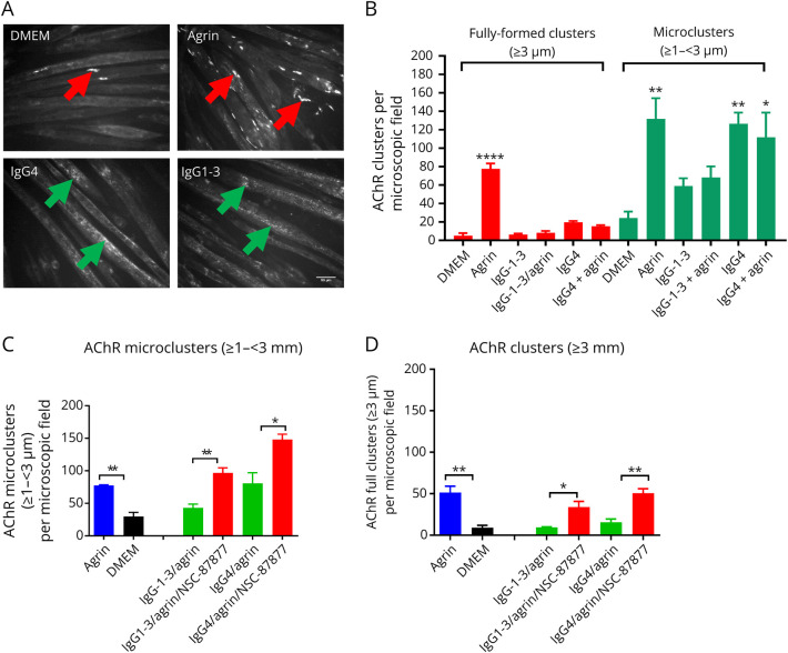
Results: IgG1-3 MuSK-Abs impaired AChR clustering without inhibiting agrin-induced MuSK phosphorylation. Moreover, the well-established pathway initiated by MuSK through DOK7, resulting in βAChR phosphorylation, was not impaired by MuSK-IgG1-3 and was agrin-independent. Nevertheless, the AChR clusters did not form, and both the number of AChR microclusters that precede full cluster formation and the myotube surface AChRs were reduced. Transcriptomic analysis did not throw light on the pathways involved. However, the SHP2 inhibitor, NSC-87877, increased the number of microclusters and led to fully formed AChR clusters.

Discussion: MuSK-IgG1-3 is pathogenic but seems to act through a noncanonical pathway. Further studies should throw light on the mechanisms involved at the neuromuscular junction.


Conflict of interest statement

A. Vincent received a proportion of royalties for a patent held by the University of Oxford for use of MuSK for antibody assays, licensed to Athena Diagnostics, that expired in 2021. The other authors report no relevant disclosures. Go to Neurology.org/NN for full disclosures.

Figures

Figure 1
Figure 1. IgG1-3 and IgG4 Both Inhibit AChR Clustering but Have Opposite Effects on MuSK and DOK7 Phosphorylation
(A) Myotubes were first exposed to MuSK-IgG1-3 and IgG4 subclasses followed 50 mins later by agrin. Agrin alone stimulated AChR clusters, but both IgG fractions inhibited the number of AChR clusters to the levels similar to those in DMEM alone. Scale bar = 50 μm. (B) To explore phosphorylation of MuSK and DOK7 in parallel, myotubes were exposed to MuSK-Abs IgG1-3 for 40 minutes, either with or without agrin. Representative Western blots of immunoprecipitated MuSK showed a strong phosphorylation band for both MuSK (left) and DOK7 (right) irrespective of the presence of agrin, with only faint signal for the IgG4-treated cells. The densities of the phosphorylated MuSK or DOK7 (upper gels) were normalized to those of the MuSK or DOK7 proteins after stripping and reprobing the blots (lower gels). (C) The results pooled from 2 independent experiments; MuSK-IgG1-3 increased both MuSK and DOK7 phosphorylation. One-way ANOVA with multiple comparisons against DMEM. Mean + SD are shown.
Figure 2
Figure 2. Time Course of Phosphorylation of MuSK, DOK7, and βAChR After Agrin or IgG1-3–Treated Myotubes
Myotubes were exposed to agrin alone or MuSK-IgG1-3 with or without agrin, and incubation stopped at different time points. MuSK (A) and DOK7 (B) were immunoprecipitated as for Figure 1, and the βAChR (C) pulled-down by streptavidin-conjugated bungarotoxin. On the left, representative Western blots showing phosphotyrosine bands (top) and corresponding expression (bottom) of MuSK (97 kDa), DOK7 (49 kDa), and βAChR (45 kDa). On the right, pooled results of 2 experiments showing MuSK, DOK7, and βAChR phosphorylation time courses. (D) In parallel, there was increasing AChR clusters over time in agrin alone and reduced clusters in the presence of MuSK-IgG1-3. Scale bar = 50 μm. (E) Summary of the results. (F) Surface AChRs were measured with 125I-α-bungarotoxin after 16 hours incubation with the agrin and MuSK-Abs, showing modest reduction in the presence of IgG1-3 compared with DMEM, agrin or IgG4 MuSK-Abs. Two-way ANOVA for time was used. Mean + SEMs are shown.
Figure 3
Figure 3. AChR Microclusters Are Reduced in IgG1-3–Treated Myotubes
Myotubes were exposed to agrin, MuSK-IgG4, and IgG1-3 for 16 hours, and AChR clusters stained with Alexa Flour 594 α-bungarotoxin. Clusters were counted with ImageJ software using different thresholds for cluster length (≥1 – <3 µm for microclusters; ≥3 µm for fully formed clusters). (A) Representative images of AChR clusters. A few fully formed clusters form spontaneously on the myotube surface in DMEM or IgG4-Abs but, as expected, are increased substantially after agrin exposure (red arrows). By contrast, only AChR microclusters (green arrows) were present in myotubes treated with IgG1-3 or IgG4 MuSK-Abs. In this case, fully formed clusters were rarely detectable. (B) However, the results of 3 experiments showed that the AChR microclusters were reduced by approximately 50% in the presence of IgG1-3 compared with either agrin alone or IgG4-treated cells. Scale bar represents 50 µm. One-Way ANOVA with multiple comparisons against DMEM. (C) SHP2 inhibition by NSC-87877 increased the number of AChR microclusters to levels comparable or greater than agrin alone and (D) also the numbers of fully formed clusters. Two-sided t tests for comparisons at each IgG concentration. One-Way ANOVA with multiple comparisons against DMEM. Mean + SEMs are shown for all experiments.

References

    1. Evoli A, Tonali PA, Padua L, et al. . Clinical correlates with anti-MuSK antibodies in generalized seronegative myasthenia gravis. Brain. 2003;126(10):2304-2311. doi:10.1093/brain/awg223 - DOI - PubMed
    1. Cao M, Koneczny I, Vincent A. Myasthenia gravis with antibodies against muscle specific kinase: an update on clinical features, pathophysiology and treatment. Front Mol Neurosci. 2020;13:159. doi:10.3389/fnmol.2020.00159 - DOI - PMC - PubMed
    1. Wallace BG, Zhican Q, Richard HL. Agrin induces phosphorylation of the nicotinic acetylcholine receptor. Neuron. 1991;6:869-878. doi:10.1016/0896-6273(91)90227-q - DOI - PubMed
    1. DeChiara TM, Bowen DC, Valenzuela DM, et al. . The receptor tyrosine kinase MuSK is required for neuromuscular junction formation in vivo. Cell. 1996;85(4):501-512. doi:10.1016/s0092-8674(00)81251-9 - DOI - PubMed
    1. Zhang B, Luo S, Wang Q, Suzuki T, Xiong WC, Mei L. LRP4 serves as a coreceptor of agrin. Neuron. 2008;60(2):285-297. doi:10.1016/j.neuron.2008.10.006 - DOI - PMC - PubMed

Publication types