Case Reports
. 2023 Aug 15;10(6):e200150.
doi: 10.1212/NXI.0000000000200150. Print 2023 Nov.

Dramatic Response to Anti-IL-6 Receptor Therapy in Children With Life-Threatening Myelin Oligodendrocyte Glycoprotein-Associated Disease

Affiliations
Case Reports

Dramatic Response to Anti-IL-6 Receptor Therapy in Children With Life-Threatening Myelin Oligodendrocyte Glycoprotein-Associated Disease

Loren A McLendon et al. Neurol Neuroimmunol Neuroinflamm. .

Abstract

Objectives: Myelin oligodendrocyte glycoprotein antibody-associated disease (MOGAD) is an immune-mediated neuroinflammatory disorder leading to demyelination of the CNS. Interleukin (IL)-6 receptor blockade is under study in relapsing MOGAD as a preventative strategy, but little is known about the role of such treatment for acute MOGAD attacks.

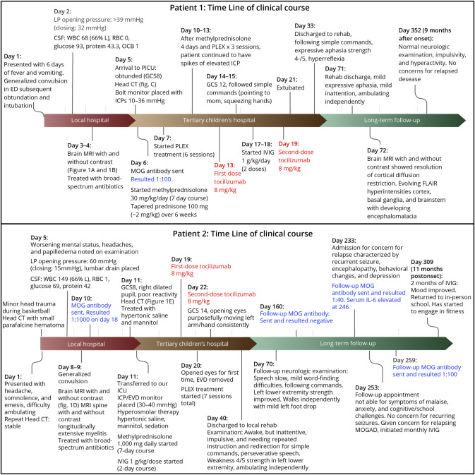
Methods: We discuss the cases of a 7-year-old boy and a 15-year-old adolescent boy with severe acute CNS demyelination and malignant cerebral edema with early brain herniation associated with clearly positive serum titers of MOG-IgG, whose symptoms were incompletely responsive to standard acute therapies (high-dose steroids, IV immunoglobulins (IVIGs), and therapeutic plasma exchange).

Results: Both boys improved quickly with IL-6 receptor inhibition, administered as tocilizumab. Both patients have experienced remarkable neurologic recovery.

Discussion: We propose that IL-6 receptor therapies might also be considered in acute severe life-threatening presentations of MOGAD.


Conflict of interest statement

J.W. Huh was funded by NIH R01NS110898 and R01NS113945 (but no relevance to this report). B. Banwell serves as a consultant to Novartis, Roche, UCB, Glaxo-Smith Kline, Teva Neuroscience, Sanofi-Genzyme, University of Texas Southwestern, and JRD pharmaceuticals. The other authors have no relevant disclosures. Go to Neurology.org/NN for full disclosures.

Figures

Figure 1
Figure 1. Clinical Course for Each Patient
Figure 2
Figure 2. Neuroimaging Features of Malignant MOGAD
Patient 1: (A) MRI performed on day 2 (at outside hospital) demonstrating multifocal areas of restricted diffusion weighted imaging on the left with apparent diffusion coefficient correlate on the right. (B) MRI multifocal areas of T2/FLAIR hyperintensity in cortical and subcortical matter, basal ganglia, and brainstem and mild leptomeningeal enhancement (not pictured). (C) Head computed tomography performed on day 5 showing diffuse cerebral edema with effacement of sulci. (D) MRI on day 39: interval evolution of the multiple extensive cortical and subcortical lesions of both cerebral hemispheres and in the brainstem as detailed. This includes developing encephalomalacia in many of the previously involved regions. The cerebrum has overall, mildly decreased in volume. Patient 2: (E) Brain MRI with T2/FLAIR hyperintensity in the frontotemporal and occipital lobes and bilateral cerebellar peduncles with diffuse leptomeningeal enhancement (not pictured). (F) Head CT obtained on day 11 on arrival to CHOP showing diffuse cerebral edema in supratentorial and infratentorial parenchyma with effacement of sulci, fissures, basal cistern, and cerebellar tonsillar descent. (G) Brain MRI showing partial improvement in T2/FLAIR hyperintensity of lesions involving both cerebral hemispheres, diencephalon, and brainstem. MOGAD = myelin oligodendrocyte glycoprotein antibody-associated disease.
Figure 3
Figure 3. Patient 2 Pathology
(A) A biopsy of the superficial cortex shows scattered neutrophilic inflammation (arrowheads, hematoxylin, and eosin stain, ×200 magnification). (B) Neutrophils within the brain parenchyma are highlighted with myeloperoxidase (myeloperoxidase immunostain, ×200 magnification). (C) Scattered T lymphocytes are present around vessels (arrows) and within the brain parenchyma (CD3 immunostain, ×200 magnification). (D) Reactive microglia are present, often in regions of neutrophilic inflammation; collections of macrophages are not seen (CD68 immunostain, ×200 magnification). (E) Reactive astrocytes within the cortex are highlighted with GFAP (GFAP immunostain, ×200 magnification). (F) A biopsy of the dura shows focal neutrophilic inflammation (arrowheads, hematoxylin, and eosin stain, ×200 magnification). Scale bar in F applies to all images.

References

    1. Banwell B, Bennett JL, Marignier R, et al. . Diagnosis of myelin oligodendrocyte glycoprotein antibody-associated disease: international MOGAD panel proposed criteria. Lancet Neurol. 2023;22(3):268-282. doi:10.1016/s1474-4422(22)00431-8 - DOI - PubMed
    1. de Mol CL, Wong Y, van Pelt ED, et al. . The clinical spectrum and incidence of anti-MOG-associated acquired demyelinating syndromes in children and adults. Mult Scler. 2020;26(7):806-814. doi:10.1177/1352458519845112 - DOI - PMC - PubMed
    1. Fadda G, Armangue T, Hacohen Y, Chitnis T, Banwell B. Paediatric multiple sclerosis and antibody-associated demyelination: clinical, imaging, and biological considerations for diagnosis and care. Lancet Neurol. 2021;20(2):136-149. doi:10.1016/s1474-4422(20)30432-4 - DOI - PubMed
    1. Ogawa R, Nakashima I, Takahashi T, et al. . MOG antibody-positive, benign, unilateral, cerebral cortical encephalitis with epilepsy. Neurol Neuroimmunol Neuroinflamm. 2017;4(2):e322. doi:10.1212/nxi.0000000000000322 - DOI - PMC - PubMed
    1. Fainberg N, Silver M, Arena J, et al. . 547: cerebral edema in MOG antibody disease treated with invasive multimodal. Crit Care Med. 2023;51(1):261. doi:10.1097/01.ccm.0000907916.29570.f1 - DOI

Publication types

Substances