. 2023 Aug 15;28(1):65.
doi: 10.1186/s11658-023-00474-5.

Astrocyte senescence-like response related to peripheral nerve injury-induced neuropathic pain

Affiliations

Astrocyte senescence-like response related to peripheral nerve injury-induced neuropathic pain

Jingyi Du et al. Cell Mol Biol Lett. .

Abstract

Background: Peripheral nerve damage causes neuroinflammation, which plays a critical role in establishing and maintaining neuropathic pain (NeP). The mechanisms contributing to neuroinflammation remain poorly elucidated, and pharmacological strategies for NeP are limited. Thus, in this study, we planned to explore the possible link between astrocyte senescence and NeP disorders following chronic sciatic nerve injury.

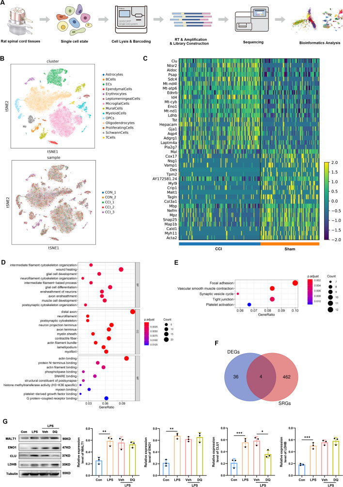
Methods: An NeP animal model was established by inducing chronic constrictive injury (CCI) to the sciatic nerve in adult rats. A senolytic drug combination of dasatinib and quercetin was gavaged daily from the first postoperative day until the end of the study. Paw mechanical withdrawal threshold (PMWT) and paw thermal withdrawal latency (PTWL) were evaluated to assess behaviors in response to pain in the experimental rats. Senescence-associated β-galactosidase staining, western blot analysis, and immunofluorescence were applied to examine the levels of proinflammatory factors and severity of the senescence-like response in the spinal cord. Lipopolysaccharide (LPS) was administered to induce senescence of spinal astrocytes in primary cultures in vitro, to explore the potential impacts of senescence on the secretion of proinflammatory factors. Furthermore, single-cell RNA sequencing (scRNA-seq) was conducted to identify senescence-related molecular responses in spinal astrocytes under neuropathic pain.

Results: Following sciatic nerve CCI, rats exhibited reduced PMWT and PTWL, increased levels of spinal proinflammatory factors, and an enhanced degree of senescence in spinal astrocytes. Treatment with dasatinib and quercetin effectively attenuated spinal neuroinflammation and mitigated the hypersensitivities of the rats subjected to sciatic nerve CCI. Mechanistically, the dasatinib-quercetin combination reversed senescence in LPS-stimulated primary cultured astrocytes and decreased the levels of proinflammatory factors. The scRNA-seq data revealed four potential senescence-related genes in the spinal astrocyte population, and the expression of clusterin (CLU) protein was validated via in vitro experiments.

Conclusion: The findings indicate the potential role of astrocyte senescence in neuroinflammation following peripheral nerve injury, and suggest that targeting CLU activation in astrocytes might provide a novel therapeutic strategy to treat NeP.

Keywords: Astrocyte; Neuroinflammation; Neuropathic pain; Peripheral nerve injury; Senescence.


Conflict of interest statement

The authors declare that the research was conducted in the absence of any commercial or financial relationships that could be construed as a potential competing interests.

Figures

Fig. 1
Fig. 1
Rats experiencing sciatic nerve CCI exhibit mechanical and thermal hypersensitivities, inflammatory cytokine overproduction, and a cellular senescence-like response. A Experimental diagram illustrating sciatic nerve injury and behavior tests. B, C Mechanical allodynia and thermal hyperalgesia developed in the paws of the CCI rats and did not develop in the paws of the sham-operation rats from the first day after CCI until at least day 13 (n = 9). D Western blot images and quantification of IL-1α, IL-1β, TNF-α, IL-6, and IL-8 in the spinal cord after sham intervention or sciatic nerve CCI in rats (n = 3). E Representative photographs of SA-β-gal staining (the positive cells show a blue precipitate) with statistical results (n = 3, scale bars 100 μm). F Western blot images and quantification for senescence-related markers P16 and γ-H2AX in the spinal cord after sham operation or sciatic nerve CCI in rats (n = 3). **P < 0.01, ***P < 0.001
Fig. 2
Fig. 2
Double staining and quantification of P16 in the spinal dorsal horn ipsilateral to sciatic nerve CCI or sham intervention. A Representative double-immunofluorescence staining images show the colocalization of P16 (green) with NeuN (neuronal marker)-, IBA1 (microglia marker)-, or GFAP (astrocyte marker)-positive cells (red). White arrows indicate colabelled cells. B Quantification analyses for the ratio of P16 immunoreactivity area to neuron/astrocyte/microglia marker immunoreactivity area in the spinal cord (n = 3, scale bars 100 μm). ***P < 0.001
Fig. 3
Fig. 3
Double staining and quantification of γ-H2AX in the spinal dorsal horn ipsilateral to sciatic nerve CCI or sham intervention. A Representative double-immunofluorescence staining images show the colocalization of γ-H2AX (green) with NeuN (neuronal marker)-, IBA1 (microglia marker)-, or GFAP (astrocyte marker)-positive cells (red). White arrows indicate colabelled cells. B Quantification analyses for the ratio of γ-H2AX immunoreactivity area to neuron/astrocyte/microglia marker immunoreactivity area in the spinal cord (n = 3, scale bars 100 μm). ***P < 0.001
Fig. 4
Fig. 4
H2O2 and LPS induce senescence and inflammatory cytokine overproduction in primary cultures of spinal astrocytes. A Representative photographs of SA-β-gal staining (the positive cells contain a blue precipitate) and statistical results (n = 3, scale bars 200 μm). B Western blot images and quantification for senescence-related markers P16 and γ-H2AX in primary cultures of spinal astrocytes (n = 3). C Western blot images and quantification for IL-1α, IL-1β, TNF-α, IL-6, and IL-8 in primary cultures of spinal astrocytes (n = 3). *P < 0.05, **P < 0.01, ***P < 0.001
Fig. 5
Fig. 5
Dasatinib and quercetin delay senescence in primary cultures of spinal astrocytes. A Representative photographs of SA-β-gal staining (the positive cells contain a blue precipitate) and statistical results (n = 3, scale bars 100 μm). B Western blot images and quantification for senescence-related markers P16 and γ-H2AX in primary cultures of spinal astrocytes (n = 3). *P < 0.05, **P < 0.01, ***P < 0.001
Fig. 6
Fig. 6
Dasatinib and quercetin reduce levels of inflammatory cytokines in senescent spinal astrocytes in vitro. A Western blot images and quantification of IL-1α, IL-1β, TNF-α, IL-6, and IL-8 in primary cultures of spinal astrocytes (n = 3). B Representative images of double staining and quantification for the coexpression of P16 (red) and inflammatory cytokines (green) in primary cultures of spinal astrocytes (n = 3, scale bars 100 μm). *P < 0.05, **P < 0.01, ***P < 0.001
Fig. 7
Fig. 7
Dasatinib and quercetin inhibit senescence, alleviating CCI-induced pain, astrocyte senescence, and spinal cord inflammation. A, B Dasatinib (5 mg/kg) and quercetin (50 mg/kg) were gavaged to the CCI rats to detect mechanical allodynia and thermal hyperalgesia. The asterisks indicate the statistical significance of differences between the CCI + Veh and CCI + DQ groups (n = 9). C Representative photographs of SA-β-gal staining (the positive cells contain a blue precipitate) and statistical results (n = 3, scale bars 100 μm). D Western blot images and quantification for senescence-related markers P16 and γ-H2AX in the spinal cord of rats (n = 3). E Representative images of double staining and quantification for the coexpression of P16 (green) and GFAP, an astrocyte marker (red), in the spinal dorsal horn ipsilateral to sciatic nerve CCI or sham intervention (n = 3, scale bars 100 μm). F Western blot images and quantification of IL-1α, IL-1β, TNF-α, IL-6, and IL-8 in the spinal cord of rats (n = 3). *P < 0.05, **P < 0.01, ***P < 0.001
Fig. 8
Fig. 8
Single-cell RNA sequencing reveals profiling and transcriptional changes in rat spinal astrocytes in a neuropathic pain model. A Schematic overview of scRNA-seq analysis workflow. B t-distributed scholastic neighbor embedding (t-SNE) plot of single cells profiled in the present work and colored by cell types and samples. C Heatmap displaying the DEGs in spinal astrocytes from sham and CCI rats. D Bar plot displaying the significantly enriched GO terms for the DEGs in spinal astrocytes from rats following sham intervention or CCI. E Bar plot showing the significantly enriched KEGG pathways for the DEGs in spinal astrocytes from rats following sham intervention or CCI. F Venn diagram identifying the intersecting genes between DEGs and SRGs as DE-SRGs. G Western blot images and quantification of DE-SRGs (MALT1, ENO1, CLU, and LDHB) in the primary cultures of spinal astrocytes (n = 3). *P < 0.05, **P < 0.01, ***P < 0.001
Fig. 9
Fig. 9
Mechanistic diagram illustrating the contribution of astrocyte senescence in neuroinflammation and neuropathic pain following peripheral nerve injury

Similar articles

Cited by

References

    1. van Hecke O, Austin SK, Khan RA, Smith BH, Torrance N. Neuropathic pain in the general population: a systematic review of epidemiological studies. Pain. 2014;155:654–662. doi: 10.1016/j.pain.2013.11.013. - DOI - PubMed
    1. Gilron I, Baron R, Jensen T. Neuropathic pain: principles of diagnosis and treatment. Mayo Clin Proc. 2015;90:532–545. doi: 10.1016/j.mayocp.2015.01.018. - DOI - PubMed
    1. Du J, Sun G, Ma H, Xiang P, Guo Y, Deng Y, Li S, Li X. Prevalence and risk factors of anxiety and depression in patients with postherpetic neuralgia: a retrospective study. Dermatology. 2021;237:891–895. doi: 10.1159/000512190. - DOI - PubMed
    1. Hamani C, Fonoff ET, Parravano DC, Silva VA, Galhardoni R, Monaco BA, Navarro J, Yeng LT, Teixeira MJ, de Andrade DC. Motor cortex stimulation for chronic neuropathic pain: results of a double-blind randomized study. Brain. 2021;144:2994–3004. doi: 10.1093/brain/awab189. - DOI - PubMed
    1. Moisset X, Page MG, Pereira B, Choiniere M. Pharmacological treatments of neuropathic pain: real-life comparisons using propensity score matching. Pain. 2022;163:964–974. doi: 10.1097/j.pain.0000000000002461. - DOI - PubMed