. 2023 Jul 31:10:1198020.
doi: 10.3389/fcvm.2023.1198020. eCollection 2023.

Fibrin deposition on bovine pericardium tissue used for bioprosthetic heart valve drives its calcification

Affiliations

Fibrin deposition on bovine pericardium tissue used for bioprosthetic heart valve drives its calcification

Bastien Poitier et al. Front Cardiovasc Med. .

Abstract

Background: Bioprosthetic heart valves (BHVs) are less thrombogenic than mechanical prostheses; however, BHV thrombosis has been proposed as a risk factor for premature BHV degeneration.

Objectives: We aimed to explore whether fibrin deposition on bovine pericardium tissue could lead to calcification.

Method: Fibrin clot was obtained by blending three reagents, namely, CRYOcheck™ Pooled Normal Plasma (4/6), tissue factor + phospholipids (Thrombinoscope BV), and 100 mM calcium (1/6), and deposited on pericardium discs. Non-treated and fibrin-treated bovine pericardium discs were inserted into the subcutaneous tissue of 12-day-old Wistar rats and sequentially explanted on days 5, 10, and 15. Calcium content was measured with acetylene flame atomic absorption spectrophotometry. Histological analysis was performed using hematoxylin-eosin staining, Von Kossa staining, and immunohistochemistry.

Results: Calcification levels were significantly higher in fibrin-treated bovine pericardium discs compared to those in non-treated bovine pericardium discs (27.45 ± 23.05 µg/mg vs. 6.34 ± 6.03 µg/mg on day 5, 64.34 ± 27.12 µg/mg vs. 34.21 ± 19.11 µg/mg on day 10, and 64.34 ± 27.12 µg/mg vs. 35.65 ± 17.84 µg/mg on day 15; p < 0.001). Von Kossa staining confirmed this finding. In hematoxylin-eosin staining, the bovine pericardium discs were more extensively and deeply colonized by inflammatory-like cells, particularly T lymphocytes (CD3+ cells), when pretreated with fibrin.

Conclusion: Fibrin deposition on bovine pericardium tissue treated with glutaraldehyde, used for BHV, led to increased calcification in a rat model. BHV thrombosis could be one of the triggers for calcification and BHV deterioration.

Keywords: bioprosthetic heart valve; calcification; fibrin; pericardium tissue; thrombosis.


Conflict of interest statement

The authors declare that the research was conducted in the absence of any commercial or financial relationships that could be construed as a potential conflict of interest.

Figures
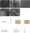
Figure 1
Figure 1
Fibrin deposition on bovine pericardium: preparation, implantation, and scanning electron microscopy analysis. (A) Non-treated pericardium tissue surface (magnification = ×600; scale bars = 20 μm). (B) Fibrin-treated pericardium tissue surface with 10 µl of fibrin solution (magnification = ×3,000; scale bars = 5 μm). (C) Fibrin-treated pericardium tissue surface with 20 µl of fibrin solution (magnification = ×600; scale bars = 20 μm). (D) Fibrin-treated pericardium tissue surface with 20 µl of fibrin solution (magnification = ×3,000; scale bars = 5 μm). (E) fibrin-treated pericardium tissue surface with 50 µl of fibrin solution (magnification = ×3,000; scale bars = 5 μm). (F) Protocol for pericardium tissue preparation and implantation, after washing.
Figure 2
Figure 2
Dosage of tissue calcium levels between fibrin-treated and non-treated bovine pericardium tissues after subcutaneous implantation in rats for 15 days.
Figure 3
Figure 3
Panel of histological analyses. Hematoxylin–eosin staining for (A) non-treated pericardium tissue at day 5, (B) non-treated pericardium tissue at day 15, (C) fibrin-treated pericardium tissue at day 5, and (D) fibrin-treated pericardium tissue at day 15. Von Kossa staining for (E) non-treated pericardium tissue at day 5, (F) non-treated pericardium tissue at day 15, (G) fibrin-treated pericardium tissue at day 5, and (H) fibrin-treated pericardium tissue at day 15.
Figure 4
Figure 4
IHC analysis with anti-CD3. (A) Non-treated pericardium tissue at day 15 (magnification = ×20; scale bars = 50 μm). (B) Fibrin-treated pericardium tissue at day 15 (magnification = ×20; scale bars = 50 μm). (C) Quantification of CD3+ cells in implanted pericardium treated or not treated with fibrin.

References

    1. Iung B, Vahanian A. Epidemiology of valvular heart disease in the adult. Nat Rev Cardiol. (2011) 8:162–72. 10.1038/nrcardio.2010.202 - DOI - PubMed
    1. Carpentier A. Lasker clinical research award. The surprising rise of nonthrombogenic valvular surgery. Nat Med. (2007) 13:1165–8. 10.1038/nm1645 - DOI - PubMed
    1. Sun JCJ, Davidson MJ, Lamy A, Eikelboom JW. Antithrombotic management of patients with prosthetic heart valves: current evidence and future trends. Lancet Lond Engl. (2009) 374:565–76. 10.1016/S0140-6736(09)60780-7 - DOI - PubMed
    1. Richez U, De Castilla H, Guerin CL, Gendron N, Luraghi G, Grimme M, et al. Hemocompatibility and safety of the Carmat Total Artifical Heart hybrid membrane. Heliyon. (2019) 5:e02914. 10.1016/j.heliyon.2019.e02914 - DOI - PMC - PubMed
    1. Jiang Z, Wu Z, Deng D, Li J, Qi X, Song M, et al. Improved cytocompatibility and reduced calcification of glutaraldehyde-crosslinked bovine pericardium by modification with glutathione. Front Bioeng Biotechnol. (2022) 10:844010. 10.3389/fbioe.2022.844010 - DOI - PMC - PubMed

LinkOut - more resources