. 2023 Dec;12(2):2249120.
doi: 10.1080/22221751.2023.2249120.

Identification of TMEM53 as a novel SADS-CoV restriction factor that targets viral RNA-dependent RNA polymerase

Affiliations

Identification of TMEM53 as a novel SADS-CoV restriction factor that targets viral RNA-dependent RNA polymerase

Yu-Lin Yao et al. Emerg Microbes Infect. 2023 Dec.

Abstract

ABSTRACTZoonotic transmission of coronaviruses (CoVs) poses a serious public health threat. Swine acute diarrhea syndrome coronavirus (SADS-CoV), originating from a bat HKU2-related CoV, causes devastating swine diseases and poses a high risk of spillover to humans. Currently, licensed therapeutics that can prevent potential human outbreaks are unavailable. Identifying the cellular proteins that restrict viral infection is imperative for developing effective interventions and therapeutics. We utilized a large-scale human cDNA screening and identified transmembrane protein 53 (TMEM53) as a novel cell-intrinsic SADS-CoV restriction factor. The inhibitory effect of TMEM53 on SADS-CoV infection was found to be independent of canonical type I interferon responses. Instead, TMEM53 interacts with non-structural protein 12 (NSP12) and disrupts viral RNA-dependent RNA polymerase (RdRp) complex assembly by interrupting NSP8-NSP12 interaction, thus suppressing viral RdRp activity and RNA synthesis. Deleting the transmembrane domain of TMEM53 resulted in the abrogation of TMEM53-NSP12 interaction and TMEM53 antiviral activity. Importantly, TMEM53 exhibited broad antiviral activity against multiple HKU2-related CoVs. Our findings reveal a novel role of TMEM53 in SADS-CoV restriction and pave the way to host-directed therapeutics against HKU2-related CoV infection.

Keywords: HKU2-related CoVs; RdRp activity; SADS-CoV; TMEM53; coronavirus zoonosis.


Conflict of interest statement

No potential conflict of interest was reported by the author(s).

Figures
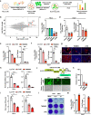
Figure 1.
Figure 1.
Identification and validation of TMEM53 as an antiviral factor against SADS-CoV. (a) Schematic of cDNA library screen. (b) Screen results from HeLa cells infected with SADS-CoV. Genes with a log2 (fold change) > 2 and reads count > 104 were highlighted in red. (c) Result validation by transient overexpression of FLAG-tagged CD14, CLU, TMEM53 and LY6E in HeLa cells followed by SADS-CoV infection (MOI = 0.1). At 48 h post-infection (hpi), the viral RNA in the supernatant was measured using RT-qPCR targeting at the SADS-CoV NSP12 gene (Table S1). (d) TMEM53 overexpression inhibited SADS-CoV infection (MOI = 0.1) in Huh7 cells. At 48 hpi, the viral RNA in the supernatant (left) and cytoplasm (right) were measured by RT-qPCR. LY6E was used as a positive control in the subsequent experiments. (e-f) Anti-SADS-CoV activity in TMEM53 stably expressing HeLa (HeLa-TMEM53) and Huh7 cells (Huh7-TMEM53). Cells were infected with SADS-CoV at a MOI of 0.1. At 48 hpi, the viral RNA in the supernatant and cytoplasm were determined by RT-qPCR. Cells transduced with lentiviral empty vector were used as control (Ctrl) in the subsequent assays unless otherwise stated. (g) HeLa-TMEM53 and Huh7-TMEM53 cells were infected with SADS-CoV (MOI = 0.1). NP proteins were assessed by immunofluorescence microscopy at 48 h post-infection. (h) Statistical analysis of Figure 1g. The normalized mean fluorescence intensity of NP protein was quantified with Image J software. N = 5 panels per group. (i) The infectious virions secreted from the HeLa– or Huh7– expressing LY6E or TMEM53 and control cells were evaluated by 50% tissue culture infectious dose (TCID50) assays in Huh7 cells. (j-k) TMEM53 inhibited the reporter virus SADS-CoV-mNG infection. Huh7-TMEM53 cells were infected with SADS-CoV-mNG (MOI = 0.1). mNG-positive cells were assessed by immunofluorescence microscopy (j) and flow cytometry (k) at 48 hpi. (l) Crystal violet staining of polyclonal cell populations infected with SADS-CoV (MOI = 0.1) in TMEM53 or LY6E stably expressing Huh7 cells at 48 hpi. (m) TMEM53 knockout promoted SADS-CoV infection in Huh7 cells. Wild type (WT) Huh7 cells and Huh7-TMEM53-KO (KO) cells were infected with SADS-CoV (MOI = 0.1) for 48 h. The viral RNA in the supernatant and cytoplasm were measured by RT-qPCR. All data were representative of three independent experiments with similar results. Data were presented as mean ± SD. *P < 0.05, **P < 0.01, ***P < 0.001, ****P < 0.0001, two-tailed unpaired Student t test.
Figure 2.
Figure 2.
TMEM53 inhibited viral RNA synthesis independent of interferon response. (a-b) Huh7-TMEM53, Huh7-LY6E or control cells were infected with SADS-CoV at a MOI of 5. Viral particles attached to the cell surface or internalized were assessed by RT-qPCR. (c) SADS-CoV growth kinetics in Huh7-TMEM53 cells. Control cells and Huh7-TMEM53 were infected with SADS-CoV (MOI = 0.1), and viral RNA in the supernatant was measured by RT-qPCR at the indicated time points. (d) TMEM53 inhibited dsRNA production. Huh7-TMEM53 and control cells were infected with SADS-CoV (MOI = 1), and dsRNA was detected at 48 hpi by immunofluorescence microscopy. Nuclei were stained with blue staining (DAPI). Scale bar, 10 μm. (e-f) Control and Huh7-TMEM53 cells were infected with SADS-CoV (MOI = 0.1), and intracellular + vRNA, – vRNA and sgRNA were measured by RT-qPCR at the indicated time. (g) TMEM53 expression inhibited SADS-CoV replicon activity. Control and Huh7-TMEM53 or Huh7-LY6E Cells were transfected with SADS-CoV replicon and Nluc activity was quantified at the indicated time. (h-k) TMEM53 induction upon SADS-CoV infection and interferon treatment. Huh7 cells were infected with SADS-CoV (MOI = 0.1) (h) or treated with universal type I IFN (500U/ml) (i-k). Cells were harvested at the indicated time and the mRNA expression levels were determined by RT-qPCR. (l) STAT1-/ – Huh7 cells transduced with TMEM53 were infected with SADS-CoV (MOI = 0.05) for 48 h. The viral RNA in the supernatant was analyzed by RT-qPCR. The experiments were independently repeated three times with similar results. Values were presented as mean ± SD. ns: non-significant, *P < 0.05, **P < 0.01, ***P < 0.001, ****P < 0.0001, two-tailed unpaired Student t test.
Figure 3.
Figure 3.
TMEM53 suppressed SADS-CoV RdRp activity. (a) Schematic of RNA immunoprecipitation. (b) Quantifying SADS-CoV RNA in TMEM53, MDA5, and IgG immunoprecipitates by RT-qPCR. FLAG-tagged TMEM53 and MDA5 in IP were shown by western blot. (c) Schematic diagram of the cell-based SADS-CoV RdRp activity assay. (d) The RdRp activity in the cell-based assay system. Huh7 cells were cotransfected with RdRp reporter plasmids and NSP12-HA for 24 or 48 h. The RdRp activity was determined by measuring the Nluc and Fluc values, and a ratio of Nluc/Fluc reflecting RdRp activity was shown. (e) Expression of NSP7 and NSP8 increased RdRp activity. Huh7 cells were co-transfected with RdRp reporter plasmids, the NSP12-HA, NSP7-HA and NSP8-HA. The Nluc and Fluc values were measured at the indicated time points. (f) TMEM53 inhibited SADS-CoV RdRp activity. Control and Huh7-TMEM53 cells were co-transfected with RdRp reporter and RdRp plasmids for 24 h or 48 h before measuring the RdRp activity. All data were representative or presented as the mean ± SD of three independent experiments. ns: non-significant, **P < 0.01, ***P < 0.001, ****P < 0.0001, two-tailed unpaired Student t test.
Figure 4.
Figure 4.
TMEM53 disrupted NSP8 and NSP12 interactions. (a-c) TMEM53 interacted with NSP12 and NSP8. HEK293T cells were transfected with TMEM53-FLAG and NSP12-HA, NSP8-HA or NSP7-HA for 48 h before harvesting for immunoprecipitation analysis. IgG was used as a control. IgG-L, IgG L chain. (d) HeLa cells were co-transfected with TMEM53-FLAG and NSP12-HA, NSP8-HA or NSP7-HA for 48 h. The colocalization of TMEM53 with NSP12, NSP8 or NSP7 was analyzed by immunofluorescence microscopy. Scale bar, 10 μm. (e) The interactions between TMEM53 and NSP12, NSP8 or NSP7 was revealed by PLA in HeLa cells. The number of PLA red fluorescent dots was counted with Image J software (right panel). N = 5 panels per group. Scale bar, 10 μm. (f) Illustration depicting truncations of NSP12. (g) Domain mapping of TMEM53 for interaction with NSP12 in HEK293T cells. The procedure was the same as in a-c. IgG-H, IgG H chain. (h) The interaction between NSP8 and NSP12 was verified by Co-IP in HEK293T cells. (i) TMEM53 impaired the recruitment of NSP8 to NSP12. HEK293T cells were co-transfected with NSP8-FLAG, NSP12-HA and TMEM53-S-tag or empty vector for 48 h before the immunoprecipitation assays. (j-k) HeLa cells were co-transfected with NSP8-FLAG, NSP12-HA and TMEM53-S-tag or empty vector for 48 h. Cells were fixed and analyzed by PLA assay (j). Scale bar, 10 μm. The number of red fluorescent dots in each panel was quantified with Image J software (k). The PLA controls are shown in Supplementary Figure S6c. The experiments were independently repeated three times with similar results. Values were presented as mean ± SD. ****P < 0.0001, two-tailed unpaired Student t test.
Figure 5.
Figure 5.
TM domain is critical for the antiviral activity of TMEM53. (a) Illustration depicting truncations of TMEM53. (b-c) Huh7 cells stably expressing TMEM53-FL or TMEM53-ΔTM were infected with SADS-CoV (MOI = 0.1) for 48 h, and the viral RNA in the supernatant (b) and cytoplasm (c) were determined by RT-qPCR. (d) TMEM53 colocalized with ER and Golgi apparatus. HeLa cells were co-transfected with FLAG-tagged TMEM53 and SecG1β-GFP (ER marker) or B4Gal-Ti-RFP (Golgi apparatus marker) for 48 h, respectively. Cells were harvested and visualized by immunofluorescence microscopy. DAPI was used for nuclei detection. (e) Colocalization of TMEM53-ΔTM with ER and Golgi apparatus. The procedure was the same as in (d). (f-g) The interaction between TMEM53-ΔTM and NSP12 (f) or NSP8 (g) in HEK293T cells. (h) TMEM53-ΔTM did not affect NSP8 and NSP12 interactions. HEK293T cells were co-transfected TMEM53-ΔTM with NSP8 and NSP12 plasmids for 48 h before harvesting for Co-IP assay. (i) TMEM53-ΔTM did not affect SADS-CoV RdRp activity. Control and Huh7– TMEM53-ΔTM cells were co-transfected with RdRp reporter plasmids and RdRp plasmids for 24 h or 48 h before measuring the RdRp activity. The data shown are representative of three independent experiments with similar results. Data were presented as mean ± SD. ns: non-significant, **P < 0.01, two-tailed unpaired Student t test.
Figure 6.
Figure 6.
TMEM53 inhibited the HKU2r-CoVs replication. (a) Huh7 cells expressing TMEM53 were infected with SADS-CoV (MOI = 0.1), WIV (MOI = 0.1), SARS-CoV-2 (MOI = 0.05), HCoV-OC43 (MOI = 0.5) and HCoV-229E (MOI = 0.5) for 48 h before harvesting for further analysis. The viral RNA was measured by RT-qPCR. (b) TMEM53 did not affect the replicon activity of MERS-CoV. Control cells and Huh7-TMEM53 cells were transfected with MERS-CoV replicon plasmids for 48 h before harvesting for Nluc value determination. (c-d) TMEM53-Huh7 cells were infected with HSV-1 (MOI = 1) and PR8 (MOI = 1) for 48 h before the harvest. The viral RNA was determined by RT-qPCR. (e-f) TMEM53 inhibited the replicon activity of bat 162140-CoV and HKU2-CoV. Huh7-TMEM53 cells were transfected with replicon plasmids for 162140-CoV or HKU2-CoV. Cells were harvested and the Nluc value was analyzed at the indicated time points. (g) Sequence identity of the NSP12 protein in coronaviruses. (h) TMEM53 interacted with NSP12 protein from HKU2-related CoVs but not SARS-CoV-2. HEK293T cells were co-transfected TMEM53-FLAG and the indicated HA-tagged NSP12 plasmids for 48 h. Cells were harvested for Co-IP assay.

References

    1. Chen B, Tian EK, He B, et al. . Overview of lethal human coronaviruses. Signal Transduct Target Ther. 2020 Jun 10;5(1):89, doi:10.1038/s41392-020-0190-2. - DOI - PMC - PubMed
    1. V'Kovski P, Kratzel A, Steiner S, et al. . Coronavirus biology and replication: implications for SARS-CoV-2. Nat Rev Microbiol. 2021 Mar;19(3):155–170. doi:10.1038/s41579-020-00468-6. - DOI - PMC - PubMed
    1. Baker RE, Mahmud AS, Miller IF, et al. . Infectious disease in an era of global change. Nat Rev Microbiol. 2022 Apr;20(4):193–205. doi:10.1038/s41579-021-00639-z. - DOI - PMC - PubMed
    1. Zhou P, Fan H, Lan T, et al. . Fatal swine acute diarrhoea syndrome caused by an HKU2-related coronavirus of bat origin. Nature. 2018 Apr;556(7700):255–258. - PMC - PubMed
    1. Yang YL, Yu JQ, Huang YW.. Swine enteric alphacoronavirus (swine acute diarrhea syndrome coronavirus): an update three years after its discovery. Virus Res. 2020 Aug;285:198024. - PMC - PubMed

Supplementary concepts