Review
. 2023 Aug 29;52(17):6191-6220.
doi: 10.1039/d3cs00202k.

Perspectives on recent advancements in energy harvesting, sensing and bio-medical applications of piezoelectric gels

Affiliations
Review

Perspectives on recent advancements in energy harvesting, sensing and bio-medical applications of piezoelectric gels

Thangavel Vijayakanth et al. Chem Soc Rev. .

Abstract

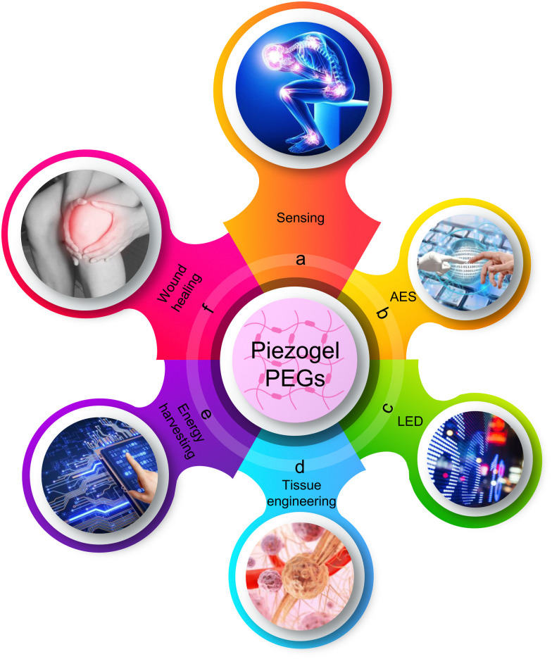
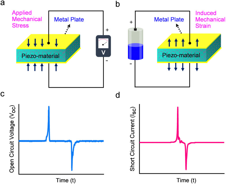
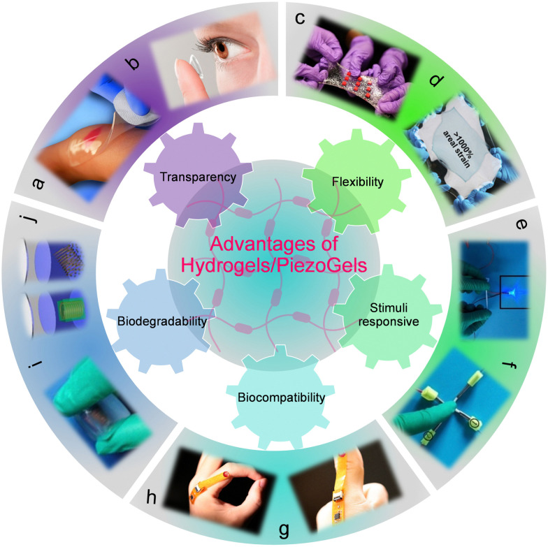
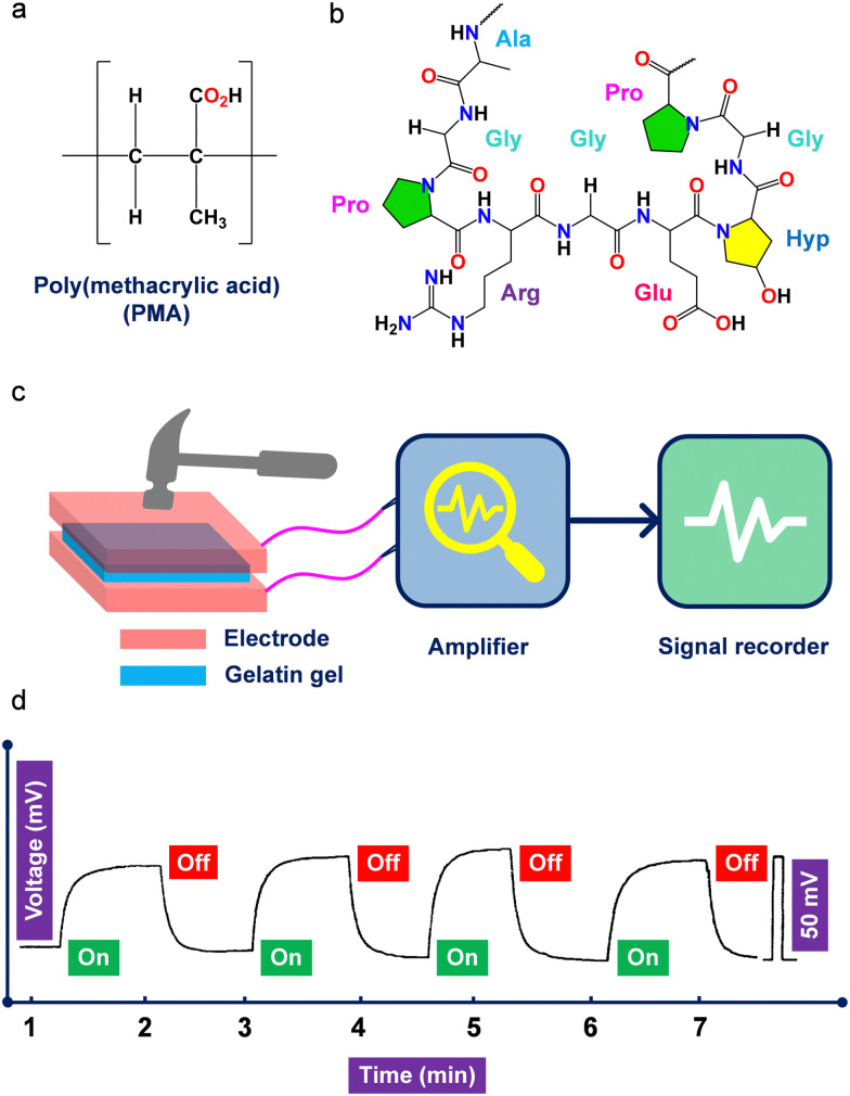
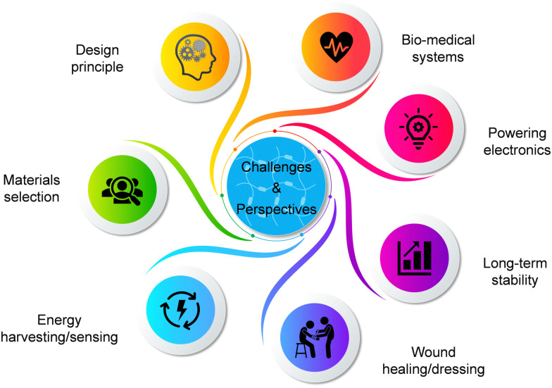
The development of next-generation bioelectronics, as well as the powering of consumer and medical devices, require power sources that are soft, flexible, extensible, and even biocompatible. Traditional energy storage devices (typically, batteries and supercapacitors) are rigid, unrecyclable, offer short-lifetime, contain hazardous chemicals and possess poor biocompatibility, hindering their utilization in wearable electronics. Therefore, there is a genuine unmet need for a new generation of innovative energy-harvesting materials that are soft, flexible, bio-compatible, and bio-degradable. Piezoelectric gels or PiezoGels are a smart crystalline form of gels with polar ordered structures that belongs to the broader family of piezoelectric material, which generate electricity in response to mechanical stress or deformation. Given that PiezoGels are structurally similar to hydrogels, they offer several advantages including intrinsic chirality, crystallinity, degree of ordered structures, mechanical flexibility, biocompatibility, and biodegradability, emphasizing their potential applications ranging from power generation to bio-medical applications. Herein, we describe recent examples of new functional PiezoGel materials employed for energy harvesting, sensing, and wound dressing applications. First, this review focuses on the principles of piezoelectric generators (PEGs) and the advantages of using hydrogels as PiezoGels in energy and biomedical applications. Next, we provide a detailed discussion on the preparation, functionalization, and fabrication of PiezoGel-PEGs (P-PEGs) for the applications of energy harvesting, sensing and wound healing/dressing. Finally, this review concludes with a discussion of the current challenges and future directions of P-PEGs.


Conflict of interest statement

There are no conflicts to declare.

Figures

Fig. 1
Fig. 1. Schematic diagram showing the development of P-PEGs. The main applications of P-PEGs are covered in this review based on the coupling of flexible electronics and piezoelectric properties. The diverse applications of P-PEGs showing sensing (a, top panel), artificial electronic skin (AES) (b, upper right panel), light-emitting diode (LED) devices (c, lower right panel), tissue engineering (d, bottom panel), energy harvesting (e, lower left panel) and wound healing (f, upper left panel).
Fig. 2
Fig. 2. Fundamentals of PEGs. (a) and (b) Schematic representation displaying the working mechanism of (a) direct and (b) converse piezoelectric effect. (c) and (d) Typical piezoelectric output (c) voltage and (d) current curves.
Fig. 3
Fig. 3. Advantages of hydrogels/PiezoGels in energy and biomedical applications. (a) Photograph of the polyvinylpyrrolidone (PVP), polyethylene oxide (PEO) and agar-based transparent hydrogel. Reproduced with permission. Copyright © 2007, HMP Communications, LLC. (b) Photograph of a contact lens-based transparent hydrogel. Reproduced with permission. Copyright © 2023, Wigram & Ware Opticians. (c) and (d) Photograph of an extremely tough, stretchable and flexible hydrogel elastomer. Reproduced with permission. Copyright © 2017, American Association for the Advancement of Science. (e) and (f) Photograph of the stimuli-responsive PVA-soluble starch composite gel. Reproduced with permission. Copyright © 2021, WILEY-VCH Verlag GmbH & Co. KGaA, Weinheim. (g) and (h) Photograph of a bio-compatible and wearable sensor based on DNA hydrogel. Reproduced with permission. Copyright © 2021, American Association for the Advancement of Science. (i) and (j) Optical images of a flexible and bio-degradable bacterial cellulose-neodymium magnet (BC-NdFeB) composite material. Reproduced with permission. Copyright © 2022, Elsevier Ltd.
Fig. 4
Fig. 4. (a) Chemical structures of poly(methacrylic acid) and (b) gelatin polymers used to develop P-PEGs. (c) Schematic representation showing the instrument used for the piezoelectric measurement of gelatin hydrogel. (d) Piezoelectric output voltage was observed using the gelatin hydrogel device under an applied load of 9.8 N. Reproduced with permission. Copyright © 1996, WILEY-VCH Verlag GmbH & Co. KGaA, Weinheim.
Fig. 5
Fig. 5. (a)–(e) Schematic representation of the formation of composite PAN-PVDF hydrogels. Acrylonitrile, AN; sodium p-styrenesulfonate, NaSS; methylene-bis-acrylamide, MBA; ammonium persulfate, APS; dimethyl sulfoxide, DMSO; water, H2O; polyacrylonitrile, PAN; polyvinylidene fluoride, PVDF. (f) and (g) Piezoelectric sensor applications of the PAN-PVDF composite hydrogel showing (f) the optical image of the sensor and (g) schematic diagram of the fabricated piezoelectric PAN-PVDF composite hydrogel. (h) Piezoelectric output voltage was observed from the device by sensing various bending angles (0°–90°). (i) Optical photo of vocal cord vibration detection. (j) and (k) Output voltage signals detection graphs for speaking different words (Hi, hobby, respectively). Reproduced with permission. Copyright © 2019, American Chemical Society.
Fig. 6
Fig. 6. (a) Schematic representation of 3D printed piezoelectric wound dressing model of ZnO NPs modified PVDF-SA piezoelectric hydrogel scaffold (ZPFSA). (b) and (c) Piezoelectric response of displaying output currents of ZPFSA (0.5) PiezoGel scaffold. (b) Simple frictional action. (c) Frictional action during swelling. (d) Wound healing rate of different piezoelectric scaffolds showing the efficacy of wound healing. (i) control; (ii) SA; (iii) ZPFSA 0; (iv) ZPFSA (0.5), * denotes p < 0.05, ** denotes p < 0.01. Reproduced with permission. Copyright © 2022, American Chemical Society.
Fig. 7
Fig. 7. (a) Schematic representation of the formation mechanism of a PVA/PVDF composite PiezoGel and its wound healing advancement mechanism in diabetic rats. (b) Representative pictures of wounds in diabetic rats treated with control, neat PVA and PVA–PVDF composite PiezoGel at timepoint 0 and after 14 days. (c) and (d) Descriptive statistics of (c) the width of granulation tissue (WGT) and (d) epithelium gap (EG) based on hematoxylin and eosin (H & E) staining assays. Reproduced with permission. Copyright © 2022, American Chemical Society.
Fig. 8
Fig. 8. (a) Schematic representation of polarization alignment under load force application. (b) Piezoelectric output voltage of the PAAN-PVDF hydrogels under an applied mechanical pressure of 40 N. (c)–(e) In vitro biocompatibility tests of 15% PAAN-PVDF hydrogels using L929 cells. (c and d) Live/dead staining images. (e) Confocal scanning microscopy images following immunohistochemistry staining using an anti-Actin antibody. (f and g) In vitro angiogenesis of human umbilical vein endothelial cells (HUVECs) demonstrating the formation of HUVEC tubular structures after 12 h. (h) The number of junctions of vascular tube formation (*p < 0.05, **p < 0.01,). Reproduced with permission. Copyright © 2022, American Chemical Society.
Fig. 9
Fig. 9. (a)–(c) Schematic structures of (a) BTO, (b) PVP, and (c) PEO. (d) Piezoelectric output voltage of poled PVP-PEO (80 : 20)-(15 wt%) (left) and PVP-PEO (80 : 20)-(15 wt%) (right) containing 1 wt% and 3 wt% of BTO-NPs. The experimental parameters used in this study were 1 N, 3 Hz, and 1 sec of mechanical stimulation, applied frequency, and time, respectively. Reproduced with permission. Copyright © 2022, Elsevier Ltd.
Fig. 10
Fig. 10. (a) Schematic illustration of stretchable and biocompatible Gel-OCS-ABTO composite hydrogels. (b) and (c) Piezoelectric energy harvesting performance of (b) output voltage as a function of different ABTO concentration under a constant stress of 4.46 kPa and (c) output voltage as a function of different applied stress levels. (e) Photographic images and piezoelectric signals of the Gel-OCS-ABTO hydrogel under different finger gesture operations. Reproduced with permission. Copyright © 2023, Elsevier Ltd.
Fig. 11
Fig. 11. (a) Schematic illustration of the fabrication and application of the piezoelectric hydrogel patch for wound healing. (b)–(d) Real-time imaging of (b) the UV-assisted 3D printing, (c) digital, and (d) optical images of the 3D-printed PiezoGel patch. (e) Statistical analysis of the number of capillary-like structures of HUVECs cultured in VEGF-patch. (f) The relative wound area curve from day 0 to day 10. Reproduced with permission. Copyright © 2023, American Association for the Advancement of Science.
Fig. 12
Fig. 12. (a) Molecular structure and (b) crystal-packing higher-order structures of DH are depicted along the crystallographic (100) plane. (c) Representation of dabco cations illustrates parallel bistable hydrogen-bonded interactions mediated by NH⋯N bonds. To allow structural clarity, the anions ReO4 were removed. (d)–(f) PUU-DH-2 optical images showing various deformations. (g) Stress–strain curves of pristine PUU gel and PUU-DH piezoelectric gel composites. (h) Piezoelectric output voltages of neat PUU and PUU-DH composite gels by applying a mechanical pressure and frequency of 25.5 kPa and 6 Hz, respectively. (i) Piezoelectric composite gel (PUU-DH-2) providing power for LED light-up via finger tapping. Reproduced with permission. Copyright © 2021, WILEY-VCH Verlag GmbH & Co. KGaA, Weinheim.
Fig. 13
Fig. 13. (a) Crystal packing diagram and (b) formation of three-dimensional higher-order structures via hydrogen-bonded interactions of Im-ClO4. The ClO4 anion shows ordered structures with four oxygen atoms occupying four vortexes of the tetrahedron, resulting in the loss of the inversion center. (c) The piezoelectric output voltage of the hybrid sensor under diverse out-of-plane mechanical pressures of 0.2, 3.75, 7.5, and 31.25 kPa. (d) Photographs of BC membrane displaying the degradation process under various time intervals. The BC membrane is completely degraded within four hours in a cellulase solution. Reproduced with permission. Copyright © 2019, American Chemical Society.
Fig. 14
Fig. 14. (a) Chemical structure of Fmoc-FF and (b) higher-order crystal packing diagram showing β-sheet hydrogen-bonded structures packed along the crystallographic ‘b’ axis (CCDC: 1027570). (c) and (d) Lateral piezoresponse force microscopy (L-PFM) analysis showing (c) amplitude and (d) phase images. (e) Representation of L-PFM amplitude curve as a function of the applied voltage. The slopes are the average local piezoelectric coefficient and piezoresponse signal for a nonpiezoelectric glass slide (red). Reproduced with permission. Copyright © 2015, American Chemical Society.
Fig. 15
Fig. 15. Challenges and future perspectives of P-PEGs.
None
Thangavel Vijayakanth
None
Sudha Shankar
None
Gal Finkelstein-Zuta
None
Sigal Rencus-Lazar
None
Sharon Gilead
None
Ehud Gazit

Similar articles

Cited by

References

    1. Cima M. J. Nat. Biotechnol. 2014;32:642–643. doi: 10.1038/nbt.2952. - DOI - PubMed
    1. Chen G. Li Y. Bick M. Chen J. Chem. Rev. 2020;120:3668–3720. doi: 10.1021/acs.chemrev.9b00821. - DOI - PubMed
    1. Kim H. Pyun K. R. Lee M. T. Lee H. B. Ko S. H. Adv. Funct. Mater. 2022;32:2110535. doi: 10.1002/adfm.202110535. - DOI
    1. Torralba A. S. Álvarez-Diduk R. Parolo C. Piper A. Merkoçi A. Chem. Rev. 2022;122:14881–14910. doi: 10.1021/acs.chemrev.1c01012. - DOI - PMC - PubMed
    1. Veletic M. Apu E. H. Simic M. Bergsland J. Balasingham I. Contag C. H. Ashammakhi N. Chem. Rev. 2022;122:16329–16363. doi: 10.1021/acs.chemrev.2c00005. - DOI - PubMed