. 2023 Aug 16;15(709):eabm5755.
doi: 10.1126/scitranslmed.abm5755. Epub 2023 Aug 16.

Endothelial FoxM1 reactivates aging-impaired endothelial regeneration for vascular repair and resolution of inflammatory lung injury

Affiliations

Endothelial FoxM1 reactivates aging-impaired endothelial regeneration for vascular repair and resolution of inflammatory lung injury

Xiaojia Huang et al. Sci Transl Med. .

Abstract

Aging is a major risk factor of high incidence and increased mortality of acute respiratory distress syndrome (ARDS). Here, we demonstrated that persistent lung injury and high mortality in aged mice after sepsis challenge were attributable to impaired endothelial regeneration and vascular repair. Genetic lineage tracing study showed that endothelial regeneration after sepsis-induced vascular injury was mediated by lung resident endothelial proliferation in young adult mice, whereas this intrinsic regenerative program was impaired in aged mice. Expression of forkhead box M1 (FoxM1), an important mediator of endothelial regeneration in young mice, was not induced in lungs of aged mice. Transgenic FOXM1 expression or in vivo endothelium-targeted nanoparticle delivery of the FOXM1 gene driven by an endothelial cell (EC)-specific promoter reactivated endothelial regeneration, normalized vascular repair and resolution of inflammation, and promoted survival in aged mice after sepsis challenge. In addition, treatment with the FDA-approved DNA demethylating agent decitabine was sufficient to reactivate FoxM1-dependent endothelial regeneration in aged mice, reverse aging-impaired resolution of inflammatory injury, and promote survival. Mechanistically, aging-induced Foxm1 promoter hypermethylation in mice, which could be inhibited by decitabine treatment, inhibited Foxm1 induction after sepsis challenge. In COVID-19 lung autopsy samples, FOXM1 was not induced in vascular ECs of elderly patients in their 80s, in contrast with middle-aged patients (aged 50 to 60 years). Thus, reactivation of FoxM1-mediated endothelial regeneration and vascular repair may represent a potential therapy for elderly patients with ARDS.


Conflict of interest statement

Competing interests: Y.Y.Z is the founder and chief scientific officer of MountView Therapeutics LLC. This project utilizes technologies subject to the following pending patents PCT/US21/70767 “Methods and compositions for the treatment of COVID-19 and associated respiratory distress and multi-organ failure, sepsis, and acute respiratory distress syndrome, and cardiovascular diseases”, and PCT/US2019/055787 “PLGA-PEG nanoparticles and methods of uses” by Zhao, Y.Y. The other authors declare no competing interests.

Figures

Fig. 1.
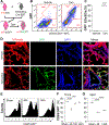
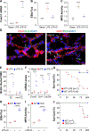
Fig. 1.. Genetic lineage tracing of ECs after polymicrobial sepsis-induced injury.
(A) Schematic illustration of the lineage-tracing strategy. Tam=Tamoxifen. (B) Flow cytometry analysis of GFP+ cells and ECs (CD45CD31+) in lungs of young adult mice. (C) Quantification of lung GFP+ ECs in young adult EndoSCL-CreERT2/mTmG mice. CD45 cells were gated for CD31+ and GFP+ analysis. (D) Representative confocal images of lungs of young adult mice showing tamoxifen treatment-induced EC labeling. Green, GFP; Red, tdTomato; Blue, DAPI. Br, bronchiole; V, vessel. Scale bar, 20 μm. (E and F) FACS analysis of GFP+ ECs at 48h and 144h in young adult mice. Lung cells were CD45 gated, and GFP+ population were quantified. (G) FACS analysis showing impaired recovery of lung GFP+ cells following CLP challenge in aged mice (19–21 mos. old). Bars represent means. ***P < 0.001, ****P < 0.0001. Unpaired two-tailed t test (C); One-way ANOVA (Dunnett) (F, G).
Fig. 2.
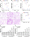
Fig. 2.. Defective endothelial proliferation and vascular repair in aged lungs following polymicrobial sepsis in mice.
(A) Representative micrographs of BrdU immunostaining showing defective EC proliferation in aged lungs. Cryosections of lungs (5 μm) collected at 96h after CLP were immunostained with anti-BrdU antibody to identify proliferating cells (green) and with anti-CD31 antibody to identify ECs (red). Nuclei were counterstained with DAPI (blue). Arrows point to proliferating ECs. Aged, 20 mos. old; young, 3 mos. old; Scale bar, 50 μm. (B) Quantification of cell proliferation in mouse lungs. Three consecutive cryosections from each mouse lung were examined, the average number of BrdU+ nuclei was used. (C) Lung vascular permeability assessed by an EBA extravasation assay. Lung tissues of aged mice were collected at indicated times after CLP for EBA assay. (D) Lung wet/dry weight ratio. At 96h after CLP, lung tissues were collected and dried at 60°C for 3 days for calculation of wet/dry ratio. (E) MPO activities in lung tissues at the indicated timepoints after CLP. MPO activity was calculated as OD460/min/g lung tissue. (F) QRT-PCR analysis showing marked increase of expression of proinflammatory genes Tnf and Il6 in lungs of aged mice at 96h after CLP compared to young mice. A.U., arbitrary units. (G) Survival rates of young (3.5 mos.) and aged (21 mos.) mice after CLP. **P < 0.01; ***P < 0.001, ****P < 0.0001. One-way ANOVA (Tukey) (B, C, E, F); Kruskal-Wallis (D). Log-rank (Mantel-Cox) test (G).
Fig. 3.
Fig. 3.. Aging impairs vascular repair and resolution of inflammatory lung injury in mice after LPS challenge.
(A) Lung vascular permeability assessed by an EBA extravasation assay of young and aged mice at the indicated timepoints after LPS challenge (i.p.). Young adult mice (3–5 mos.) were challenged with 2.5 mg/kg of LPS whereas aged mice (19–21 mos.) were challenged with 1.0 mg/kg of LPS. (B) Lung wet/dry ratios in young adult and aged mice at 72h after LPS. (C) MPO activity in young adult and aged lungs at the indicated timepoints after LPS challenge. (D) Representative micrographs of H &E staining and quantification of lung injury. Based on inflammatory cell infiltration, alveolus edema and septal thickening, each section area was assigned to an injury score from 1(normal) to 4 (severe injury) and the size of the area was quantified. Lung injury score was calculated by the sum of the percentage of the area size × its respective injury score. Scale bar 40 μm. (E) EBA flux assay in the lungs of mice at different ages, before and after LPS challenge. WT mice at indicated ages were challenged with LPS (mice at age of 3–9 mos. with 2.5 mg/kg, and at age of 12–21 mos. with 1.25 mg/kg LPS). At 72h after LPS, lungs were collected for EBA extravasation assay. (F) Lung MPO activity in mice at various ages at 72h after LPS challenge. *P < 0.05; **P < 0.01; ****P < 0.0001. Kruskal-Wallis (A); One-way ANOVA (B-D); Two-way ANOVA (Sidak) (E-F).
Fig. 4.
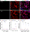
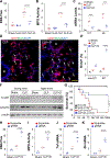
Fig. 4.. Reduced endothelial proliferation and FoxM1 induction in aged lungs after LPS challenge in mice.
(A) Representative micrographs of immunostaining showing inhibited endothelial proliferation in lungs of aged mice (20 mos.) at 72h post-LPS. Lung cryosections were immunostained with anti-BrdU (green), and anti-CD31 (red, ECs). Nuclei were counterstained with DAPI (blue). White arrows point to proliferating ECs. Br = bronchiole. Scale bar, 50 μm. (B) Quantification of cell proliferation in mouse lungs at baseline (Ctl) and 72h after LPS. (C) QRT-PCR analysis of Foxm1 expression in mouse lungs at indicated times after LPS. (D) QRT-PCR analysis of expression of genes Cdc25c, Ccna2, and Ccnb1 at 72h after LPS in the lungs of young mice and aged mice. **P < 0.01; ***P < 0.001; ****P < 0.0001. One-way ANOVA (Tukey).
Fig. 5.
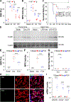
Fig. 5.. Forced expression of FoxM1 improves resolution of inflammatory lung injury and promotes survival of aged mice.
(A) EBA flux assay in 20-month-old WT and FOXM1Tg mice challenged with LPS (1 mg/kg, i.p.). (B) Lung MPO activity in aged WT and aged FOXM1Tg mice. (C) Kaplan-Meier survival curves after LPS challenge. WT mice at age of 3–5 (Young WT), or 21 mos. (Aged WT), and aged FOXM1Tg mice (21 mos.) were challenged with 1.5 mg/kg of LPS (i.p.). Survival rates were recorded for 7 days. (D) Representative Western blotting demonstrating FoxM1 expression in lungs of aged WT mice administered with FOXM1 plasmid DNA (FOX) compared to empty vector (Vec). Mixture of nanoparticles with FOXM1 plasmid DNA (CDH5 promoter) or empty vector DNA (Vec) were administered retro-orbitally to aged WT mice at 24h after LPS (1mg/kg, i.p.). Lungs were collected at 72h after LPS. Basal, mice without plasmid DNA and LPS. (EEBA flux in FOXM1 plasmid-administered mice at 72h after LPS compared to vector mice. (F) MPO activity assay in vector- and FOXM1 plasmid-adminsitered aged mice at 72h after LPS. (G) Quantification of BrdU+ nuclei of CD31+ (ECs) and CD31 (non-ECs) cells in mouse lungs at baseline and 72h after LPS. (H) Representative micrographs of anti-BrdU immunostaining (green) of mouse lungs collected at 72h after LPS. ECs were immunostained with anti-CD31 (red). Nuclei were counterstained with DAPI (blue). White arrows point to proliferating ECs. Scale bar, 50 μm. (I) QRT-PCR analysis of FoxM1 target genes Cdc25c, Ccna2, and Ccnf. *P < 0.05; **P < 0.01; ***P < 0.001; ****P < 0.0001. One-way ANOVA (Tukey) (A, B, E-I). Log-rank (Mantel-Cox) test (C).
Fig. 6.
Fig. 6.. Therapeutic activation of FoxM1-dependent endothelial regeneration by decitabine improves resolution of inflammatory lung injury and promotes survival of aged WT mice.
(A) QRT-PCR analysis demonstrating induction of FOXM1 expression in lungs of aged WT mice after decitabine treatment. 21 mos. old WT mice were challenged with LPS (1.0 mg/kg, i.p.) and then treated with either decitabine (0.2mg/kg, oral gavage) (LPS+D) or PBS (LPS) once daily at 24 and 48h after LPS. Lung tissues were collected at 96h after LPS for assays. (B) EBA flux assay in aged WT mice treated with decitabine or PBS after exposure to LPS. (C) Lung MPO activity assessment. (D) Representative micrographs of anti-BrdU staining. Lung sections were immunostained with anti-CD31 (red) and anti-BrdU (green). Nuclei were counterstained with DAPI (blue). Arrows point to BrdU+ ECs. Scale bar, 20 μm. (E) Quantification of BrdU+ ECs and non-ECs in mouse lungs. (F) QRT-CPR analysis of FoxM1 target genes in mouse lungs. (G) Kaplan-Meier survival curves of aged WT mice (21 mos.) after LPS (1.5 mg/kg, i.p.) and treated with either PBS or decitabine. (H, I) Lung vascular permeability assessed by an EBA extravasation assay (H) and lung MPO activities (I) in aged Foxm1 CKO (CKO) mice. 24 mos. old WT or Foxm1 CKO mice were challenged with 0.3 mg/kg LPS followed by Decitabine treatment at 24 and 48h. Lung tissues were collected at 96h post-LPS for EBA (H) and MPO (I) assays. (J) Kaplan-Meier survival curves of aged WT and Foxm1 CKO (CKO) mice after LPS challenge. 20 mos. old WT and Foxm1 CKO mice were challenged with LPS (1.5 mg/kg, i.p.) and then treated with decitabine at 24 and 48h post-LPS. *P < 0.05; **P < 0.01; ***P < 0.001; ****P < 0.0001. One-way ANOVA (Tukey) (A-C, H, I); Unpaired two-tailed t test (E, F); Log-rank (Mantel-Cox) test (G, J).
Fig. 7.
Fig. 7.. Decitabine treatment promotes FoxM1-mediated vascular repair and resolution of inflammatory lung injury after polymicrobial sepsis in mice.
20–21 mos. old WT mice were challenged with either CLP or sham surgery and then treated with decitabine (D) or PBS at 24 and 48h post-CLP. Lung tissues were collected at 96h after CLP. S+D=Sham+decitabine. (A) Lung vascular permeability assessed by an EBA extravasation assay. (B) lung MPO activities. (C) Quantitative RT-PCR analysis of Tnf and Il6 gene expression. (D) Representative micrographs of anti-BrdU immunostaining (green) of lung cryosections. ECs were immunostained with anti-CD31 (red). Nuclei were counterstained with DAPI (blue). White arrows point to proliferating ECs. Br = Bronchiole. Scale bar, 50 μm. (E) Quantification of BrdU+ nuclei. (F) Western blotting demonstrating expression of FoxM1 and Cdc25C in lungs of aged mice after sham surgery or CLP and treatment with decitabine. (G) Survival rates of aged WT mice after CLP. S=Sham, S+D=Sham+D. (H) Lung vascular permeability assessed by an EBA extravasation. 20 months old WT mice were administered retro-orbitally with mixture of nanoparticle:plasmid DNA expressing Cas9 under the control of CDH5 promoter and guide RNA (gRNA) or scrambled RNA (Scramble) driven by U6 promoter and 7 days later, the mice were subjected to Sham or CLP followed by decitabine treatment at 24 and 48h post-CLP (CLP+D). At 96h post-CLP, lung tissues were collected for EBA assay. (I-K) Lung MPO activity (I) and expression of Tnf and Il6 analyzed by qRT-PCR (J, K). ***P < 0.001; ****P < 0.0001. One-way ANOVA (Tukey) (A-C, E, F, H-K); Log-rank (Mantel-Cox) test (G).
Fig. 8.
Fig. 8.. Aging impaired FoxM1 expression in mice and human through hypermethylation of the promoter.
(A) Diagram presentation of MethPrimer analysis of human FOXM1 and mouse Foxm1 promoter showing a predicted conserved CpG island near the transcription start site (TSS). O/E, Observed versus Expected. (B) Methylation-specific high-resolution DNA melting assay of the Foxm1 promoter in lungs of aged mice compared to young mice at baseline. (C) Methylation-specific high-resolution DNA melting assay of the Foxm1 promoter in lungs of aged mice (21 mos.) challenged with LPS. (D) Representative micrographs of RNAscope in situ hybridization staining of human lung sections showing induction of FOXM1 expression in vascular ECs of middle-aged (50–60 years of age) COVID-19 patients but not in elderly (over 80 years old) patients. Lung autopsy tissues were collected from patients with COVID-19 and unused healthy donor lungs (normal) for paraffin-sectioning and immunostaining. Anti-CD31 antibody was used to immunostain ECs (green). FOXM1 mRNA expression (purple) was detected by RNAscope in situ hybridization. Nuclei were counterstained with DAPI (blue). White arrows point to FOXM1 expressing ECs. V, vessel. Scale bar, 50 μm. (E) Quantification of endothelial expression of FOXM1. FOXM1 expressing ECs per vessel (endothelial FOXM1+ nuclei/total endothelial nuclei of each vessel) was quantified in 15–33 vessels of each sample. Bars represent means. *P < 0.05; **P < 0.01; ****P < 0.0001, One-way ANOVA (Tukey) (B, C, E).

References

    1. Matthay MA, Ware LB, and Zimmerman GA, The acute respiratory distress syndrome. J Clin Invest. 122, 2731–2740 (2012). - PMC - PubMed
    1. Matthay MA, Zemans RL, Zimmerman GA, Arabi YM, Beitler JR, Mercat A, Herridge M, Randolph AG, and Calfee CS, Acute respiratory distress syndrome. Nat Rev Dis Primers. 5, 18 (2019). - PMC - PubMed
    1. Rubenfeld GD, Caldwell E, Peabody E, Weaver J, Martin DP, Neff M, Stern EJ, and Hudson LD, Incidence and outcomes of acute lung injury. N Engl J Med. 353, 1685–93 (2005). - PubMed
    1. Aird WC, The role of the endothelium in severe sepsis and multiple organ dysfunction syndrome. Blood. 101, 3765–77 (2003). - PubMed
    1. Goldenberg NM, Steinberg BE, Slutsky AS, and Lee WL, Broken barriers: a new take on sepsis pathogenesis. Sci Transl Med. 3, 88ps25 (2011). - PubMed

Publication types