. 2023 Aug 16;15(709):eabq0603.
doi: 10.1126/scitranslmed.abq0603. Epub 2023 Aug 16.

Polymer nanoparticles deliver mRNA to the lung for mucosal vaccination

Affiliations

Polymer nanoparticles deliver mRNA to the lung for mucosal vaccination

Alexandra Suberi et al. Sci Transl Med. .

Abstract

An inhalable platform for messenger RNA (mRNA) therapeutics would enable minimally invasive and lung-targeted delivery for a host of pulmonary diseases. Development of lung-targeted mRNA therapeutics has been limited by poor transfection efficiency and risk of vehicle-induced pathology. Here, we report an inhalable polymer-based vehicle for delivery of therapeutic mRNAs to the lung. We optimized biodegradable poly(amine-co-ester) (PACE) polyplexes for mRNA delivery using end-group modifications and polyethylene glycol. These polyplexes achieved high transfection of mRNA throughout the lung, particularly in epithelial and antigen-presenting cells. We applied this technology to develop a mucosal vaccine for severe acute respiratory syndrome coronavirus 2 and found that intranasal vaccination with spike protein-encoding mRNA polyplexes induced potent cellular and humoral adaptive immunity and protected susceptible mice from lethal viral challenge. Together, these results demonstrate the translational potential of PACE polyplexes for therapeutic delivery of mRNA to the lungs.


Conflict of interest statement

Competing interests: A.S.P.-D., A.I., and W.M.S. are cofounders of Xanadu Bio, and B.I. and T.M. serve as consultants for Xanadu Bio. A.I., B.I., T.M., W.M.S., A.S., M.R., A.S.P.-D., and H.-W.S. are listed as inventors on patent applications relating to intranasal PACE SARS-CoV-2 vaccines filed by Yale University (Nanoparticle immunogenic compositions and vaccination methods, PCT/US2022/081199). W.M.S. is listed as inventor on patent applications relating to end group–modified PACE NPs filed by Yale University [Poly(amine-co-ester) polymers and polyplexes with modified end groups and methods of use thereof, 16/862,491]. A.S., M.K.G., and W.M.S. are listed as inventors on patent applications relating to PACE-mRNA delivery to the lung filed by Yale University [Poly(amine-co-ester) polymers with modified end groups and enhanced pulmonary delivery, 18/002,241].

Figures

Fig. 1.
Fig. 1.
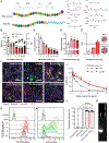
Characterization of PACE-mRNA polyplexes and in vitro activity. (A) Schematic of end group–modified and PEGylated PACE polymer composition with chemical structures of base monomers and end groups. PDL, 15-pentadecanolide; MDEA, N-methyl diethanolamine; SA, sebacic acid. (B to E) Shown are size and polydispersity index (PDI) (B), zeta potential (C), mRNA loading (D), and PEG conformation on the surface of PACE-E14 polyplexes encapsulating EGFP mRNA with varying PACE-PEG content (E). RF represents PEG Flory radius, and D represents the distance between PEG chains. Asterisks indicate statistical difference in size from non-PEGylated polyplexes. Data were analyzed using two-way (A and B) or one-way (C and D) analysis of variance (ANOVA) with Dunnet’s multiple comparisons test. (F) Representative mRNA uptake and transfection efficiency of cyanine 5 (Cy5)–conjugated EGFP mRNA delivered with PACE-PEG–blended polyplexes or LipoMM (scale bars, 75 μm; Hoechst in blue, phalloidin in gray, Cy5-mRNA in red, and EGFP in green). Naked mRNA delivery to cells is shown as a control. (G and H) Uptake of Cy5-conjugated mRNA (G) and transfection efficiency of EGFP mRNA (H) are shown for human alveolar epithelial cells (A549) incubated with PEGylated PACE-E14. mRNA indicates cells treated with naked mRNA. (I) In vitro cytotoxicity of PEGylated PACE-E14 polyplexes was compared with LipoMM. Asterisks indicate differences between all PACE polyplexes and LipoMM. Data were analyzed using two-way ANOVA with Tukey’s multiple comparisons test. (J and K) Shown are transfection efficiency of EGFP mRNA PACE-E14 polyplexes in human embryonic kidney–293 cells with or without coincubation of RNase (J) and a gel run with either naked mRNA or naked mRNA and RNase showing degradation of mRNA by the enzyme (K). Data were analyzed using one-way ANOVA with Tukey’s multiple comparisons test. *P ≤ 0.05, **P ≤ 0.01, ***P ≤ 0.001, and ****P ≤ 0.0001; ns, not significant. Data are presented as means ± SD. Data are pooled from three independent experiments with one batch of polyplexes per triplicate measurement and n = 2 wells per treatment group.
Fig. 2.
Fig. 2.
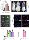
IT mRNA delivery with PACE-mRNA polyplexes drives protein expression and mRNA distribution throughout the airways and parenchyma. (A) Luciferase protein expression was measured in lung tissue after delivery of 5 μg of FLuc mRNA with PEGylated PACE-E14 and in vivo-jetPEI. RLU, relative light units. (B and C) Representative luciferase expression by IVIS 24 hours after IT delivery of PACE-E14 10% PACE-PEG FLuc mRNA polyplexes in animals (B) and in explanted organs (C). Average radiance units of photons per second per square centimeter per steradian (p/s/cm2/sr). (D) Distribution of Cy5-conjugated mRNA in the lung 30 min after delivery with PACE-E14 polyplexes (scale bars, 150 μm). (E) Luciferase protein expression was measured in lung tissue after delivery of FLuc mRNA with polyplexes of various end group–modified PACE and 10% PACE-PEG content, untreated controls, naked mRNA controls, and base polymer with no end-group controls (n = 3 to 5 per group). (F) Quantification of luciferase protein extracted from lungs 24 hours after treatment with either E14 or E27 polyplexes with 10% PACE-PEG. Data were analyzed by one-way ANOVA with Tukey’s multiple comparisons test of log-transformed sample mean values. Data are presented as means ± SD. Data are pooled from two independent experiments (n = 3 to 6 mice per group). **P ≤ 0.01 and ****P ≤ 0.0001.
Fig. 3.
Fig. 3.
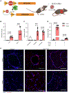
mRNA expression after IT PACE-mRNA delivery occurs in epithelial cells and antigen-presenting cells. (A) Schematic of Cre-mediated recombination in Ai14 mice, resulting in expression of tdTomato protein in transfected cells. (B to D) Shown are the percent of all live cells in the lung and BALF (B); endothelial, epithelial, or leukocyte cells in the lung (C); and antigen-presenting cells (D) in the lung that expressed tdTomato 24 hours after administration of PACE-E14 polyplexes (10% PACE-PEG) loaded with 10 μg of Cre mRNA. Data are presented as means ± SD. Statistical significance was calculated by multiple unpaired t test with Holm-Sidak method. ***P ≤ 0.001 and ****P ≤ 0.0001. Data are pooled from two independent experiments (n = 4 to 6 per group). (E) Shown are representative images of control (untreated) and PACE-E14 polyplex (10% PACE-PEG)–treated lungs from Ai14 mice by fluorescence microscopy [scale bars, 100 μm; 4′,6-diamidino-2-phenylindole (DAPI) in blue; tdTomato in red].
Fig. 4.
Fig. 4.
Intranasal PACE-mRNA vaccination induces antigen-specific T and B cell responses in the draining lymph node. (A) Schematic of PACE-mRNA vaccination in K18-hACE2 mice. Mice were primed (day 0) and boosted (day 28) with a 10-μg dose of spike protein–encoding mRNA encapsulated in PACE-E14 polyplexes with 10% PACE-PEG (n = 10 to 12 mice per cohort). MLNs were harvested on day 42 for analysis. (B) Quantification of extravascular (IV) SARS-CoV-2 spike protein–specific (Tetramer+) CD8+ T cells in MLNs. (C) Quantification of extravascular (IV) CXCR5+PD1+ TFH cells in MLNs. (D to I) Quantification of various extravascular B cell subsets, including RBD tetramer-binding B cells (Tetramer+ B cells) (D), class-switched B cells (IgDIgM CS B cells) (E), IgA+ memory B cells (F), IgG+ memory B cells (G), activated germinal center B cells (GL7+ GC B cells) (H), and antibody-secreting cells in MLNs (CD138+ ASC) (I). Data are presented as means ± SEM. Statistical significance was calculated by Mann-Whitney test. **P ≤ 0.01, ***P ≤ 0.001, and ****P ≤ 0.0001. Data are pooled from two independent experiments.
Fig. 5.
Fig. 5.
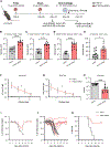
Intranasal PACE-mRNA vaccination induces protective cellular and humoral immunity. (A) Schematic of PACE-mRNA vaccination in K18-hACE2 mice. Mice were primed (day 0) and boosted (day 28) with a 10-μg dose of spike protein–encoding mRNA encapsulated in PACE-E14 polyplexes with 10% PACE-PEG. The lungs, sera, and BALF were harvested on day 42 for analysis (n = 10 to 12 mice per group). Additional groups of vaccinated animals (n = 8 to 10 mice per group) were challenged with 6 × 10³ PFU of SARS-CoV-2 on day 56. One group was used for viral titer measurement in lung tissues at 3 DPI. A second group was used to evaluate weight loss and survival over 2 weeks compared with untreated naïve mice. (B to D) Shown is quantification of extravascular (IV) SARS-CoV-2 spike protein–specific (Tetramer+) CD8+ T cells (B), CD69+CD103Tetramer+ CD8+ T cells (C), and CD69+CD103+Tetramer+ CD8+ T cells in the lung (D). (E) Quantification of circulating (IV+) SARS-CoV-2 spike protein–specific, Tetramer+ CD8+ T cells from the lung vasculature. (F and G) Shown is the serum (F) and BALF (G) abundance of SARS-CoV-2 S1 subunit–specific IgG. O.D., optical density. (H) Shown are infectious viral titers in lung tissues at 3 DPI as measured by plaque assay. (I and J) Shown are average (I) and individual (J) weight measurements after viral challenge in naïve and PACE-mRNA–vaccinated mice. (K) Shown is survival of naïve and vaccinated mice from 1 to 14 DPI. Data are presented as means ± SEM. Statistical significance was calculated by Mann-Whitney test (B to H) or log-rank Mantel-Cox test (K). *P ≤ 0.05, **P ≤ 0.01, ***P ≤ 0.001, and ****P ≤ 0.0001. Data are pooled from two independent experiments.

Update of

Comment in

  • Delivering mRNA to the lungs.
    Crunkhorn S. Crunkhorn S. Nat Rev Drug Discov. 2023 Oct;22(10):787. doi: 10.1038/d41573-023-00142-5. Nat Rev Drug Discov. 2023. PMID: 37666973 No abstract available.

References

    1. Polack FP, Thomas SJ, Kitchin N, Absalon J, Gurtman A, Lockhart S, Perez JL, Pérez Marc G, Moreira ED, Zerbini C, Bailey R, Swanson KA, Roychoudhury S, Koury K, Li P, Kalina WV, Cooper D, Frenck RW Jr., Hammitt LL, Türeci Ö, Nell H, Schaefer A, Ünal S, Tresnan DB, Mather S, Dormitzer PR, Şahin U, Jansen KU, Gruber WC, Safety and efficacy of the BNT162b2 mRNA Covid-19 vaccine. N. Engl. J. Med. 383, 2603–2615 (2020). - PMC - PubMed
    1. Baden LR, El Sahly HM, Essink B, Kotloff K, Frey S, Novak R, Diemert D, Spector SA, Rouphael N, Creech CB, McGettigan J, Khetan S, Segall N, Solis J, Brosz A, Fierro C, Schwartz H, Neuzil K, Corey L, Gilbert P, Janes H, Follmann D, Marovich M, Mascola J, Polakowski L, Ledgerwood J, Graham BS, Bennett H, Pajon R, Knightly C, Leav B, Deng W, Zhou H, Han S, Ivarsson M, Miller J, Zaks T, Efficacy and safety of the mRNA-1273 SARS-CoV-2 vaccine. N. Engl. J. Med. 384, 403–416 (2021). - PMC - PubMed
    1. Pardi N, Hogan MJ, Porter FW, Weissman D, mRNA vaccines - a new era in vaccinology. Nat. Rev. Drug Discov. 17, 261–279 (2018). - PMC - PubMed
    1. Kowalski PS, Rudra A, Miao L, Anderson DG, Delivering the messenger: Advances in technologies for therapeutic mRNA delivery. Mol. Ther. 27, 710–728 (2019). - PMC - PubMed
    1. Piotrowski-Daspit AS, Kauffman AC, Bracaglia LG, Saltzman WM, Polymeric vehicles for nucleic acid delivery. Adv. Drug Deliv. Rev. 156, 119–132 (2020). - PMC - PubMed

Publication types