. 2023 Aug 1;164(9):bqad124.
doi: 10.1210/endocr/bqad124.

Luteal Lipid Droplets: A Novel Platform for Steroid Synthesis

Affiliations

Luteal Lipid Droplets: A Novel Platform for Steroid Synthesis

Michele R Plewes et al. Endocrinology. .

Abstract

Progesterone is an essential steroid hormone that is required to initiate and maintain pregnancy in mammals and serves as a metabolic intermediate in the synthesis of endogenously produced steroids, including sex hormones and corticosteroids. Steroidogenic luteal cells of the corpus luteum have the tremendous capacity to synthesize progesterone. These specialized cells are highly enriched with lipid droplets that store lipid substrate, which can be used for the synthesis of steroids. We recently reported that hormone-stimulated progesterone synthesis by luteal cells requires protein kinase A-dependent mobilization of cholesterol substrate from lipid droplets to mitochondria. We hypothesize that luteal lipid droplets are enriched with steroidogenic enzymes and facilitate the synthesis of steroids in the corpus luteum. In the present study, we analyzed the lipid droplet proteome, conducted the first proteomic analysis of lipid droplets under acute cyclic adenosine monophosphate (cAMP)-stimulated conditions, and determined how specific lipid droplet proteins affect steroidogenesis. Steroidogenic enzymes, cytochrome P450 family 11 subfamily A member 1 and 3 beta-hydroxysteroid dehydrogenase (HSD3B), were highly abundant on lipid droplets of the bovine corpus luteum. High-resolution confocal microscopy confirmed the presence of active HSD3B on the surface of luteal lipid droplets. We report that luteal lipid droplets have the capacity to synthesize progesterone from pregnenolone. Lastly, we analyzed the lipid droplet proteome following acute stimulation with cAMP analog, 8-Br-cAMP, and report increased association of HSD3B with luteal lipid droplets following stimulation. These findings provide novel insights into the role of luteal lipid droplets in steroid synthesis.

Keywords: bovine; corpus luteum; lipid droplets; mitochondria; progesterone; protein kinase A; steroidogenesis.


Figures

Figure 1.
Figure 1.
Proteome of the luteal lipid droplet. Lipid droplet proteins associated with the corpus luteum were grouped into molecular processes based on GO analysis and UNIPROT functional analysis using ShinyGO analysis. Proteins are grouped into functional modules. Unmapped represent proteins that did not group into representative functional modules.
Figure 2.
Figure 2.
Validation of proteins associated with the lipid droplets of the bovine corpus luteum. Lipid droplets from punch biopsies obtained from bovine corpora lutea were isolated by gradient ultracentrifugation and subject to Western blotting. A, Representative transmission electron micrograph of isolated lipid droplet fraction following successful enrichment using ultracentrifugation. B, Representative Western blotting of essential steroidogenic proteins expressed in enriched bovine luteal lipid droplets. Micron bar represents 2 µm. Perilipin 2 (PLIN2); perilipin 3 (PLIN3); steroidogenic acute regulatory protein (STAR); vimentin (VIM); cholesterol side-chain cleavage enzyme (CYP11A1); 3beta-hydroxysteroid dehydrogenase (HSD3B); cytochrome c oxidase subunit 4 (COX IV); heat shock protein 47 (HSP47); beta tubulin (TUBB; loading control).
Figure 3.
Figure 3.
Subcellular localization of 3beta-hydroxysteroid dehydrogenase (HSD3B) in steroidogenic small luteal cells. Populations of small luteal cells were enriched from bovine luteal tissue and visualized using confocal microscopy. A, Representative confocal micrograph of HSD3B (red) and luteal lipid droplets (green), calnexin (endoplasmic reticulum; green), or TOM20 (mitochondria; green). White boxes represent an area of 10× enlargement. White dashed lines represent the area of intensity overlap. B, Representative quantitative analysis of intensity overlap. Arrows represent spatial location of lipid droplet. TOM20 (streptavidin, Alexa Fluor 647); calnexin (Alexa Fluor 594); lipid droplet (BODIPY493/503), and HSD3B (DyLight 405). Micron bar represents 20 and 2 µm, respectively.
Figure 4.
Figure 4.
Subcellular localization of 3beta-hydroxysteroid dehydrogenase (HSD3B) in steroidogenic large luteal cells. Enriched populations of large luteal cells from bovine luteal tissue were visualized using confocal microscopy. A, Representative confocal micrograph of HSD3B (red) and luteal lipid droplets (green), calnexin (endoplasmic reticulum; green), or TOM20 (mitochondria; green). White boxes represent an area of 10× enlargement. White dashed lines represent the area of intensity overlap. B, Representative quantitative analysis of intensity overlap. Arrows represent spatial location of lipid droplet. TOM20 (streptavidin, Alexa Fluor 647); calnexin (Alexa Fluor 594); lipid droplet (BODIPY493/503), and HSD3B (DyLight 405). Micron bar represents 20 and 2 µm, respectively.
Figure 5.
Figure 5.
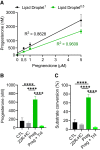
Isolated lipid droplets can synthesize the steroid hormone progesterone. Lipid droplets from tissue obtained from bovine corpora lutea were isolated by gradient ultracentrifugation. Lipid droplets were harvested and aliquoted into equal volumes (500 μL) for immediate treatment following centrifugation. Following incubation, samples were immediately placed at −80 °C until further isolation of steroids. A, Progesterone obtained from isolated lipid droplets (Lipid droplet1 and Lipid droplet0.5 [50% of Lipid droplet1]) incubated with either control buffer (TE buffer supplemented with nicotinamide adenine dinucleotide (NAD+; final concentration of 1 mM) and increasing concentrations of pregnenolone (final concentration of 0.5-5 µM). B, Progesterone obtained from isolated luteal lipid droplets incubated with either control buffer, 22-hydroxycholesterol (final concentration of 1 µM), NADPH (1 mM), and NAD+ (1 mM); pregnenolone (final concentration of 1 µM) and NAD+ (1 mM), or pregnenolone (1 µM), Trilostane (inhibitor of HSD3B; 1 µM) and NAD+ (1 µM). Bars represent means ± SEM, n = 3-6. Statistically significant difference between treatments, ****P less than .0001.
Figure 6.
Figure 6.
Proteomic analysis of luteal lipid droplets following acute stimulation with 8-Br-cAMP. Tissue punch biopsies obtained from bovine corpora lutea were stimulated with 8-Br-cAMP (1 mM) for 30 minutes (n = 3). Following stimulation, lipid droplets from each set of punch biopsies were isolated by gradient ultracentrifugation and subject to mass spectrometry. A, Diagram of experimental design. B, Number of identified upregulated or downregulated proteins. Upregulated (red) and downregulated (blue). C, Top enriched canonical pathways analyzed by ingenuity pathway analysis (IPA) of lipid droplet-associated proteins modulated by acute stimulation with 8-Br-cAMP. Protein annotation through evolutionary relationship (PANTHER) classification system was used to classify biological processes, molecular functions, and protein groups. D, Upregulated protein classes (red bars) in 8-Br-cAMP–stimulated lipid droplets. E, Downregulated protein classes (blue bars) in 8-Br-cAMP–stimulated lipid droplets. F, ShinyGO analysis of top molecular functions following stimulation with 8-Br-cAMP treatment. Lipid droplets from punch biopsies treated with 8-Br-cAMP were isolated by gradient ultracentrifugation and subject to Western blotting. G, Representative Western blotting of PLIN2 and HSD3B expressed in enriched bovine luteal lipid droplets following 30 minutes’ stimulation with 8-Br-cAMP. Postnuclear supernatant depleted of lipid droplets (PNS-LD). H, Densitometric analyses of HSD3B. Data are means ± SE, n = 3. Bars represent means ± SEM, n = 3. Statistically significant difference between treatment, **P less than .01.

References

    1. Kolatorova L, Vitku J, Suchopar J, Hill M, Parizek A. Progesterone: a steroid with wide range of effects in physiology as well as human medicine. Int J Mol Sci. 2022;23(14):7989. - PMC - PubMed
    1. Quinkler M, Meyer B, Bumke-Vogt C, et al. . Agonistic and antagonistic properties of progesterone metabolites at the human mineralocorticoid receptor. Eur J Endocrinol. 2002;146(6):789‐799. - PubMed
    1. Azeez JM, Susmi TR, Remadevi V, Ravindran V, Sujatha AS, Sreeja S. New insights into the functions of progesterone receptor (PR) isoforms and progesterone signaling. Am J Cancer Res. 2021;11(11):5214. - PMC - PubMed
    1. Takao Y, Honda T, Ueda M, et al. . Immunohistochemical localization of the LH/HCG receptor in human ovary: HCG enhances cell surface expression of LH/HCG receptor on luteinizing granulosa cells in vitro. Mol Hum Reprod. 1997;3(7):569‐578. - PubMed
    1. Marsh JM. The role of cyclic AMP in gonadal steroidogenesis. Biol Reprod. 1976;14(1):30‐53. - PubMed

Publication types