. 2023 Aug 15;4(8):101156.
doi: 10.1016/j.xcrm.2023.101156.

Bioengineered human tissue regeneration and repair using endogenous stem cells

Affiliations

Bioengineered human tissue regeneration and repair using endogenous stem cells

Jiao Wei et al. Cell Rep Med. .

Abstract

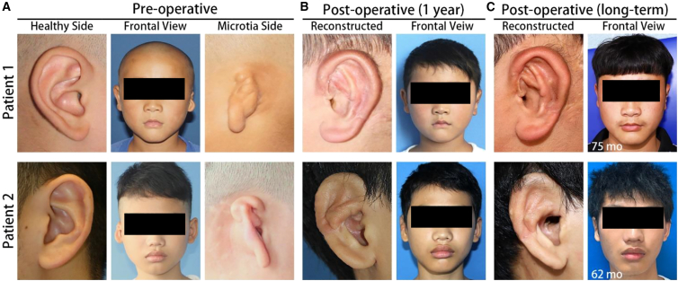
We describe a general approach to produce bone and cartilaginous structures utilizing the self-regenerative capacity of the intercostal rib space to treat a deformed metacarpophalangeal joint and microtia. Anatomically precise 3D molds were positioned on the perichondro-periosteal or perichondral flap of the intercostal rib without any other exogenous elements. We find anatomically precise metacarpal head and auricle constructs within the implanted molds after 6 months. The regenerated metacarpal head was used successfully to surgically repair the deformed metacarpophalangeal joint. Auricle reconstructive surgery in five unilateral microtia patients yielded good aesthetic and functional results. Long-term follow-up revealed the auricle constructs were safe and stable. Single-cell RNA sequencing analysis reveal early infiltration of a cell population consistent with mesenchymal stem cells, followed by IL-8-stimulated differentiation into chondrocytes. Our results demonstrate the repair and regeneration of tissues using only endogenous factors and a viable treatment strategy for bone and tissue structural defects.

Keywords: auricular reconstruction; cartilage regeneration; endogenous stem cells; joint reconstruction; regeneration medicine; tissue regeneration.


Conflict of interest statement

Declaration of interests The authors declare no competing interests.

Figures

None
Graphical abstract
Figure 1
Figure 1
Auricle cartilaginous construct regeneration strategy and timeline (A) Schematic illustration of MP joint construct fabrication and auricular reconstruction. (1) osteochondral flap was elevated from the 7th to 8th costal cartilage. (2) A finger-shaped HA mold was sutured on the cambial surface of the osteochondral flap to initiate tissue regeneration within the mold. (3) The regenerated joint construct was used for MP joint reconstructive surgery. (B) Images show (from left to right) the HA molds for the metacarpal head and phalanx base, the elevated and interconnected osteochondral flap in the patient, and the HA molds sutured onto the osteochondral flap in situ. (C) MRI scan of the HA molds in situ within the patient’s chest cavity.
Figure 2
Figure 2
MP regeneration and reconstruction (A) Transplantation of the head of the third metacarpal construct into the middle finger of the patient. Existing bone is connected to the MP construct using a titanium plate. (B) CT scan of patient’s middle MP joint before and after surgery, demonstrating good alignment of the MP construct. (C) Functional characterization of the middle finger MP joint. Prior to surgery, the middle finger MP joint was able to flex 5 degrees. Following successful transplantation of the MP construct, the flexion angle was increased to 32 degrees. (D) The base of the proximal phalanx (not used in the surgery) in situ within the chest cavity. H&E stain of sections of the base of the third proximal phalanx shows articular cartilaginous tissue in the head of the bone, fused to subchondral bone characterized by a hollow center filled with bone marrow.
Figure 3
Figure 3
Auricle cartilaginous constructs (A–C) Images (A) show ear-shaped cartilaginous constructs in patient chest wall 6 to 8 months following implantation of the HA mold. Images are taken following removal of the mold. H&E (B) and Alcian blue (C) histological analysis of excess cartilage trimmed from regenerated cartilaginous constructs prior to surgery. (D) Boxplot of histological grading of native and regenerated cartilage. The boxplot was constructed using median (line), interquartile range (box), and minimum/maximum (error bars). A Mann-Whitney U-test found a statistically significant reduction in the histological score in the regenerated cartilage (p = 0.041, n = 5). (E) Graph shows mean (±SEM) of the modulus of elasticity for regenerated cartilage versus native cartilage. There was no statistically significant difference in elasticity (p = 0.094, t test).
Figure 4
Figure 4
Clinical outcomes of the reconstructed ears in patients 1 and 2 (A) Pre-operative images of the healthy ears, microtia ears, and the frontal view of two of the participants in this study. (B) 12-month post-operative images of the reconstructed ears and the frontal view of the patients. (C) Post-operative images of the reconstructed ears and the frontal view of the patients at the last follow-up.
Figure 5
Figure 5
Swine chondrogenesis model (A) H&E staining of perichondrium and neo-tissue between 1 day and 6 months. Middle panel shows enlarged area indicated by the box. Bottom panel is Masson staining of the same area marked by the box. Scale bars: 100 μm. (B) Elasticity of the recovered cartilaginous tissue at 1, 3, and 6 months. Graph shows mean (±SEM) of the modulus of elasticity, normalized to that of the contralateral control cartilage tissue. There was a statistically significant increase in the modulus of elasticity between 6 months and 1 month (one-way ANOVA p = 0.011; post hoc Bonferroni’s comparison between 1 month and 6 months p < 0.05). It is also noteworthy that the normalized modulus of elasticity at 6 months is close to 100%.
Figure 6
Figure 6
Model of cartilage self-regeneration Our data suggest that guided endogenous cartilage regeneration involved an initiation of inflammation and recruitment of immune cells, followed by recruitment of mesenchymal stem cells into the engineered space. These then differentiate into proliferating chondrogenic progenitor cells and ultimately chondrocytes, which lay down the extracellular matrices consistent with cartilage tissue.

References

    1. Brooks P.M. The burden of musculoskeletal disease--a global perspective. Clin. Rheumatol. 2006;25:778–781. - PubMed
    1. Lawrence R.C., Felson D.T., Helmick C.G., Arnold L.M., Choi H., Deyo R.A., Gabriel S., Hirsch R., Hochberg M.C., Hunder G.G., et al. Estimates of the prevalence of arthritis and other rheumatic conditions in the United States. Part II. Arthritis Rheum. 2008;58:26–35. - PMC - PubMed
    1. Helmick C.G., Felson D.T., Lawrence R.C., Gabriel S., Hirsch R., Kwoh C.K., Liang M.H., Kremers H.M., Mayes M.D., Merkel P.A., et al. Estimates of the prevalence of arthritis and other rheumatic conditions in the United States. Part I. Arthritis Rheum. 2008;58:15–25. - PubMed
    1. Klein J. Chemistry. Repair or replacement--a joint perspective. Science. 2009;323:47–48. - PubMed
    1. Atala A., Bauer S.B., Soker S., Yoo J.J., Retik A.B. Tissue-engineered autologous bladders for patients needing cystoplasty. Lancet. 2006;367:1241–1246. - PubMed

Publication types

LinkOut - more resources